During endochondral ossification, chondrocytes
Chondrocyte
Chondrocytes are the only cells found in healthy cartilage. They produce and maintain the cartilaginous matrix, which consists mainly of collagen and proteoglycans. Although the word chondroblast is commonly used to describe an immature chondrocyte, the term is imprecise, since the progenitor of chondrocytes can differentiate into various cell types, including osteoblasts.
What happens to chondrocytes during ossification?
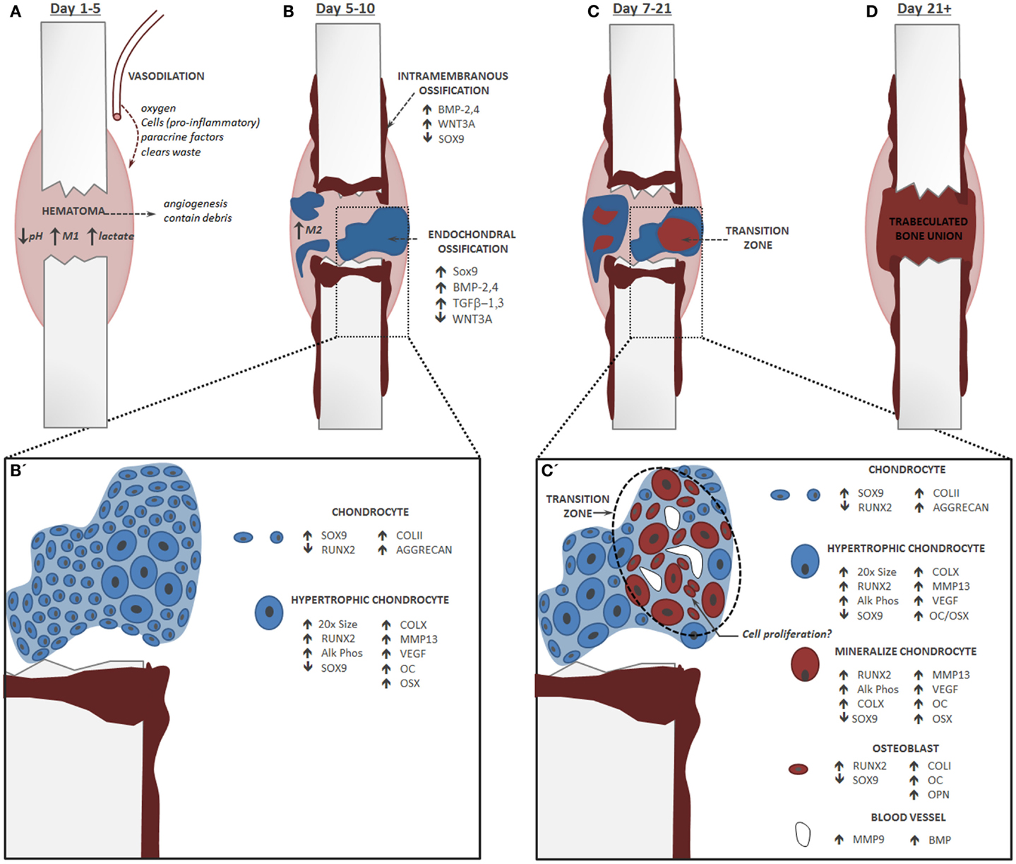
May 22, 2020 · During endochondral ossification, chondrocytes proliferate, undergo hypertrophy and die; the cartilage extracellular matrix they construct is then invaded by blood vessels, osteoclasts, bone marrow cells and osteoblasts, the last of which deposit bone on remnants of cartilage matrix.
What happens during endochondral ossification?
蠟 Click to see full answer. Accordingly, what occurs during endochondral ossification? 蠟 Endochondral ossification is the process by which growing cartilage is systematically replaced by bone to form the growing skeleton. The chondrocyte columns are then invaded by metaphyseal blood vessels, and bone forms on the residual columns of calcified cartilage.
What factors regulate the behaviour of chondrocytes in cartilage?
During endochondral ossification, chondrocytes proliferate, undergo hypertrophy and die; the cartilage extracellular matrix they construct is then invaded by blood vessels, osteoclasts, bone marrow cells and osteoblasts, the last of which deposit bone on remnants of cartilage matrix. The sequential changes in chondrocyte behaviour are tightly regulated by both systemic factors …
Are chondrocytes terminally differentiated cells?
Mar 21, 2022 · During endochondral ossification, terminally differentiated hypertrophic chondrocytes are eventually completely removed from the initial cartilage template or growth plates. ... To evaluate whether the growth plate chondrocytes during postnatal growth also contribute to the osteoblast pool in the primary spongiosa as is the case during ...
What happens to chondrocytes during ossification?
What happens to the chondrocytes after they proliferate?
What are the 5 steps of endochondral ossification?
- Cartilage enlarges; Chondrocytes die.
- blood vessels grow into perichondrium; cells convert to osteoblasts; shaft becomes covered with superficial bone.
- more blood supply and osteoblasts; produces spongy bone; formation spreads on shaft.
- Osteoclasts create medullary cavity; appositional growth.
How does endochondral ossification occur?
This process involves the replacement of hyaline cartilage with bone. It begins when mesoderm-derived mesenchymal cells differentiate into chondrocytes. Chondrocytes proliferate rapidly and secrete an extracellular matrix to form the cartilage model for bone.May 8, 2021
What is the difference between Endochondral and intramembranous ossification?
What happens during ossification?
What is the correct order of events for endochondral ossification?
What happens during bone remodeling?
Problem 22 Medium Difficulty
In endochondral ossification, what happens to the chondrocytes? a. They develop into osteocytes. b. They die in the calcified matrix that surrounds them and form the medullary cavity. c. They grow and form the periosteum. d. They group together to form the primary ossification center.
Video Transcript
So this question is asking what happens to Contra sites in endo Conjugal ossification and conjure sites are these cartilage cells and endo contre la ossification is the redevelopment of bone via the replacement of highland cartilage.
What is the process of endochondral ossification?
Endochondral ossification is one of the two essential processes during fetal development of the mammalian skeletal system by which bone tissue is created. Unlike intramembranous ossification, which is the other process by which bone tissue is created, cartilage is present during endochondral ossification.
Is cartilage present during endochondral ossification?
Unlike intramembranous ossification, which is the other process by which bone tissue is created, cartilage is present during endochondral ossification. Endochondral ossification is also an essential process during the rudimentary formation of long bones, the growth of the length of long bones, and the natural healing of bone fractures.
Where does ossification occur?
The first site of ossification occurs in the primary center of ossification, which is in the middle of diaphysis (shaft). Then: The perichondrium becomes the periosteum. The periosteum contains a layer of undifferentiated cells (osteoprogenitor cells) which later become osteoblasts. The osteoblasts secrete osteoid against the shaft ...
Where is the secondary ossification center?
Secondary center of ossification. About the time of birth in mammals, a secondary ossification center appears in each end (epiphysis) of long bones. Periosteal buds carry mesenchyme and blood vessels in and the process is similar to that occurring in a primary ossification center.
What is the point of union of the primary and secondary ossification centers?
The point of union of the primary and secondary ossification centers is called the epiphyseal line.
What is the zone of ossification?
Zone of ossification. Osteoprogenitor cells invade the area and differentiate into osteoblasts, which elaborate matrix that becomes calcified on the surface of calcified cartilage. This is followed by resorption of the calcified cartilage/calcified bone complex.
How does cartilage grow?
The cartilage model will grow in length by continuous cell division of chondrocytes, which is accompanied by further secretion of extracellular matrix. This is called interstitial growth. The process of appositional growth occurs when the cartilage model also grows in thickness due to the addition of more extracellular matrix on ...
What happens to chondrocytes during endochondral ossification?
During endochondral ossification, chondrocytes proliferate, undergo hypertrophy and die; the cartilage extracellular matrix they construct is then invaded by blood vessels, osteoclasts, bone marrow cells and osteoblasts, the last of which deposit bone on remnants of cartilage matrix.
What is endochondral ossification?
Endochondral ossification is the process by which the embryonic cartilaginous model of most bones contributes to longitudinal growth and is gradually replaced by bone. During endochondral ossification, chondrocytes proliferate, undergo hypertrophy and die; the cartilage extracellular matrix they construct is then invaded by blood vessels, ...
Which factors regulate chondrocytes?
The sequential changes in chondrocyte behaviour are tightly regulated by both systemic factors and locally secreted factors, which act on receptors to effect intracellular signalling and activation of chondrocyte-selective transcription factors.
What are the factors that regulate the behaviour of chondrocytes in cartilage?
Systemic factors that regulate the behaviour of chondrocytes in growth cartilage include growth hormone and thyroid hormone, and the local secreted factors include Indian hedgehog, parathyroid hormone-related peptide, fibroblast growth factors and components of the cartilage extracellular matrix.
Which family of proteins is responsible for the invasion of cartilage?
The invasion of cartilage matrix by the ossification front is dependent on its resorption by members of the matrix metalloproteinase family, as well as the presence of blood vessels and bone-resorbing osteoclasts.
What is the function of the notch signaling pathway?
Like, the canonical Wnt pathway, the functional roles of Notch signaling suggest it as a candidate regulator of chondrocyte-to-osteoblast transformation. Activation of this pathway begins when the Notch transmembrane receptor binds to membrane-bound ligands (Delta or Jagged) on the surface of neighboring cells. This triggers the proteolytic cleavage of the Notch intracellular domain (NICD) by y-secretase. NICD then translocates to the nucleus where it forms a complex with and activates the transcription factor CSL, which recruits its co-activator Mastermind-like (MAML) and initiates transcription of target genes ( Lin and Hankenson, 2011) (Figure 1C ).
What is the WNT pathway?
Wnt signaling is traditionally categorized into the β-catenin-dependent canonical pathway and the β-catenin-independent non-canonical pathways (planar cell polarity and Ca 2+ -mediated pathways), as recently reviewed ( Gammons and Bienz, 2018 ). While some evidence suggests that the non-canonical pathways may play a role in regulating osteogenesis ( Chen et al., 2007 ), the canonical Wnt/β-catenin pathway is the most studied and has been shown to play a dominant role in bone development and fracture repair. Thus, this review focuses on the canonical Wnt pathway.
What is hedgehog signaling?
This results in the proteolytic processing of Gli transcription factors into a repressor form (GliR). GliR then enters the nucleus and prevents Hedgehog target gene expression. Hedgehog signaling is activated by the binding of Hh ligands to Patched, thus relieving Patched-mediated suppression of Smoothened through Patched endocytosis. Smoothened enters the primary cilia where it prevents Gli transcription factors from being processed. Thus, Gli remains in its full-length, active form (GliA), which translocates to the nucleus and activates expression of Hedgehog target genes ( Lin and Hankenson, 2011) (Figure 1D ).
Overview
Growth of the cartilage model
The cartilage model will grow in length by continuous cell division of chondrocytes, which is accompanied by further secretion of extracellular matrix. This is called interstitial growth. The process of appositional growth occurs when the cartilage model also grows in thickness due to the addition of more extracellular matrix on the peripheral cartilage surface, which is accompanied by new chondroblasts that develop from the perichondrium.
Primary center of ossification
The first site of ossification occurs in the primary center of ossification, which is in the middle of diaphysis (shaft). Then:
1. Formation of periosteum
2. Formation of bone collar
3. Calcification of matrix
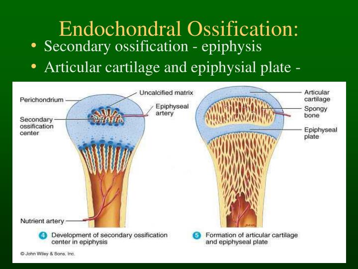
Secondary center of ossification
About the time of birth in mammals, a secondary ossification center appears in each end (epiphysis) of long bones. Periosteal buds carry mesenchyme and blood vessels in and the process is similar to that occurring in a primary ossification center. The cartilage between the primary and secondary ossification centers is called the epiphyseal plate, and it continues to form new cartilage, which is replaced by bone, a process that results in an increase in length of the bo…
Appositional bone growth
The growth in diameter of bones around the diaphysis occurs by deposition of bone beneath the periosteum. Osteoclasts in the interior cavity continue to resorb bone until its ultimate thickness is achieved, at which point the rate of formation on the outside and degradation from the inside is constant.
Fracture healing
During fracture healing, cartilage is often formed and is called callus. This cartilage ultimately develops into new bone tissue through the process of endochondral ossification. Recently it has been shown that biomimetic bone like apatite inhibits formation of bone through endochondral ossification pathway via hyperstimulation of extracellular calcium sensing receptor (CaSR).
Examples in human body
Tubular and flat bones, vertebrae, the skull base, ethmoids, and the ends of the clavicles are formed by endochondral ossification.
Additional images
• Masson Goldner trichrome stain of growth plate in a rabbit tibia.
• Section of fetal bone of cat. ir. Irruption of the subperiosteal tissue. p. Fibrous layer of the periosteum. o. Layer of osteoblasts. im. Subperiosteal bony deposit. (From Quain’s “Anatomy,” E. A. Schäfer.)