Cells make up more than 8 million species, each unique because of their different functions: Chameleons have camouflage, fireflies have butts that glow, plants have flowers that open to the sun. The amazing micrographs in this gallery show the tiny building blocks of life in rich, strange detail.
What is a micrograph?
Most photographs of cells are taken using a microscope, and these pictures can also be called micrographs. From the definition above, it might sound like a microscope is just a kind of magnifying glass.
What do bacteria look like under a microscope?
The bacteria show up as tiny purple dots in the light microscope image, whereas in the electron micrograph, you can clearly see their shape and surface texture, as well as details of the human cells they’re trying to invade. Image of an electron microscope. It is very large, roughly the size of an industrial stove. Image credit: OpenStax Biology.
What is the function of a microscope?
A microscope is an instrument that magnifies objects otherwise too small to be seen, producing an image in which the object appears larger. Most photographs of cells are taken using a microscope, and these pictures can also be called micrographs.
How can electron microscopes be used to examine cells?
Electron microscopes can be used to examine not just whole cells, but also the subcellular structures and compartments within them. One limitation, however, is that electron microscopy samples must be placed under vacuum in electron microscopy (and typically are prepared via an extensive fixation process).
What does a micrograph show?
A micrograph or photomicrograph is a photograph or digital image taken through a microscope or similar device to show a magnified image of an object. This is opposed to a macrograph or photomacrograph, an image which is also taken on a microscope but is only slightly magnified, usually less than 10 times.
What cellular structures were visible in the transmission electron micrographs?
The cell wall, nucleus, vacuoles, mitochondria, endoplasmic reticulum, Golgi apparatus, and ribosomes are easily visible in this transmission electron micrograph.
What is a micrograph of cells?
A micrograph is a photo or digital image taken through a microscope to show a magnified image of a specimen. While organelles have identifying structures, specific shapes may vary depending on the location of cross-sections. Prokaryotic Cell Features.
What are micrographs in biology?
A micrograph is an image taken with a camera attached to a microscope or other magnifying technology. Micrographs can show distinct detail on the nanometer scale, meaning that even a cell's organelles can be captured with high clarity. It also is known as a photomicrograph.
How do you identify electron micrographs?
0:125:121.2 Skill: Interpretation of electron micrographs - YouTubeYouTubeStart of suggested clipEnd of suggested clipHere we will identify structures from electron micrographs. First to be labeled here are theMoreHere we will identify structures from electron micrographs. First to be labeled here are the ribosomes which you can identify as tiny black dots that would be present in the cytoplasm.
What cell organelles are not visible with light microscope?
Light microscopes cannot be used to view certain cell organelles such as endoplasmic reticulum, ribosomes, centrioles, golgi bodies, lysosomes etc. This is because the required magnification to view these parts cannot be achieved under these microscopes, which are relatively tinier.
How do you describe a cell under a microscope?
Under a low-power microscope, the cell membrane is observed as a thin line, while the cytoplasm is completely stained. The cell organelles are seen as tiny dots throughout the cytoplasm, whereas the nucleus is seen as a thick drop.
How can you identify a cell under a microscope?
4:556:51Animal Cells Under a Microscope - YouTubeYouTubeStart of suggested clipEnd of suggested clipAll right so um if you can see my pointer this one is kind of on top of these others but the littleMoreAll right so um if you can see my pointer this one is kind of on top of these others but the little dark spots inside them the small round dark spots that's the nucleus of those cells.
How do you identify parts of a cell?
2:345:58All About Cells and Cell Structure: Parts of the Cell for Kids - FreeSchoolYouTubeStart of suggested clipEnd of suggested clipFirst the nucleus is the control center of the cell. And acts kind of like the brain the nucleusMoreFirst the nucleus is the control center of the cell. And acts kind of like the brain the nucleus contains the dna or genetic material that determines everything about the cell.
What is an electron micrograph biology?
Electron micrograph. (Science: microscopy) a photographic reproduction of an image formed by the action of an electron beam.
Why is photomicrography important?
Photomicrography allowed dissemination of scientific literature related to plant pathology and with the advent of satellites and power geographical informative system software, it became possible to use remote sensing for diagnostic applications.
How do you make a micrograph?
0:333:57How to make a simple microscope at home - YouTubeYouTubeStart of suggested clipEnd of suggested clipOkay now let's start you need to have a small focal length convex lens you can find it from manyMoreOkay now let's start you need to have a small focal length convex lens you can find it from many places I find it out from a laser pointer pen I cut it out now.
What does Micrography mean?
noun. the description or delineation of microscopic objects. examination or study with the microscope (opposed to macrography). the technique or practice of using the microscope. the art or practice of writing in very small characters.
What are microscope images called?
Most photographs of cells are taken using a microscope, and these pictures can also be called micrographs.
How do you find the actual size of a micrograph?
To calculate the actual size of a magnified specimen, the equation is simply rearranged: Actual Size = Image size (with ruler) ÷ Magnification.
How many different types of cells are there in every living thing?
That single cell hung around for 24 hours before dividing in two, then proliferating like mad. During this time, the cells developed into about 200 different types. But every living thing starts as a single cell—the biggest single cell being an ostrich egg—and the range of cell life is breathtaking.
What is the biggest single cell in the world?
But every living thing starts as a single cell—the biggest single cell being an ostrich egg —and the range of cell life is breathtaking. Using 250 illustrations and microscope photographs (micrographs), Challoner takes readers through the history of cell biology and explores incredible cell machinery and diversity.
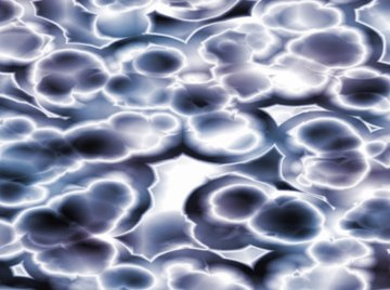
What do scientists study in freshwater?
Scientists study these glowing, freshwater organisms as models for how living creatures develop specialized cells and tissue. Strands of cytoplasm connect neighboring cells, allowing them to communicate, and slender flagella propel the colony through the water. High school biology didn’t do much to make cells seem like fascinating, ...
Answer
The micrographs of cells shown in figure 8-3. what information about cells do these micrographs suggest is explained below in details.
New questions in Biology
Too mule deer lock antlers as they demonstrate strength and worthiness to a female mule deer. The winner of this battle will make with the female type …
What is a student microscope?
Most student microscopes are classified as light microscopes. In a light microscope, visible light passes through the specimen (the biological sample you are looking at) and is bent through the lens system, allowing the user to see a magnified image. A benefit of light microscopy is that it can often be performed on living cells, so it’s possible to watch cells carrying out their normal behaviors (e.g., migrating or dividing) under the microscope.
How to tell if salmonella is in a microscope?
In the image above, you can compare how Salmonella bacteria look in a light micrograph (left) versus an image taken with an electron microscope (right). The bacteria show up as tiny purple dots in the light microscope image, whereas in the electron micrograph, you can clearly see their shape and surface texture, as well as details of the human cells they’re trying to invade.
What is the microscope used to take leaf pictures?
The leaf picture at the start of the article was taken using a specialized kind of fluorescence microscopy called confocal microscopy. A confocal microscope uses a laser to excite a thin layer of the sample and collects only the emitted light coming from the target layer, producing a sharp image without interference from fluorescent molecules in the surrounding layers.
What is a brightfield microscope?
Student lab microscopes tend to be brightfield microscopes, meaning that visible light is passed through the sample and used to form an image directly, without any modifications. Slightly more sophisticated forms of light microscopy use optical tricks to enhance contrast, making details of cells and tissues easier to see.
How do electron microscopes differ from light microscopes?
Electron microscopes differ from light microscopes in that they produce an image of a specimen by using a beam of electrons rather than a beam of light . Electrons have much a shorter wavelength than visible light, and this allows electron microscopes to produce higher-resolution images than standard light microscopes. Electron microscopes can be used to examine not just whole cells, but also the subcellular structures and compartments within them.
Why does a compound microscope not produce an inverted image?
For example, if you were looking at a piece of newsprint with the letter “e” on it, the image you saw through the microscope would be “ə." More complex compound microscopes may not produce an inverted image because they include an additional lens that “re-inverts” the image back to its normal state.
What type of microscope can produce high resolution images?
However, if you want to see something very tiny at very high resolution, you may want to use a different, tried-and-true technique: electron micro scopy.
What are the characteristics of eukaryotic cells?
A defining characteristic of eukaryotic cells is that they enclose their DNA in a nuclei. They are mainly bigger and more complex than prokaryotic cells. Plants, animals, and fungi are all types of mulicellular organisms that contain eukaryotic cells.
Why are mircographs black and white?
Electron mircographs are only black and white since they don't come in colors. Scienctists will frequently use various computer techniques to add "false-colors'' to make certain structures stand out, making it significantly less complicated to anaylze.
What is the accurate question to ask in an investigation when observing a structure under the microscope to determine whether it is?
The accurate question to ask in an investigation when observing a structure under the microscope to determine whether it is living is if it has a nucleus in eukaryotic or nuckeoid if prokaryotic.
Who is responsible for the cell theory?
In this situation, I would argue Schwann and Schleiden are responsible for the cell theory. They both orginally presented the cell theory, making them accountable for the devlopment of the cell theory.
When were cells discovered?
Cells were discovered in 1665 when Robert Hooke was analyzing a thin slice of cork under a mircoscope. Hooke saw "thousands of tiny chambers" which he named cells. Over time more scientists like Anton Van Leeuwemhoek, Matthias Schleiden, Theodor Schwann, Rudolf Virchow, and many more, made impactful discoveries which developed into a cell theory.
When to use a light microscope?
Considering light microscope are commonly used to analyze cells and cell structures,a scientist would use a light microscope when studying a structure found on the surface of yeast.
What did Robert Hooke discover about cells?
Hooke's work contributed to the cell theory in that in 1665, Robert Hooke saw "thousands of tiny chambers, which he named cells. His incredible discovery led many other scientists to develop the cell theory.
