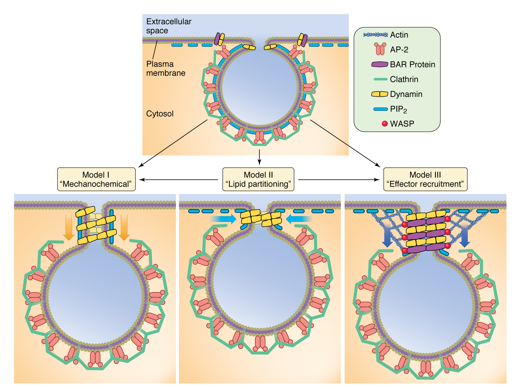
Clathrin
Clathrin is a protein that plays a major role in the formation of coated vesicles. Clathrin was first isolated and named by Barbara Pearse in 1975. It forms a triskelion shape composed of three clathrin heavy chains and three light chains. When the triskelia interact they form a polyhedral lattic…
Full Answer
What does coated vesicles mean?
Coated Vesicle Coated vesicles are derived from internalization of membrane constituents and macromolecules that have been taken up from the extracellular fluid by receptor-mediated endocytosis. From: Clinical Neuroscience, 2014 Download as PDF About this page Complementary Strategies to Understand Virus Structure and Function
What would a vesicle be in a city?
Killingsworth 79 described the various deposits as coated membrane-bound (CMB) bodies, and their fragments have been called coated vesicle-like (CVL) bodies, fibrous-banded material, and focal areas of mineralization. CMB bodies have a uniform electron-dense coat covering their single membrane and contain fine granular material, electron-lucent droplets, dense particles, …
Is a vesicle a plant or animal cell?
Apr 02, 2020 · What is a coated vesicle? coat·ed ves·i·cle. a vesicle that has its biomembrane coated with the protein clathrin. It is involved in the transport of proteins from one membrane site to …
What is the structure of a vesicle?
coat·ed ves·i·cle. a vesicle that has its biomembrane coated with the protein clathrin. It is involved in the transport of proteins from one membrane site to another. Farlex Partner Medical Dictionary © Farlex 2012. Want to thank TFD for its existence?

What does a coated vesicle do?
The transport of proteins and lipids between distinct cellular compartments is conducted by coated vesicles. These vesicles are formed by the self-assembly of coat proteins on a membrane, leading to collection of the vesicle cargo and membrane bending to form a bud.
Are all vesicles coated?
There are three types of vesicle coats: clathrin, COPI and COPII. The various types of coat proteins help with sorting of vesicles to their final destination. Clathrin coats are found on vesicles trafficking between the Golgi and plasma membrane, the Golgi and endosomes and the plasma membrane and endosomes.
What is a clathrin coated vesicle?
Definition. Clathrin coated vesicles (CCVs) mediate the vesicular transport of cargo such as proteins between organelles in the post-Golgi network connecting the trans-Golgi network, endosomes, lysosomes and the cell membrane.
What is coated in biology?
a covering; a layer of some substance spread over a surface. Last updated on July 21st, 2021.Jul 21, 2021
Where do clathrin-coated vesicles go?
Clathrin-coated vesicles (CCV) selectively sort cargo at the cell membrane, trans-Golgi network, and endosomal compartments for multiple membrane traffic pathways.
What proteins are involved in coating vesicles?
The three types of coated vesicles recognize specific sets of cargo and have different protein and lipid compositions. CCVs are composed of two cytosolic protein complexes, clathrin triskelia and heterotetrameric adaptor complexes (AP-1 and AP-2).
What do coat proteins do?
Abstract. Coat proteins allow the selective transfer of macromolecules from one membrane-enclosed compartment to another by concentrating macromolecules into specialized membrane patches and then deforming these patches into small coated vesicles.May 1, 2003
What are the components of coated vesicles?
Coated vesicles, first described by Gray (1961), are involved in receptor-mediated endocytotic processes (Goldstein, Anderson and Brown, 1979). The major constituent of these vesicles is a 180,000-dalton protein called clathrin (Pearse, 1975).
What happens with coated vesicles after they lose their coat?
When the vesicle is fully intracellular it loses its clathrin coat and becomes an endosome, which fuses with primary lysosomes that have a high content of acid hydrolases and other proteases. These lead to degradation of the ingested material, and further processing depending on the cell type.
What do clathrin coated vesicles carry?
Clathrin-coated vesicles were the first discovered and remain the most extensively characterized transport vesicles. They mediate endocytosis of transmembrane receptors and transport of newly synthesized lysosomal hydrolases from the trans-Golgi network to the lysosome.
What happens to the coat proteins after a vesicle buds from a membrane?
There Are Various Types of Coated Vesicles They bud off as coated vesicles that have a distinctive cage of proteins covering their cytosolic surface. Before the vesicle fuses with a target membrane, the coat is discarded, as is required to allow the two cytosolic membrane surfaces to interact directly and fuse.
Where are secretory vesicles produced?
Secretory vesicles are synthesized in the Golgi apparatus so the correct answer is A. The smooth endoplasmic reticulum synthesizes lipids.
Complementary Strategies to Understand Virus Structure and Function
Coated vesicles and enveloped viruses share certain similar mechanisms of structural assembly and can be considered to perform homologous processes. Both are higher-order pleomorphic structures, assembled to transport their selectively recruited cargo to distinct target locations.
Platelet Structure
Small, smooth, and coated vesicles the sizes of those budding from the trans-Golgi face of the Golgi zone in the parent megakaryocyte are difficult to identify in normal circulating platelets. Smooth vesicles could easily be mistaken for cross-sectional channels of the OCS or DTS.
Organelles
One of the best-characterized examples of vesicle formation occurs during receptor-mediated endocytosis. Clathrin-coated pits on the cytoplasmic side of the plasma membrane develop into clathrin-coated vesicles that carry material from the plasma membrane to endosomes.
G Protein Pathways
ARF is found to copurify with coatomer-coated vesicles that are generated in vitro by the incubation of Golgi-enriched membranes with cytosol and GTP γ S. 8 The ARF dependence of coatomer recruitment can be assayed in vitro with purified myrARF, ARF-free cytosol, and Golgi-enriched membranes.
Exocytosis
The pathway of exocytosis starts with the synthesis of a protein on the rough endoplasmic reticulum (RER). From there, the protein must travel through each of the compartments of the Golgi complex, finally arriving at the trans Golgi network (TGN) before being transported to the plasma membrane, lysosome, or vacuole.
Cells
Exocytosis principally involves the budding off of membrane-coated vesicles from the terminal cisterna of the Golgi apparatus and their passage toward either the apical or lateral surface of a cell. These vesicles, which contain either proteins or carbohydrates designed for export, are not clathrin-coated.
Hedgehog Signaling
MPs are a heterogeneous population of small membrane- coated vesicles virtually generated from any cell types during activation or apoptosis. Their release is a well-regulated process, although these vesicles are highly variable in size, composition, and function.
Platelet Structure
Small, smooth and coated vesicles the sizes of those budding from the trans-Golgi face of the Golgi zone in the parent megakaryocyte are difficult to identify in normal circulating platelets. Smooth vesicles could easily be mistaken for cross-sectional channels of the OCS or dense tubular system.
The Role of Platelets in Diabetes Mellitus
Francesca Santilli, ... Rossella Liani, in Platelets (Fourth Edition), 2019
Biology of the cell
Constitutive exocytosis of proteins is a nonregulated process that occurs in all cells. Transport vesicles involved in the default pathway of secretion have a cytosolic coat of the protein coatamer. A GTPase regulates the assembly and disassembly of coatamer-coated vesicles. The coatamer coat is removed after the vesicle reaches its destination.
Medical Retina
The initial age-related change in Bruch's membrane, as seen by electron microscopy, is the accumulation of vesicles and granular and filamentous material. 45,77 These deposits are seen initially in the ICZ but with time occupy both collagenous zones.
Release of Neurotransmitters
Robert S. Zucker, ... Thomas L. Schwarz, in From Molecules to Networks, 2004
Placental Function in Maternofetal Exchange
In women, passive immunity is conferred on the fetus by the selective transfer of immunoglobulin G (IgG) by the placenta.
Biosynthesis, Processing, and Secretion of the Islet Hormones
Donald F. Steiner, ... Shu Jin Chan, in Endocrinology (Sixth Edition), 2010
Biochemistry and cell biology
John V. Forrester MB ChB MD FRCS (Ed) FRCP (Glasg) (Hon) FRCOphth (Hon) FMedSci FRSE FARVO, ... Eric Pearlman BSc PhD, in The Eye (Fourth Edition), 2016
Liver Physiology and Energy Metabolism
Mark Feldman MD, in Sleisenger and Fordtran's Gastrointestinal and Liver Disease, 2021
Endocytosis and the Endosomal Membrane System
Clathrin-dependent endocytosis ( Fig. 22.8) occurs on specialized patches of the plasma membrane, called coated pits, formed by a protein lattice of clathrin and adapter molecules on their cytoplasmic surface (see Fig. 21.12 for details about clathrin structure and mechanism).
Cell structure and function
Drugs that modify membrane receptors can be used in the treatment of disease. One example of this is the use of trastuzumab in the treatment of some breast cancers. Normal breast epithelium expresses a signalling molecule called human epidermal growth factor type 2 ( Her2, also known as Her2/neu or ErbB-2) on the plasma membrane.
HIV Immune Evasion
Elizabeth R. Wonderlich, ... Kathleen L. Collins, in Advances in Virus Research, 2011
Endocytosis in Neurons
CCVs are formed by the coordinated assembly of clathrin triskelia, formed from three tightly linked heavy chains and their associated light chains on the plasma membrane.
Endocytosis and Presynaptic Scaffolds
CCVs are formed by the coordinated assembly of clathrin triskelia built from three tightly linked heavy and associated light chains onto the plasma membrane.
Intracellular Trafficking
Gustavo Pigino, ... Scott T. Brady, in Basic Neurochemistry (Eighth Edition), 2012
Protein Synthesis, Processing, and Trafficking
Cargo proteins exit the ER in COPII-coated vesicles that enter the ERGIC and are ultimately delivered to the cis -Golgi either in vesicles or along extended tubules. However, the mechanism whereby cargo proteins move across the Golgi complex from cis to trans remains controversial. Two models have been proposed.
Trafficking of GPCRs
Guangyu Wu, ... Maoxiang Zhang, in Progress in Molecular Biology and Translational Science, 2015
Intracellular Trafficking of G Protein-Coupled Receptors to the Plasma Membrane in Health and Disease
Alfredo Ulloa-Aguirre, P. Michael Conn, in Cellular Endocrinology in Health and Disease, 2014
Protein Quality Control, Retention, and Degradation at the Endoplasmic Reticulum
Ron Benyair, ... Gerardo Z. Lederkremer, in International Review of Cell and Molecular Biology, 2011
Anterograde Trafficking of Nascent α2B-Adrenergic Receptor: Structural Basis, Roles of Small GTPases
The small GTPase Sar1 and the heterodimeric Sec23/24 and Sec13/31 complexes are the components of COPII-coated transport vesicles, which exclusively mediate export of newly synthesized cargoes from the ER.
Biology of the Endoplasmic Reticulum
Egress of the mature virion represents the final step in the viral life cycle. All viruses exit the host cell by being released into the extracellular environment. Viruses which assemble and mature in the ER take advantage of the ER-associated biosynthetic machinery that enables the synthesis of viral structural proteins.
