What are glomangiomas and what are they for?
What are glomangiomas Glomus bodies are a normal part of the skin circulation that help regulate body temperature by constricting blood flow to the skin to preserve body heat, or relaxing to increase skin circulation to release body heat.
What is the histopathology of glomangioma?
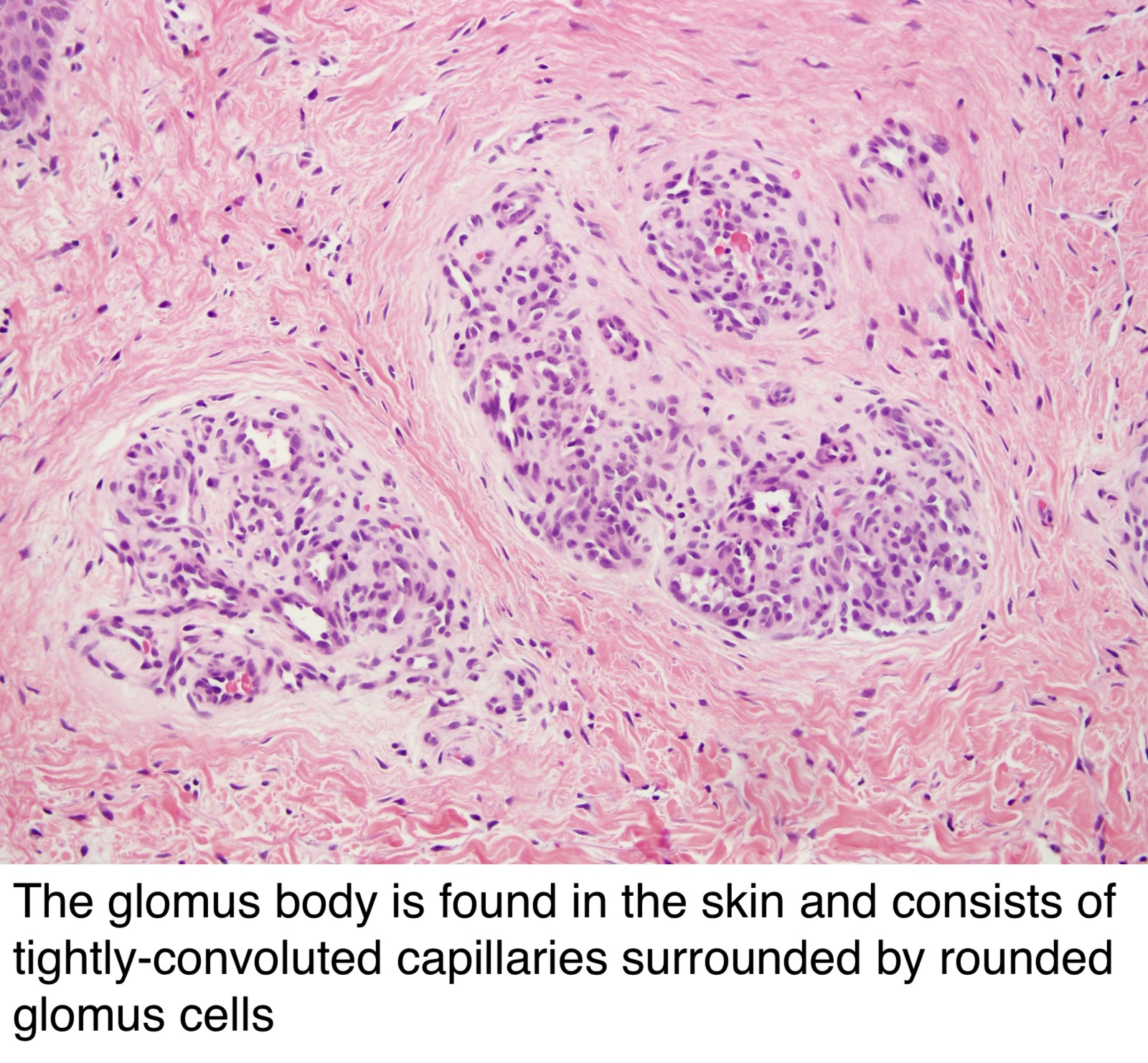
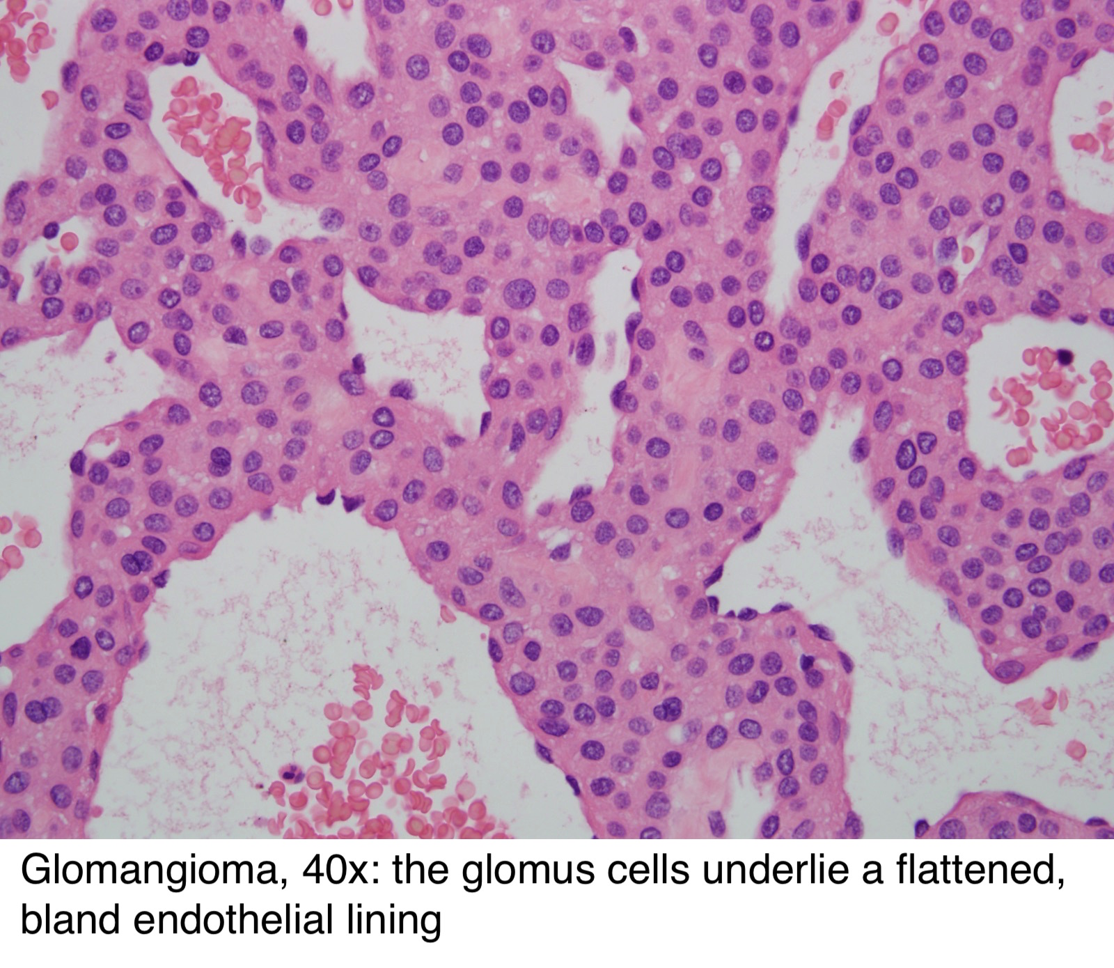
Multiple glomangiomas are rare and comprise about 10 percent of all glomus tumours. In glomangioma, the histopathology shows dilated venous channels that resemble venous malformations (figures 1, 2). Unlike venous malformations, they demonstrate single to multiple rows of surrounding cuboidal glomus cells (figures 3,4).
What is the difference between glomangioma and glomus?
June 2018. Glomangiomas (also called glomuvenous malformation) differ clinically from glomus tumors in that they occur in childhood and adolescence, are usually asymptomatic, do not have a predilection for the subungal region, and often are multifocal.
What are the symptoms of glomangiomas?
The clusters of glomus bodies typical of glomangiomas are blue or purple and visible underneath the skin. They can also become painful and are very sensitive to touch or pressure. What are glomangiomas care options?
What causes Glomangioma?
What causes glomangiomas? In most cases, the cause of glomangiomas are unknown. However, some may have a genetic component as they can be passed along from parents to their children.
What is Glomangioma medical term?
Glomangioma or glomuvenous malformation is a rare cutaneous venous malformation that shows glomus cells (undifferentiated smooth muscle cells which are thermoregulatory units) along with the venous system in histology. Glomus cells are specialized smooth muscle cells who regulates the temperature in the body.
What causes glomus tumors?
The cause of a glomus jugulare tumor is unknown. In most cases, there are no known risk factors. Glomus tumors have been associated with changes (mutations) in a gene responsible for the enzyme succinate dehydrogenase (SDHD).
Can a glomus tumor be cancerous?
Malignant glomus tumors are exceedingly rare, comprising as high as 2.9% of all glomus tumors [5], and are often reported in the literature as single case reports from various institutions. Previously, glomus tumors that displayed unusual features were characterized as “atypical” or “malignant” [6].
Is glomus tumor benign or malignant?
Glomus tumors, or paragangliomas, are slow-growing, usually benign tumors in the carotid arteries (major blood vessels in your neck), the middle ear or the area below the middle ear (jugular bulb). Glomus tumors are most often benign; however, they can cause significant damage to surrounding tissues as they grow.
What is an Angiolipoma?
An angiolipoma is a small, benign, rubbery tumor that contains blood vessels and grows under your skin. Angiolipomas usually develop in young adults between the ages of 20 and 30. They most often appear in your forearms, and they can be painful if touched.
Should glomus tumor be removed?
A glomus tumor is typically treated with surgical intervention. Because this condition can affect the patient's quality of life, a definitive diagnosis is necessary to evaluate the extent of the condition. However, because this condition is benign, recurrence is uncommon. Multiple growths in the area may occur.
How do you get rid of a glomus tumor?
How are Glomus Tumors treated? Surgical excision of the tumor is the only treatment modality. During a 15-minute outpatient procedure the nail is removed, an incision is made into the nail bed exposing the tumor, and the tumor is removed.
What are symptoms of glomus tumor?
Symptoms of glomus tumors depend on their location: middle ear, jugular bulb, deep neck, or carotid artery. Your ear may perceive the flow of blood as a pulsating sound or ringing. Bleeding from the ear....Glomus jugulare (jugular bulb)Pulsations in the ear.Hearing loss.Ear pain.Bleeding from the ear.
Is a glomus tumor a brain tumor?
Glomus tumors (also called paragangliomas) are a rare, slow-growing, and usually benign type of skull base tumor that often develop near the inner ear. Without treatment, they can harm surrounding tissue, damage nerves, and cause other serious problems.
Do glomus tumors spread?
Malignant glomus tumor, or glomangiosarcoma, is a very rare mesenchymal neoplasm that, when seen, occurs in visceral organs. Despite having histologic features of malignancy, these tumors usually do not metastasize. However, when metastasis occurs, this disease is often fatal.
Can you feel a glomus tumor?
Symptoms of a glomus jugulare tumor include: Hearing loss. Ear fullness. Ear pain.
What is a glomangioma?
Glomus bodies are a normal part of the skin circulation that help regulate body temperature by constricting blood flow to the skin to preserve body heat, or relaxing to increase skin circulation to release body heat. Abnormal collections of glomus cells, called glomangiomas or glomus cell tumors, ...
What is a benign glomus cell?
Abnormal collections of glomus cells, called glomangiomas or glomus cell tumors, are benign clusters of bluish-purple nodules that are often painful and sensitive to pressure. Glomangiomas are benign but can be annoying, and patients may develop clusters in any part of the body. They may have a dominant inheritance pattern.
Can laser treatment be used for glomangiomas?
Treatment. Laser treatment is not usually effective in the treatment of glomangiomas because of the depth of involvement in the skin, and surgical excision is usually recommended for symptomatic lesions.
Where do glomus tumors come from?
Glomus tumors originate from the neuromyoarterial plexus: modified smooth muscle cells of the glomus body. They are best thought of as hamartomas rather than true tumors. There are two main components on microscopy:
What is a benign vascular tumor that is typically seen at the distal extremities?
Glomangioma. Glomangiomas , also known as glomus tumors , are benign vascular tumors typically seen at the distal extremities. On imaging, they characteristically present as small hypervascular nodules under the fingernail.
Where is the glomus tumor?
a blue-red, extremely painful chemodectoma involving an arteriovenous anastomosis or cluster of blood cells, which may be found anywhere in the skin, most often in the distal portion of the fingers and toes, especially beneath the nail. Such tumors may also occur in the stomach and nasal cavity.
What is a giant condyloma?
Called also giant condyloma. carcinoid tumor carcinoid (def. 1). carotid body tumor a chemodectoma of a carotid body, found as a firm round mass at the bifurcation of the common carotid artery. connective tissue tumor any tumor arising from a connective tissue structure, such as a fibroma or sarcoma.
What is a heterologous tumor?
heterologous tumor one made up of tissue differing from that in which it grows. homoiotypic tumor ( homologous tumor) one made up of tissue resembling that in which it grows. Hürthle cell tumor see hürthle cell tumor. islet cell tumor a tumor of the islands of Langerhans; many secrete excessive amounts of hormones.
Why do tumors grow in a disorganized manner?
They grow in a disorganized manner and so rapidly that nutrition of the cells becomes a problem. For this reason necrosis and ulceration are characteristic of malignant tumors. They also invade surrounding tissues and are metastatic, initiating the growth of similar tumors in distant organs. (See also cancer .)
What is a melanoameloblastoma?
melanotic neuroectodermal tumor a benign, rapidly growing, dark tumor of the jaw or occasionally some other site, almost always seen in infants; called also melanoameloblastoma. mixed tumor one composed of more than one type of neoplastic tissue.
What is a giant cell tumor?
giant cell tumor. 1. a benign or malignant tumor containing giant cells; see under carcinoma, granuloma, and sarcoma. 2. a bone tumor, ranging from benign to frankly malignant, composed of cellular spindle cell stroma containing multinucleated giant cells resembling osteoclasts.
What is a false tumor?
false tumor pseudotumor. fibroid tumor. 1. fibroma. 2. leiomyoma uteri. germ cell tumor any of a group of tumors arising from primitive germ cells, usually of the testis or ovum; they range from benign to highly malignant.
What are the other diagnoses of glomangioma?
Other diagnoses to be considered include: Vascular malformation — these don't have the perivascular glomus cell accumulations seen in glomangioma. Myopericytoma, angioleiomyoma, myofibroma — these all have varying degrees of perivascular muscle cells but these cells lack the classic cuboidal glomus cells. These tumours usually show ...
What is a glomuvenous malformation?
Glomangiomas (also called glomuvenous malformation) differ clinically from glomus tumors in that they occur in childhood and adolescence, are usually asymptomatic, do not have a predilection for the subungal region, and often are multifocal.
Are You Confident of the Diagnosis?
Glomuvenous malformations (GVMs) are of two distinct types: the single lesion and the multiple variant type.
Treatment Options
The treatment of choice for isolated cutaneous GVMs is surgical excision. The typical method is nail avulsion, followed by excision. The “trap door” technique has been recently reported as a nail-plate-conserving surgical approach with good results. The surgical approach may, however, be impractical in cases of multiple or large segmental lesions.
Patient Management
For single tumors, if the lesion is completely excised, the risk of recurrence is low. Malignant transformation is rare. Nail plate dystrophy is a common complication of surgical excision.
What is the Evidence?
Henning, JS. “Glomovenous malformations”. Dermatol Online J. vol. 13. 2007 Jan 27. pp. 17 (Case report and review of current literature on multiple GVM)
Where does a malignant glomus tumor come from?
Another report of a malignant glomus tumor ( glomangiosarcoma) with metastases from the skin. A malignant glomus tumor one arose from the kidneys.
Where are glomus tumors found?
Glomus tumors are usually solitary and small lesions. The vast majority are found in the hand, wrist, foot, and under the fingernails. They are often painful, and the pain is reproduced when the lesion is placed in cold water. Multiple tumors are less likely to be painful.
What are the criteria for glomus tumor diagnosis?
Criteria for the diagnosis of malignancy in glomus tumors are: Tumor size of more than 2 centimeters and subfascial or visceral location. Atypical mitotic figures. Marked nuclear atypia and any level of mitotic activity. Pericytes of Zimmerman.
How often do glomus tumors occur?
Solitary glomus tumors are more frequent in adults than in others. Multiple glomus tumors develop 11–15 years earlier than single lesions; about one third of the cases of multiple tumors occur in those younger than 20 years. Congenital glomus tumors are rare; they are plaguelike in appearance and are considered a variant of multiple glomus tumors.
What is the size of a glomus tumor?
The tumor is the translucent oblate spheroid in the center of the incision, approximate horizontal dimension is 4 millimeters. Surgical excision is the preferred treatment for benign glomus tumors.
Is a glomus tumor benign?
The majority of glomus tumors are benign, but they can also show malignant features. Glomus tumors were first described by Hoyer in 1877 while the first complete clinical description was given by Masson in 1924. Histologically, glomus tumors are made up of an afferent arteriole, anastomotic vessel, and collecting venule.
Is a glomangiosarcoma fatal?
However, metastases do occur and are usually fatal.
Terminology
Epidemiology
- It classically presents in young to middle-aged (4th to 5th decades 7). There is a recognized female predilection. They can be multiple in ~ 10% of cases. Glomus tumors account for 1-5% of the soft-tissue tumors in the hand 4.
Clinical Presentation
- The lesion usually presents as a small firm red-blue nodule under the fingernail and is exquisitely painful and sensitive to cold temperature and touch. Sometimes the pain is worse at night; it may disappear when a tourniquet is applied. May also present as hemorrhage under the nail. The presence of the Hildreth sign (pain relief following the application of a tourniquet proximally) is c…
Pathology
- Glomus tumors originate from the neuromyoarterial plexus: modified smooth muscle cells of the glomus body. They are best thought of as hamartomas rather than true tumors. There are two main components on microscopy: 1. branching vascular channels 2. aggregates of specialized glomus cells Approximately 75% occur in hand 4; asubungual position is cha...
Radiographic Features
- the tumor is difficult if not impossible to identify, rarely can be seen as a subtle soft tissue density
- may show a marginated osseous erosion or thinning of the adjacent cortical bone
Differential Diagnosis
- General imaging differential considerations include: 1. hemangioma 2. epidermal inclusion cyst 3. angioleiomyomas 4. tenosynovial giant cell tumor