Symptoms
Pulmonary embolism remains a common and potentially deadly disease, despite advances in diagnostic imaging, treatment and prevention. Managing pulmonary embolism requires a multifactorial approach involving risk stratification, determining appropriate diagnostics and selecting individualised therapy.
Causes
This can happen:
- after an operation or a serious limb injury
- after long periods of bed rest
- during a long-haul flight or a long train or car journey lasting more than 4 hours
Prevention
Who is most at risk for pulmonary embolism? People at risk for PE are those who: Have been inactive or immobile for long periods of time. Have certain inherited conditions, such as blood clotting disorders or factor V Leiden. Are having surgery or have broken a bone (the risk is higher weeks following a surgery or injury).
Complications
This would be followed by an increased in breathing rate, heart rate and severe chest pain. The pain would get worse as the person takes deeper breaths. The patient would also cough up blood, lose consciousness and be dead within minutes.
Is pulmonary embolism a deadly disease?
What are some causes of pulmonary embolism?
Who is most at risk for pulmonary embolism?
Would death from a pulmonary embolism be painful?
Can you survive a massive pulmonary embolism?
Pulmonary embolism can be life-threatening. About one-third of people with undiagnosed and untreated pulmonary embolism don't survive. When the condition is diagnosed and treated promptly, however, that number drops dramatically.
What causes massive pulmonary embolism?
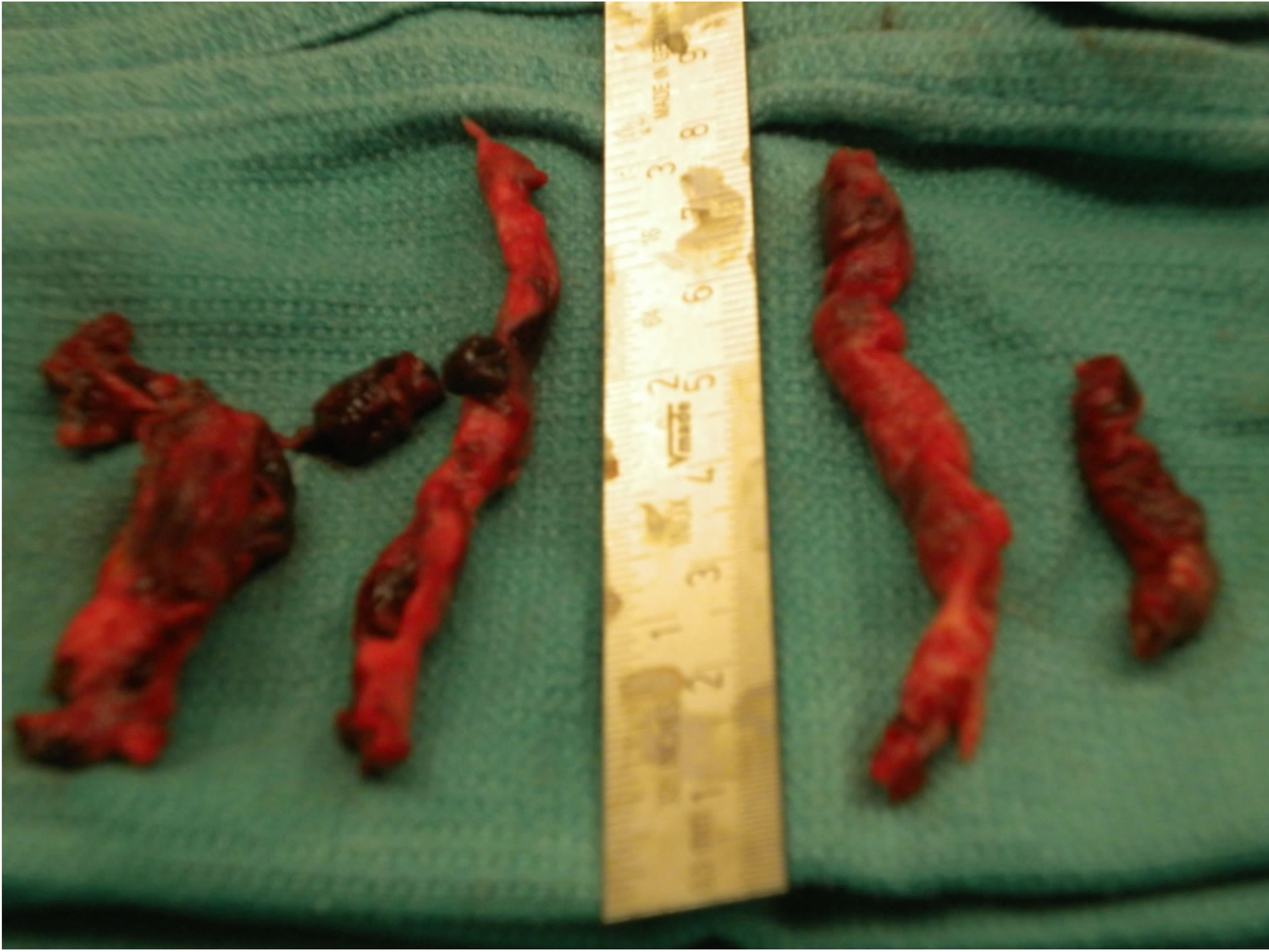
Pulmonary embolism is caused by a blocked artery in the lungs. The most common cause of such a blockage is a blood clot that forms in a deep vein in the leg and travels to the lungs, where it gets lodged in a smaller lung artery. Almost all blood clots that cause pulmonary embolism are formed in the deep leg veins.
What is considered massive PE?
As a cause of sudden death, massive pulmonary embolism is second only to sudden cardiac death. Massive pulmonary embolism is defined as presenting with a systolic arterial pressure less than 90 mm Hg. The mortality for patients with massive pulmonary embolism is between 30% and 60%, depending on the study cited.
What is the survival rate of a pulmonary embolism?
However, reported survival after venous thromboembolism varies widely, with "short-term" survival ranging from 95% to 97% for deep vein thrombosis8,9 and from 77% to 94% for pulmonary embolism,4,6,8,9 while "long-term" survival ranges from 61% to 75% for both deep vein thrombosis and pulmonary embolism.
What are the warning signs of a pulmonary embolism?
The most common symptoms are:Shortness of breath.Chest pain that may become worse when breathing in.Cough, which may contain blood.Leg pain or swelling.Pain in your back.Excessive sweating.Lightheadedness, dizziness or passing out.Blueish lips or nails.
How do you treat a massive pulmonary embolism?
TreatmentBlood thinners (anticoagulants). These drugs prevent existing clots from enlarging and new clots from forming while your body works to break up the clots. ... Clot dissolvers (thrombolytics). While clots usually dissolve on their own, sometimes thrombolytics given through the vein can dissolve clots quickly.
How quick is death from pulmonary embolism?
Of 162 patients, 44 suffered sudden death (within 24 hours of onset). Among these, 28 patients died within 1 hour and 9 within 1 to 24 hours. In the remaining seven patients, the time until death could not be determined because the subject was detected postmortem.
How common is sudden death from pulmonary embolism?
Sudden death is the first symptom in about one-quarter (25%) of people who have a PE. 10% – 30% of people will die within one month of diagnosis.
How common is massive pulmonary embolism?
In the United States, deep venous thrombosis and pulmonary embolism are associated with approximately 250,000 hospitalizations each year, and as many as 50,000 individuals die each year as a result of pulmonary embolism.
Is pulmonary embolism a stroke?
stroke – where the blood supply to the brain is cut off. pulmonary embolism – where a foreign body blocks the artery that carries blood from the heart to the lungs (the pulmonary artery)
How long does it take for a blood clot to go away with blood thinners?
After being stopped, warfarin takes 5–7 days to clear the body. Takes 24 to 48 h to clear after being stopped. There are proven reversal methods in case of excessive bleeding on warfarin.
How long do you stay in the hospital for a blood clot in the lung?
People who present to hospital with blood clots in the legs or lungs should be offered treatment within 4 hours and have their investigative tests including scans within 24 hours, according to latest guidelines.
Who is at risk for pulmonary embolism?
People at risk for PE are those who: Have been inactive or immobile for long periods of time. Have certain inherited conditions, such as blood clotting disorders or factor V Leiden. Are having surgery or have broken a bone (the risk is higher weeks following a surgery or injury).
Can drugs cause pulmonary embolism?
Septic pulmonary emboli due to the injection of contaminated illicit drugs via infected needles is another infectious complication of injection illicit drug use. Smoking crack cocaine and other illicit drugs has been implicated in an increased prevalence of pulmonary tuberculosis.
Can stress cause blood clots in lungs?
Effect of Stress on Blood Vessels But anxiety can also increase blood pressure, putting additional stress on the blood vessel walls, making them stiffer and decreasing the amount of blood that flows through the body. Combined these forces can lead to serious blood clots that can cause blockages in the heart and lungs.
Is pulmonary embolism fatal?
Pulmonary embolism is the third-leading cause of cardiovascular death.
What is the treatment for massive PE?
Massive pulmonary embolism (PE) is a potentially lethal condition, with death usually caused by right ventricular (RV) failure and cardiogenic shock. Systemic thrombolysis (unless contraindicated) is recommended as the first-line treatment of massive PE to decrease the thromboembolic burden on the RV and increase pulmonary perfusion. Surgical pulmonary embolectomy or catheter-directed thrombectomy should be considered in patients with contraindications to fibrinolysis, or those with persistent hemodynamic compromise or RV dysfunction despite fibrinolytic therapy. Critical care management predominantly involves supporting the RV, by optimizing preload, RV contractility, and coronary perfusion pressure and minimizing afterload. Despite these interventions, mortality remains high.
Is pulmonary embolectomy considered fibrinolysis?
Surgical pulmonary embolectomy or catheter-directed thrombectomy should be considered in patients with contraindications to fibrinolysis, or those with persistent hemodynamic compromise or RV dysfunction despite fibrinolytic therapy.
What are the symptoms of a pulmonary embolism?
Symptoms of a pulmonary embolism include sudden shortness of breath, pain in and around the chest and coughing. Caused by a blood clot, a pulmonary embolism is a serious but very treatable condition if done immediately. Appointments & Access. Contact Us.
How to reduce the risk of pulmonary embolism?
Be sure you discuss and understand your follow- up care with your doctor. Follow your doctor’s recommendations to reduce the risk of another pulmonary embolism. Keep all appointments with your doctor and the laboratory so your response to prescribed treatments can be monitored.
What is the term for a blood clot in the lung?
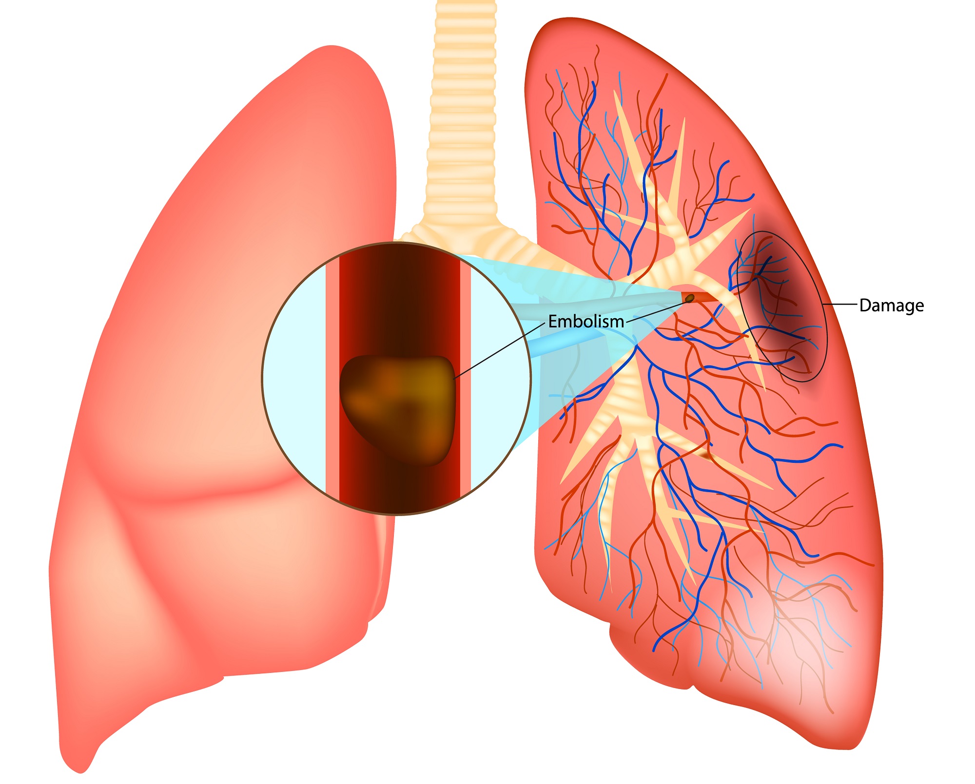
A pulmonary embolism is a blood clot in the lung that occurs when a clot in another part of the body (often the leg or arm) moves through the bloodstream and becomes lodged in the blood vessels of the lung. This restricts blood flow to the lungs, lowers oxygen levels in the lungs and increases blood pressure in the pulmonary arteries.
What is it called when a clot develops in a vein?
If a clot develops in a vein and it stays there, it’s called a thrombus. If the clot detaches from the wall of the vein and travels to another part of your body, it’s called an embolus. If PEs are not treated quickly, they can cause heart or lung damage and even death. Cleveland Clinic is a non-profit academic medical center.
Can pulmonary embolism cause shortness of breath?
Although most people with a pulmonary embolism experience symptoms, some will not. The first signs are usually shortness of breath and chest pains that get worse if you exert yourself. You may cough up bloody sputum. If you have these symptoms get medical attention right away.
Can a pulmonary embolism dissolve on its own?
A pulmonary embolism may dissolve on its own; it is seldom fatal when diagnosed and treated properly. However, if left untreated, it can be serious, leading to other medical complications, including death. A pulmonary embolism can: Cause heart damage. Be life-threatening, depending on the size of the clot.
Who is at risk for developing a blood clot?
Who is at risk of developing a blood clot? People at risk for developing a blood clot are those who: Have been inactive or immobile for long periods of time due to bed rest or surgery. Have a personal or family history of a blood clotting disorder, such as deep vein thrombosis (DVT) or pulmonary embolism (PE).
Where does pulmonary embolism occur?
Pulmonary embolism occurs when a clump of material, most often a blood clot, gets wedged into an artery in your lungs. These blood clots most commonly come from the deep veins of your legs, a condition known as deep vein thrombosis (DVT).
What are the symptoms of pulmonary embolism?
Other signs and symptoms that can occur with pulmonary embolism include: Rapid or irregular heartbeat. Lightheadedness or dizziness. Excessive sweating. Fever. Leg pain or swelling, or both, usually in the calf caused by a deep vein thrombosis.
What are the risks of having a blood clot?
You're at higher risk if you or any of your family members have had venous blood clots or pulmonary embolism in the past. In addition, some medical conditions and treatments put you at risk, such as: Heart disease. Cardiovascular disease, specifically heart failure, makes clot formation more likely. Cancer.
What is PE in a lung?
Pulmonary embolism (PE) occurs when a blood clot gets lodged in an artery in the lung, blocking blood flow to part of the lung. Blood clots most often start in the legs and travel up through the right side of the heart and into the lungs. This is called DVT. However, PE sometimes can occur without any evidence of DVT.
What is it called when you have multiple clots in your lungs?
The portions of lung served by each blocked artery are robbed of blood and may die. This is known as pulmonary infarction. This makes it more difficult for your lungs to provide oxygen to the rest of your body.
What is the best treatment for pulmonary embolism?
For this reason, most hospitals are aggressive about taking measures to prevent blood clots, including: Blood thinners (anticoagulants).
How to get rid of pulmonary embolism?
Elevating your legs when possible and during the night also can be very effective. Raise the bottom of your bed 4 to 6 inches (10 to 15 cm) with blocks or books. Physical activity. Moving as soon as possible after surgery can help prevent pulmonary embolism and hasten recovery overall.
What is a massive pulmonary embolism?
Massive pulmonary embolism can be defined anatomically as a greater than 50% thrombotic obstruction of the pulmonary vasculature or the occlusion of two or more lobar arteries. 2 However, the clinical impact of this obstruction depends on the size of the embolus and on the patient’s underlying cardiopulmonary function.
Where does pulmonary embolism occur?
Pulmonary embolism usually occurs secondary to venous thrombosis in the deep veins of the lower limbs, proximal to but including the popliteal veins. 1 Risk factors for venous thrombosis are listed in Table 23-1.
What is the effect of pulmonary embolism on vascular resistance?
Massive pulmonary embolism results in an acute, dramatic increase in pulmonary vascular resistance. This increase occurs not only because of mechanical obstruction but also as a result of pulmonary arteriolar vasoconstriction secondary to hypoxemia and neurohumoral activation.
What is a V/Q lung scan?
V/Q lung scintigraphy is a noninvasive test for pulmonary embolism that was, until recently, the most commonly performed diagnostic imaging modality. A normal V/Q scan effectively excludes pulmonary embolus. A high-probability scan, as evidenced by multiple segmental mismatches between ventilation and perfusion in the context of a high clinical suspicion, identifies more than 90% of patients with pulmonary embolism. 12 However, the utility of V/Q scans is limited by difficulties in interpreting them, especially in patients with underlying lung disease in whom regional variations in ventilation and perfusion confound interpretation of the image. Thus, although agreement among observers is high for normal and high-probability scans, it is substantially lower for intermediate scans, and these are the kinds of scans most commonly encountered in everyday practice. Scans should be performed within 24 hours of clinical suspicion of pulmonary embolus because the changes may revert to normal within a week.
What is troponin T?
Elevated troponin levels (troponin T >0.01 ng/ml) are predictive of a complicated in-hospital course and increased mortality rates in patients with pulmonary embolism. 10 High levels of B-type natriuretic peptide (BNP) are also predictive of adverse outcome; a BNP level below 50 pg/ml identifies 95% of patients with benign clinical courses. 11 However, the clinical utility of both troponin and BNP in patients with massive pulmonary embolism has yet to be determined.
Why is the right ventricle occluded?
The right ventricle is a thin-walled structure that is better suited to volume work than pressure work, and an acute increase in pulmonary vascular resistance sufficient ...
Is lobar oligemia normal on a chest radiograph?
Segmental or lobar oligemia is usual in massive (as opposed to submassive) pulmonary embolism, although the chest radiograph can sometimes appear normal. Other common findings include atelectasis, consolidation, pleural effusion, and right-sided cardiac enlargement. The so-called Hampton hump refers to the uncommonly seen wedge-shaped pulmonary infarct.
What is the Difference Between Massive and Submassive Pulmonary Embolism?
The key difference between massive and submassive pulmonary embolism is the presence of hypotension to bring out the symptoms. During massive pulmonary embolism, hypotension causes severe cardiopulmonary failure derived from right ventricular overload, whilst during submassive pulmonary embolism, hypotension does not cause right ventricular dysfunction or myocardial necrosis. Moreover, massive pulmonary embolism has a higher mortality rate than submassive pulmonary embolism.
What is Submassive Pulmonary Embolism?
Submassive pulmonary embolism is the condition where individuals suffer from pulmonary embolism with right ventricular dysfunction or myocardial necrosis but without systemic hypotension. Submassive pulmonary embolism causes organ failure. Compared to massive pulmonary embolism, submassive pulmonary embolism causes minor risks in severity and mortality. Even though patients develop organ failure, they are hemodynamically stable while presenting symptoms.
Hemodynamic stability
A submassive PE is hemodynamically stable. This means that a person’s heart rate and blood pressure remain steady.
Right ventricular dysfunction
Another feature of submassive PE is right ventricular dysfunction (RVD). The right ventricle is the chamber of the heart that sends blood with low oxygen into the lungs to receive fresh oxygen.
High troponins
Elevated troponin levels are another potential finding in submassive PE. Troponins are proteins that are released when damage to the heart has occurred.
Anticoagulation
One of the main treatments for submassive PE is anticoagulant therapy. Anticoagulant drugs are also called blood thinners.
Systemic thrombolytic therapy
Another potential treatment option is systemic thrombolytic therapy. Thrombolytic drugs work to dissolve clots quickly. However, their use with submassive PE is controversial, according to a 2019 consensus paper.
Catheter-directed thrombolysis
A catheter is a thin, flexible tube inserted into blood vessels. In catheter-directed thrombolysis, doctors use a catheter to deliver low doses of thrombolytic drugs at the location of the PE.
Embolectomy
An embolectomy involves removing the blood clot from the body. Doctors can do this either using a catheter or through a surgical procedure.
What is a PE in the lung?
Cavan Images/Getty Images. A pulmonary embolism (PE) is when a blood clot becomes stuck in the blood vessels of your lung. These clots typically begin in the leg and then break free and travel to the lung. The American Lung Association estimates that about 1 in 1,000 people in the United States experience a PE each year.
What percentage of people have shortness of breath after a PE?
A 2019 study looked at quality of life in 101 people who’d had a PE. It found that 6 months after a PE, 47 percent of participants reported lingering shortness of breath and 25.3 percent reported some type of impairment or difficulty in functioning.
How long after PE can you breathe?
The American Lung Association recommends making an appointment with your doctor to be tested for pulmonary hypertension if you still have trouble with breathing 6 months after your PE.
What is the recovery for PE?
A big part of the recovery for PE aims to prevent additional blood clots from forming. There are several risk factors that can increase your risk for blood clots, such as:
What causes blood clots?
thrombophilia, a condition that causes blood clots
How long does it take for chest pain to go away after PE?
However, it’s not uncommon to continue to have shortness of breath or chest pain for weeks, months, or even years after a PE. A 2019 study. Trusted Source.
What happens if you have a PE?
In some people who have had a PE, scar tissue can form in nearby arteries, causing them to become narrower. This can lead to a condition called pulmonary hypertension.
What is the classification of pulmonary embolism?
Classification of a pulmonary embolism may be based upon: the presence or absence of hemodynamic compromise. temporal pattern of occurrence. the presence or absence of symptoms. the vessel which is occluded.
How long does it take for 80% of pulmonary embolism to resolve?
Several studies report around 80% emboli resolving at around 30 days 20,21. According to one study, residual pulmonary obstruction at 6 months after the first episode of pulmonary embolism was shown to be an independent predictor of recurrent venous thromboembolism and/or chronic thromboembolic pulmonary hypertension 28.
What are the features of chronic pulmonary emboli?
Features noted with chronic pulmonary emboli include: webs or bands, intimal irregularities 3. abrupt narrowing or complete obstruction of the pulmonary arteries 3. “ pouching defects ” which are defined as chronic thromboembolism organized in a concave shape that “points” toward the vessel lumen 3.
What is CTPA in pulmonary angiography?
CT pulmonary angiography (CTPA) will show filling defects within the pulmonary vasculature with acute pulmonary emboli. When the artery is viewed in its axial plane the central filling defect from the thrombus is surrounded by a thin rim of contrast, which has been called the Polo Mint sign.
What is PE in medical terms?
Pulmonary embolism (PE) refers to embolic occlusion of the pulmonary arterial system . The majority of cases result from thrombotic occlusion, and therefore the condition is frequently termed pulmonary thromboembolism which is what this article mainly covers. Non-thrombotic pulmonary embolus sources include 30 :
What causes pulmonary hypertension?
Cumulative damage from repeated embolic insults is a common cause of chronic thromboembolic pulmonary hypertension, which demonstrates a variable degree of the aforementioned signs, but with significantly higher right ventricular pressures, right ventricular hypertrophy and diastolic dysfunction, and a higher degree of tricuspid regurgitation .
What is a chronic thromboemboli?
In contrast to acute pulmonary embolism, chronic thromboemboli are often complete occlusions or non-occlusive filling defects in the periphery of the affected vessel which form obtuse angles with the vessel wall 9. The thrombus may be calcified.
What is PE in medical terms?
Pulmonary embolism (PE) presents a spectrum of hemodynamic consequences, ranging from being asymptomatic to a life-threatening medical emergency. Management of submassive and massive PE often involves clinicians from multiple specialties, which can potentially delay the development of a unified trea …
Can submassive PE deteriorate?
In addition, patients with submassive PE can deteriorate after their presentation and require escalation of care. Underlying comorbidities such as chronic obstructive pulmonary disease, cancer, congestive heart failure, and interstitial lung disease can impact the patient's hemodynamic ability to tolerate submassive PE.

Overview
Symptoms
Causes
Risk Factors
Complications

Prevention
- Pulmonary embolism symptoms can vary greatly, depending on how much of your lung is involved, the size of the clots, and whether you have underlying lung or heart disease. Common signs and symptoms include: 1. Shortness of breath.This symptom typically appears suddenly and always gets worse with exertion. 2. Chest pain.You may feel like you're having a heart attack…