What and how do they do a mylogram?
A myelogram is a diagnostic imaging test generally done by a radiologist. It uses a contrast dye and X-rays (fluoroscopy) or computed tomography (CT) to look for problems in the spinal canal. Problems can develop in the spinal cord, nerve roots, and other tissues. This test is also called myelography. The contrast dye is injected into the ...
What are the side effects of a myelogram?
Side effects are summarized here:
- Forty-three patients had headache on the day of examination and 22 the next day. ...
- Thirty-one of the patients complained of muscular pain in various parts of the body. ...
- Two patients noticed a high-frequency tone, and one patient heard bell-ringing for several hours.
- The symptoms varied considerably. ...
What is the procedure of a myelogram?
Myelography, which is often termed as a myelogram, is a radiographic imaging process that is done to inspect the spine and the outer membrane surrounding it. The process involves the injection of a radiographic contrast media or dye into the cerebrospinal fluid that surrounds the spine.
What does an arthrogram show?
An arthrogram may be more useful than a regular X-ray because it shows the surface of soft tissues lining the joint as well as the joint bones. A regular X-ray only shows the bones of the joint. Your radiologist will see tendons, ligaments, cartilage and your joint capsule.
See 7 key topics from this page & related content

What can myelogram diagnose?
A myelogram can detect conditions affecting the spinal cord and nerves within the spinal canal, including disc herniations, bone spurs, spinal stenosis, tumors, and infection.
What is a myelogram and is it painful?
How does it feel? You will feel a quick sting from a small needle that has medicine to numb the skin on your back. You will also feel some pressure as the long, thin spinal needle is put into your spinal canal. You may feel a quick, sharp pain down your buttock or leg when the needle is moved in your spine.
Will a myelogram show nerve damage?
A myelogram is able to show your spinal cord, spinal nerves, nerve roots, and bones in the spine by injecting contrast into your spinal fluid. As a result, it will also reveal whether anything is pressing against your spinal cord or nerves.
Is a myelogram better than an MRI?
Myelograms are usually accompanied by CT/CAT scans, which often have higher resolution than MRI. That means the images show more detail, much like a higher resolution digital camera produces sharper pictures. Consequently, more subtle signs of disease may be identified.
How long do I have to lay flat after a myelogram?
It's normal to have a mild headache for up to 24 hours after the test. Take a pain reliever such as acetaminophen (Tylenol). Follow the instructions on the bottle. Do not lie flat or let your head be lower than the rest of your body for the first 24 hours after the test.
Are you awake during a myelogram?
What is a myelogram like? You will be awake during the procedure. You will lie on your stomach. You will be given a numbing injection that may sting for a few seconds.
What is the most common clinical indication for a myelogram?
As a result, one of the most common indications for CT myelography is to help evaluate the spinal canal and neural foramina in degenerative disease when the patient cannot undergo MRI because of an MRI-incompatible implanted device or when MR images would be nondiagnostic because of extensive artifact (1).
How will I feel after myelogram?
A myelogram may increase your risk for a headache, neck or back pain, nausea, or vomiting. You may have bleeding or spinal fluid may leak from the injection site. The procedure may cause injury to a disc, nerves, or your spinal cord. The dye used during the procedure may cause and allergy, seizure, or brain problems.
How long is a myelogram procedure?
The duration of the procedure will vary, but the average is about 1 hour. The technologist will position you on the exam table, usually on your stomach. A CT scan will be done following the procedure. The technologist and radiologist will be available to answer any questions.
How do you prepare for a myelogram?
Before Your Myelogram You should not eat any solid food after midnight the day before your exam. You can drink clear liquids as instructed. You should let your healthcare provider know if you are or may be pregnant, have any bleeding issues, take any medications, have allergies or have had back surgery or pain.
Is a myelogram the same as a spinal tap?
A myelogram is performed first in a separate procedure. This is similar to a lumbar puncture, or spinal tap, where the fluid space around the spinal cord (within the spinal canal) is accessed with local anesthesia and contrast (usually 12cc non-ionic iodinated contrast) is administered.
What kind of sedation is used for a myelogram?
Anesthesia. There is usually no anesthesia with this procedure. Your doctor may give you a mild sedative. You will have local anesthetic to reduce the pain of the needle.
How long is a myelogram procedure?
The duration of the procedure will vary, but the average is about 1 hour. The technologist will position you on the exam table, usually on your stomach. A CT scan will be done following the procedure. The technologist and radiologist will be available to answer any questions.
What are the after effects of a myelogram?
Common side effects include headache, aches or discomfort in the arms or legs, nausea, vomiting, and dizziness. Most patients do not experience any side effects, and when side effects do occur, they usually disappear within 24 hours.
Where is contrast injected for a myelogram?
The contrast material usually is injected into the lower lumbar spinal canal, because it is considered easier and safer. Occasionally, if it is deemed safer or more useful, the contrast material will be injected into the upper cervical spine.
What Conditions Are Diagnosed With a CT Myelogram?
The CT myelogram is helpful in diagnosing the following conditions: - Brain tumors. - Bone spurs. - Spinal nerve root injuries. - Spinal cord infec...
Can CT Myelogram Cause Any Side Effect?
After a CT myelogram procedure, the following side effects may occur: - Headache. - Nausea. - Vomiting. - Fever. - Numbness of the legs. - Pain at...
What Is the Time Taken for a CT Myelogram?
The CT myelogram procedure involves administering contrast material to visualize spine and brain disorders. Usually, after the myelogram, the speci...
What Are the Benefits of a CT Myelogram Over an MRI?
CT myelogram has various benefits over magnetic resonance imaging (MRI) in the following aspects: - CT myelogram shows images with high resolution....
When Can I Recover From a Myelogram?
After the myelogram procedure, you may need to stay in the radiologist's room for an hour, and then you may return home. However, the recovery time...
How Do a CT Scan and a Myelogram Differ?
Both myelogram and computed tomography (CT) are different radiological modalities. They are explained as follows: - A myelogram is mainly indicated...
Why Is a Post-Myelogram CT of the Spine Performed?
The specialist may indicate a computed tomography (CT) following a myelogram for the following reasons: Spinal stenosis. - Cerebrospinal fluid (CSF...
How Is a CT Myelogram of the Lumbar Spine Explained?
CT myelogram of the lumbar spine is usually taken when a patient complains of persistent back pain and if other modalities like MRI (magnetic reson...
Why Is a Myelogram Painful?
A myelogram is done to evaluate the spinal nerves, bones, and other parts of the spinal column. The initial step in the process is to inject a loca...
What Is a CT Myelogram?
A CT (computed tomography) myelogram helps evaluate the vertebral disk and other regions of the spinal cord. The procedure is also helpful in detec...
What is the purpose of myelography?
It is particularly useful for assessing the spine following surgery and for assessing disc abnormalities in patients who cannot undergo MRI.
Why is myelography not recommended for MR?
In patients with spinal instrumentation (screws, plates, rods, etc.), MR imaging may not be optimal because of artifacts generated by these instrument s. In these cases your doctor may decide to order CT myelography.
What are the limitations of Myelography?
The most significant limitation of myelography is that it only sees inside the spinal canal and the adjacent spinal nerve roots. Abnormalities outside these areas may be better imaged with MRI or CT. MR is superior to myelography when assessing intrinsic spinal cord disease.
What are some common uses of the procedure?
Magnetic resonance imaging (MRI) is often the first imaging exam done to evaluate the spinal cord and nerve roots. However, on occasion, a patient has a medical device, such as a cardiac pacemaker, that may prevent him or her from undergoing MRI. In such cases, myelography and/or a CT scan, in lieu of MRI, is performed to better define abnormalities.
How does the procedure work?
X-rays are a form of radiation like light or radio waves. X-rays pass through most objects, including the body. The technologist carefully aims the x-ray beam at the area of interest. The machine produces a small burst of radiation that passes through your body. The radiation records an image on photographic film or a special detector.
Who interprets the results and how do I get them?
A radiologist, a doctor trained to supervise and interpret radiology examinations, will analyze the images. The radiologist will send a signed report to your primary care or referring physician who will discuss the results with you.
What is the term for the injection of contrast material into the spinal canal?
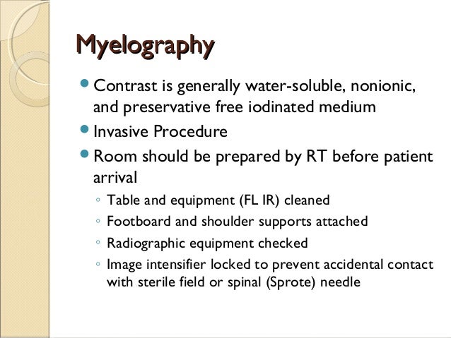
Myelography is an imaging examination that involves the introduction of a spinal needle into the spinal canal and the injection of contrast material in the space around the spinal cord and nerve roots (the subarachnoid space) using a real-time form of x-ray called fluoroscopy.
What is a myelogram?
A myelogram is a diagnostic imaging test generally done by a radiologist. It uses a contrast dye and X-rays (fluoroscopy) or computed tomography (CT) to look for problems in the spinal canal. Problems can develop in the spinal cord, nerve roots, and other tissues. This test is also called myelography.
Why might I need a myelogram?
A myelogram may be done to assess the spinal cord, subarachnoid space, or other structures for changes or abnormalities. It may be used when another type of exam, such as a standard X-ray, doesn't give clear answers about the cause of back or spine problems. Myelograms may be used to evaluate many diseases, including:
What happens during a myelogram?
The procedure takes about an hour, but may vary depending on your condition and the clinic's practices.
What is the term for a disk that bulges and presses on nerves and the spinal cord?
Herniated disks. These are disks that bulge and press on nerves or the spinal cord. Spinal cord tumors. Infection or inflammation of tissues around the spinal cord. Spinal stenosis. This is a breakdown and swelling of the bones and tissues around the spinal cord. This breakdown makes the canal narrow.
What is myelography?
Myelography, also called a myelogram, is an imaging test that checks for problems in your spinal canal. The spinal canal contains your spinal cord, nerve roots, and the subarachnoid space. The subarachnoid space is a fluid-filled space between the spinal cord and the membrane that covers it. During the test, contrast dye is injected into the spinal canal. Contrast dye is a substance that makes specific organs, blood vessels, and tissue show up more clearly on an x-ray.
What happens during myelography?
A myelography may be done at a radiology center or in the radiology department of a hospital. The procedure usually includes the following steps:
Why is the xray table tilted?
Your x-ray table will be tilted in different directions to allow the contrast dye to move to different areas of the spinal cord.
What is the difference between a CT scan and a myelogram?
Myelography involves using one of these two imaging procedures: Fluoroscopy, a type of x-ray that shows internal tissues, structures, and organs moving in real time. CT scan (computerized tomography), a procedure that combines a series of x-ray images taken from different angles around the body. Other names: myelogram.
Is there anything else I need to know about myelography?
MRIs use a magnetic field and radio waves to create images of organs and structures inside the body. But myelography can be more useful in diagnosing some conditions , such as certain spinal tumors and spinal disk problems. It's also used for people who are unable to have an MRI because they have metal or electronic devices in their bodies. These include a pacemaker, surgical screws, and cochlear implants.
What is a myelogram?
A myelogram is a procedure that involves multiple images obtained after an iodine containing contrast agent was injected into the spinal canal and the sac that contains the spinal cord and nerve roots.
How is a myelogram performed?
A myelogram is performed by placing a needle in the lower back or occasionally in the neck. The needle placement is localized using imaging guidance. Although local anesthesia is used, the needle tip is in a location near the nerve roots, so pain or an electric shock sensation may be felt down the leg; if this happens, the needle position can be adjusted. A small amount of cerebrospinal fluid is removed and contrast is injected, and the needle is removed. A series of x-ray images are obtained. A CT scan is routinely performed after the myelogram in order to provide cross sectional information. Much of the procedure is performed with the patient lying face down. The entire procedure from start to finish lasts approximately one hour and the patient is awake during the procedure.
What medications should be withheld for a myelogram?
In order to prepare for a myelogram certain medications should be withheld because they lower the seizure threshold; e.g. anti-depressant medications, Zyban (for smoking cessation), anti-psychotic medications, CNS stimulants, muscle relaxants or any other medication that lowers the seizure threshold. The physician and the radiologist should be informed in advance so that these medications can be stopped at least 48 hours before the procedure. Patients should not have any caffeine or alcohol on the day of the procedure. A nurse will take vital signs, start intravenous line and answer any questions just prior to the procedure. Routine radiographs are usually performed prior to a myelogram.
What happens when the test is performed?
Patients usually wear a hospital gown. Typically, you lie on your side with your knees curled up against your chest. In some cases, the doctor asks you to sit on the bed or a table instead, leaning forward against some pillows.
What is the test?
A myelogram is an x-ray test in which dye is injected directly into your spinal canal to help show places where the vertebrae in your back may be pinching the spinal cord. It is sometimes used to help diagnose back or leg pain problems, especially if surgery is being planned.
How is medicine injected into the spinal canal?
Medicine is injected through a small needle to numb the skin and the tissue underneath the skin in the area. This causes some very brief stinging. A different needle is then placed in the same area and moved forward until fluid can be injected through it into the spinal canal.
MRI vs. Myelogram
In the world of spinal imaging, there are many forms of procedures and imaging available for radiologists, or doctors who specialize in x-ray, to diagnose diseases. Each procedure acts as a tool a radiologist can select at any time to complete a specific task.
What Is an MRI?
MRI, or magnetic resonance imaging, is a widespread imaging technique most often used to examine and diagnose medical conditions for internal aspects of the body.
What Is a Myelogram?
The least common of the two procedures is called a myelogram. A myelogram is a dynamic form of x-ray that helps physicians and technologists diagnose and start treating medical conditions thanks to their top-notch image quality.
Get Your Spinal Imaging Done at Envision
Do you need an MRI, myelogram or other type of medical imaging? At Envision Imaging, we offer a wide variety of imaging services like MRIs, CT scans, x-rays and myelograms at an affordable price with compassionate care.
What is the difference between a myelogram and an EMG?
The test helps identify nerve compression caused by herniated disks or fractures. EMG: An electromyogram allows doctors to check muscle activity.
What tests can help with pain?
Your doctor will also examine you and may order blood tests or X-rays. Among the tests that can help pinpoint the cause of your pain are: CT scan: Computed tomography scans use X-rays and computers to produce an image of a cross-section of the body.
What is the purpose of a bone scan?
Bone scans: These help diagnose and track infection, fracture, or other disorders in the bone . A doctor injects a small amount of radioactive material into your bloodstream.
What is the name of the test that uses sound waves to get images of the inside of the body?
Ultrasound imaging: Also called ultrasound scanning or sonography, this test uses high-frequency sound waves to get images of the inside of the body. The sound wave echoes are recorded and displayed as a real-time image.
What is a discography test?
Discography: This test is for people who are considering surgery for their back pain. Doctors also use it when they want to do tests before deciding on a treatment. During this test, a dye is injected into the disk that’s thought to be causing the pain. The dye outlines damaged areas on X-rays.
Do you need contrast for MRI?
For certain MRIs, you’ll need a shot of a contrast material to help make clearer images. Because an MRI uses magnets, some people, such as people who have pacemakers, shouldn’t have one. Nerve blocks: These tests can treat and diagnose the cause of your pain.
