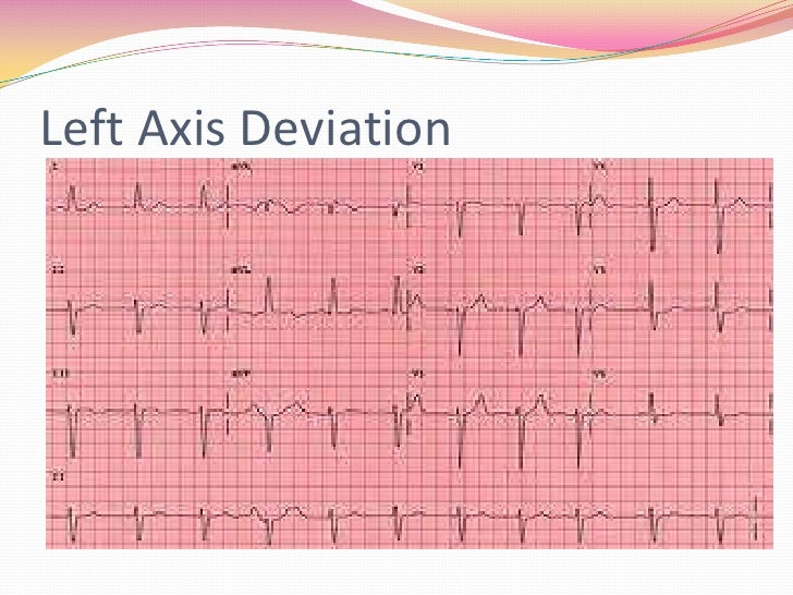
The normal QRS axis should be between -30 and +90 degrees. Left axis deviation is defined as the major QRS vector, falling between -30 and -90 degrees. Right axis deviation occurs with the QRS axis and is between +90 and +180 degrees.
The P wave and PR segment is an integral part of an electrocardiogram (ECG). It represents the electrical depolarization of the atria of the heart. It is typically a small positive deflection from the isoelectric baseline that occurs just before the QRS complex.
How to determine Axis on ECG?
the axis. In order to determine the axis, the electrocardiograph makes one electrode positive and one or more electrodes negative. The instrument then calculates the direction of depolarization, or the vector, for each lead and averages those results to yield the heart's axis. Interpretation of a 12-lead ECG depends on precise placement of the ...
What are normal ranges for ECG results?
Normal range up to 120 ms (3 small squares on ECG paper). QT interval (measured from first deflection of QRS complex to end of T wave at isoelectric line). Normal range up to 440 ms (though varies with heart rate and may be slightly longer in females) Heart rate estimation from the ECG. Standard ECG paper allows an approximate estimation of the heart rate (HR) from an ECG recording.
What is the normal speed of an ECG?
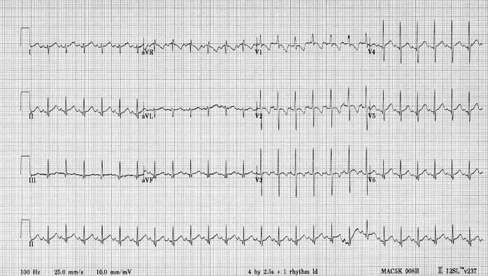
A standard ECG is recorded at 25mm/sec and with a frequency cut off of no lower than 150Hz in adults, and 250Hz in children. On the standard ECG paper, with standard calibration, the squares represent: The standard calibration signal will look like this: This will be present at the beginning or end of all four rows of the trace, and shows:
What is unusual P axis?
- Limb lead misplacement (most common cause)
- Dextrocardia
- Ectopic atrial rhythm
- Ventricular to atrial (V-A) conduction - abnormal P wave axis that occurs after the QRS interval
What is abnormal P axis?
An abnormal P-wave axis (aPWA) obtained in the routine 12-lead electrocardiogram (ECG) is thought to be a marker of left atrial fibrosis and delayed conduction. 14, 15. Consistent with this, aPWA has been linked to the development of atrial fibrillation, stroke and total mortality.
What is normal P value in ECG?
Normal ECG values for waves and intervals are as follows: RR interval: 0.6-1.2 seconds. P wave: 80 milliseconds. PR interval: 120-200 milliseconds.
What is normal P QRS axis?
Baseline ECG axes were automatically measured with normal values defined as follows: P-wave axis 0° to 75°, QRS axis -30° to 90°, and T axis 15° to 75°.
What does P indicate in ECG?
The P wave and PR segment is an integral part of an electrocardiogram (ECG). It represents the electrical depolarization of the atria of the heart.
What is an abnormal ECG reading?
Abnormal results can signify several issues. These include: Defects or abnormalities in the heart's shape and size: An abnormal ECG can signal that one or more aspects of the heart's walls are larger than another meaning that the heart is working harder than normal to pump blood.
What is an abnormal ECG report?
An abnormal EKG can mean many things. Sometimes an EKG abnormality is a normal variation of a heart's rhythm, which does not affect your health. Other times, an abnormal EKG can signal a medical emergency, such as a myocardial infarction (heart attack) or a dangerous arrhythmia.
What are good ECG values?
The normal range of the ECG differed between men and women: heart rate 49 to 100 bpm vs. 55 to 108 bpm, P wave duration 81 to 130 ms vs. 84 to 130 ms, PR interval 119 to 210 ms vs. 120 to 202 ms, QRS duration 74 to 110 ms vs.
How do you identify a normal ECG vs an abnormal ECG?
The signals with heart rates outside the range of 60–100 beats per minute and QRS durations outside the range of 0.08 sec to 0.12 sec are considered as abnormal signals. The ECG signals with heart rates and QRS durations within the range are considered as normal signals.
What is an abnormal T-wave axis?
T-wave axis was considered abnormal if >75° or <15° and QRS-T angle if ≥73° in men and ≥67° in woman. Associations were assessed by bivariate tests and multivariate logistic regressions. One hundred and four (20.9 %) patients had abnormal T-wave axis, and 84 (14 %) had increased QRS-T angle.
Do you have P waves in AFIB?
Diagnosis – Atrial Fibrillation. The diagnosis of atrial fibrillation is confirmed with a standard 12-lead ECG. P waves are absent, coarse “fibrillatory waves” can frequently be seen and sometimes no atrial activity can be identified. The QRS complexes are “irregularly irregular”, with varying R-R intervals.
What does a high P wave mean?
The presence of tall, peaked P waves in lead II is a sign of right atrial enlargement, usually due to pulmonary hypertension (e.g. cor pulmonale from chronic respiratory disease).
What does depressed P wave mean?
Elevation or depression of the PTa segment (the part between the p wave and the beginning of the QRS complex) can result from atrial infarction or pericarditis. If the p-wave is enlarged, the atria are enlarged. If the P wave is inverted, it is most likely an ectopic atrial rhythm not originating from the sinus node.
What are good ECG values?
The normal range of the ECG differed between men and women: heart rate 49 to 100 bpm vs. 55 to 108 bpm, P wave duration 81 to 130 ms vs. 84 to 130 ms, PR interval 119 to 210 ms vs. 120 to 202 ms, QRS duration 74 to 110 ms vs.
Why does ECG start with P?
When Einthoven used the letter P, he was undoubtedly thinking about Descartes' use of the letter P to designate a point on a curve. As will be shown, this eventually led to the use of PQRST to designate the waves in the ECG.
How do you read a normal and abnormal ECG?
The signals with heart rates outside the range of 60–100 beats per minute and QRS durations outside the range of 0.08 sec to 0.12 sec are considered as abnormal signals. The ECG signals with heart rates and QRS durations within the range are considered as normal signals.
Can ECG detect heart failure?
Tests for heart failure Tests you may have to diagnose heart failure include: blood tests – to check whether there's anything in your blood that might indicate heart failure or another illness. an electrocardiogram (ECG) – this records the electrical activity of your heart to check for problems.
What does in an ekg a p axis 26 and qrs axis -23?
Electrical angle: They are angles of electrical vectors of the upper (p) and lower (qrs) chambers of the heart. P wave axis is within normal range. The qrs is leftward ... Read More
What is an EKG?
Ekg : An EKG is a report of the electrical signals for only 6 seconds of the heart. Those numbers seem to be within accepted physiological ranges. Normal i ... Read More
Is EKG normal if heart rate is not too bad?
EKG: Other than the heart rate not too bad the axis and intervals are normal.
What is the cardiac axis?
Cardiac axis represents the sum of depolarisation vectors generated by individual cardiac myocytes. Clinically is is reflected by the ventricular axis, and interpretation relies on determining the relationship between the QRS axis and limb leads of the ECG (below diagram)
Which direction does the cardiac axis go?
Since the left ventricle makes up most of the heart muscle under normal circumstances, normal cardiac axis is directed downward and slightly to the left:
What direction does the QRS point?
If the QRS is POSITIVE in any given lead, the axis points in roughly the same direction as this lead.
What does a positive QRS in lead I mean?
A positive QRS in Lead I puts the axis in roughly the same direction as lead I.
What is the axis of QRS?
If the QRS is ISOELECTRIC (equiphasic) in any given lead ( positive deflection = negative deflection ), the axis is at 90° to this lead.
What quadrant is the axis in?
This puts the axis in the upper right quadrant, between -90° and 180°, i.e. extreme axis deviation.
What is the axis of lead I and II?
We can then combine both coloured areas and the area of overlap determines the axis. So If Lead I and II are both positive, the axis is between -30° and +90° (i.e. normal axis).
What is the P wave axis?
P wave axis is one of the routinely reported variables on the printout of most electrocardiograms (ECGs) that may be the most practical clinical tool for evaluation of cardiovascular disease and may provide better predictive value than the currently used P wave indexes. The P wave is the first positive deflection on the ECG and represents atrial depolarisation. Normal P wave axis is between 0° and +75°. However, the relationship between P wave abnormalities and the cardiovascular death has been determined before.3
How fast is a standard 12 lead surface ECG?
Methods:Standard 12-lead surface ECGs were recorded by at a paper speed of 25 m/s and an amplifier gain of 10 mm/mV. The heart rate (HR), the duration of PR, QRS, QTd (dispersion), the axis of P wave were measured by ECG machine automatically.
What is the speed of a Burdick Atria 6100 EKG machine?
Standard 12-lead surface ECGs were recorded by ‘The Cardiac Science Burdick Atria 6100 EKG Machine’ at a paper speed of 25 m/s and an amplifier gain of 10 mm/mV. The heart rate (HR), the duration of PR, QRS, QTd (dispersion), the axis of P wave were measured by ECG machine automatically.
Does SLE have a positive correlation with P wave axis?
SLE disease activity score was found positively correlated with the P wave axis degrees which means that; as the SLE activity getting impaired, P wave axis degree increases that may also increase the risk of all-cause and cardiovascular mortality in patients with SLE.
Do SLE patients have abnormal P wave axis?
Second, the long term monitoring to detect the possible arrhythmia or the risk of mortality is lacking. Altough SLE patients did not have abnormal P wave axis, we should emphasize that they were in the remission period. The P wave axis degrees are greater than the control group even in the lack of disease activity. The most important limitation of our study is, we did not compare them with the SLE patients in the active period.
Does the P wave axis increase with SLE?
Conclusion:P wave axis was found significantly increased in patients with SLE and positively correlated with SELENA-SLEDAI score. As the risk score increases in patients with SLE, P wave axis changes which may predict the risk of all-cause and cardiovascular mortality.
Does the P wave axis affect LA volume?
According to a study by Win et al, there was an association between p wave duration and increase in LA volume and decrease in LA emptying fraction and reservoir function but they did not find any association with P wave axis.16Likewise, we could not find any association between the P wave axis and the LA volume and LA peak systolic strain.
