
Ray Amputations. In a finger example, ray amputations are the removal of an entire finger along with the corresponding metacarpal bones in the hand. They are same-day surgeries with the patient going home with a bulky soft dressing. The recoveries can vary, but light use of the hand is almost immediate.
What is a ray amputation?
In a finger example, ray amputations are the removal of an entire finger along with the corresponding metacarpal bones in the hand. They are same-day surgeries with the patient going home with a bulky soft dressing.
When is Ray resection indicated in the treatment of finger amputation?
Today, ray resection results in good cosmetic and functional outcomes when preservation of a functional digit is unattainable or when the presence of an abnormal, unreconstructable digit interferes with the overall hand function. Keywords: amputation; finger; ray resection.
What happens when you have your finger amputated?
While the amputation of a finger is a relatively minor amputation, it is still an operation. An amputation affects not only the function of your hand, but its appearance as well. Initially, the situation will be unusual for you - you have to learn what you can still do after the amputation...
What is RayRay resection of the finger?
Ray resection of the finger is performed to reduce pain and improve function by removing the finger using metacarpal resection.
Is a ray resection and amputation?
The indications for ray resection are ischemic necrosis involving the metacarpal, severe dysfunction of the proximal interphalangeal joint (PIPJ) and amputations at the level of the proximal phalanx (6, 7).
What is first ray amputation?
We defined the partial first ray amputation as occurring distal to the first metatarsocuneiform joint, including the phalanges of the hallux, with primary closure at the time of surgery.
How painful is a finger amputation?
Your fingertips have many nerves and are very sensitive, so the injury may be very painful. Recovery can take several weeks. Your finger may be sensitive to cold and painful for a year or more.
What is a third ray amputation?
Ray amputation The removal of a single metatarsal in the middle of the foot (ie, the second, third, or fourth metatarsal) results in a V-shaped wedge, which again maintains good function.
How do you do a ray amputation?
The technique of central ray amputation involves the use of a circumferential incision at the midproximal phalanx in conjunction with a dorsal longitudinal incision. The dorsal incision is extended through the extensor. The periosteum is scored at the level of the metacarpal base.
What is partial 1st ray amputation?
Partial first-ray resections are used to help salvage the foot and maintain bipedal ambulation. Losing the first metatarsophalangeal joint has biomechanical consequences that lead to further foot deformities and result in more proximal amputations of the ipsilateral limb, such as a transmetatarsal amputation.
How long are you in the hospital after a finger amputation?
Average Hospital Stay Leg: 2 days to 2 weeks or more. Arm: 7 to 12 days. Finger: 0 to 1 day.
What happens after finger amputation?
After an amputation, pain, swelling, and hand stiffness can be problems at first, but gradually improve with time. Scar sensitivity is common and can be improved with scar massage and hand therapy. Some people report an increase in symptoms during cold weather.
Is finger amputation a disability?
Losing a finger certainly can qualify as a disability, as you clearly would not have all of the same physical skills as someone with all of their digits. No matter which finger is lost, you may be able to qualify for compensation and assistance.
Why is Ray amputation?
Ray amputation was first described in the early 20th century [3-6], and it was performed on those with proximal interphalangeal joint dysfunction or loss of proximal phalangeal skeleton; it was also used for the treatment of traumatic hand injuries, infections, and tumors [7].
What is 5th ray amputation?
Traditional Partial Fifth Ray Amputation The traditional approach to partial fifth ray amputation involves a standard tennis racket incision consisting of dorsal and plantar soft tissue flaps with midline proximal extension along the lateral border of the foot (Fig. 18.13).
What are the types of amputations?
Common types of amputation involve:Above-knee amputation, removing part of the thigh, knee, shin, foot and toes.Below-knee amputation, removing the lower leg, foot and toes.Arm amputation.Hand amputation.Finger amputation.Foot amputation, removing part of the foot.Toe amputation.
What is the first ray of the foot?
The first ray is the segment of the foot composed of the first metatarsal and first cuneiform bones. The location of this joint is important as it intersects the transverse and medial longitudinal arches. This segment serves as a critical element in the structural integrity of the foot.
What is 5th ray amputation?
Traditional Partial Fifth Ray Amputation The traditional approach to partial fifth ray amputation involves a standard tennis racket incision consisting of dorsal and plantar soft tissue flaps with midline proximal extension along the lateral border of the foot (Fig. 18.13).
What is Ray amputation of toe?
Ray amputation, which involves the excision of the toe and part of the metatarsal, provides a more viable option of ensuring an adequate surgical debridement of the septic margins.
Where is the 1st metatarsal?
The first metatarsal bone is the bone in the foot just behind the big toe. The first metatarsal bone is the shortest of the metatarsal bones and by far the thickest and strongest of them.
What is digit amputation?
Digit amputations are most commonly performed for bite wounds to the digit or for traumatic degloving, shearing, or crush injuries. Initial management following an injury should be conservative, as many injuries will resolve with treatment with NSAIDs and antibiotics, and marmosets can function well with osteoarthritis and ankylosis of the interphalangeal joints. Digital wounds with severe soft tissue injury and cellulitis, however, may require amputation. Prior to surgery, local anesthetic is applied within the joint at the site of amputation and in the soft tissues. The level of amputation is determined by the extent of the injury and health of the soft tissue, as these will determine if closure of the wound can be achieved. Due to the small size of the digits, dissection of the deeper tissues (flexor and extensor tendons) is limited. For amputation, a circumferential skin incision should be made at a point distal to the joint to allow for disarticulation of the interphalangeal joint via transection of the flexor and extensor tendons, as well as collateral ligaments and joint capsule. Digital arteries and veins can be difficult to identify and ligate, so hemostasis is generally managed by compression and cautery. Closure of the surgical site can include an interrupted subcutaneous suture with simple interrupted skin sutures. A light bandage can be applied over the surgical site but must allow for use of the remaining digits for ambulation around the cage. The authors recommend a small bandage of a nonadherent dressing (Telfa, Coviden, Mansfield, MA, USA) trimmed to cover only the immediate surgical site followed by an adherent dressing retention sheet (Hypafix, BSN Medical, Charlotte, NC, USA or Tegaderm, 3M Health care, St. Paul, MN, USA). Analgesia should include an NSAID to help reduce inflammation at the surgical site.
Where is revision finger amputation done?
In general, revision finger amputations are done through the bony shaft, rather than at joint level. Knowing the anatomy of the fingers is important for maintaining attachments of flexor and extensor tendons if possible, as well as contouring bone appropriately for the revision stump (Fig. 3.2A and B ).
What is lumbrical plus digit?
Lumbrical plus digit (paradoxical extension) occurs when the interphalangeal (IP) joints of the injured finger extend when the patient attempts to actively perform a composite fist. When an amputation occurs in which the FDP integrity is disrupted, the FDP muscle retracts, thus pulling the lumbrical origin proximally, which increases tension on its insertion at the radial lateral band. Thus, with FDP contraction, force is transmitted through the tightened lumbrical to the extensor mechanism, causing an extension force of the IP joints. 11,18,19
What is the term for a shortening of the FDP tendon?
Following a trauma, such as an amputation, quadrigia may occur if the lacerated proximal FDP tendon is surgically secured to the wound or pulley (to achieve tenodesis) more distally than its natural position. The common FDP muscle belly to the middle, ring, and small fingers would allow only as much excursion in each digit as that which occurs in the shortest tendon. Therefore, quadrigia would prevent the amputated and uninvolved fingers from being able to achieve full composite flexion and cause a decrease in grasp strength. 11,18
How many members of Boryoku Dan have had their fingers cut off?
It was found that 194 of 741 (or 26%) members of Boryoku-Dan and 95 of 355 (or 27%) members of Boryoku-Josho-Sha have had their fingers cut off. Digital amputation is usually done at the middle phalanx of the left little finger.
What is the treatment for sharp amputation?
For sharp amputations where the removed digit is in a clean and fresh state, immediate re-implantation is considered the treatment of choice, which involves microsurgical anastomosis of nerves, blood vessels, bone, and tendon.
Why are digit prostheses not suitable for pediatric patients?
These prosthetic devices are often associated with complications, such as erosion, infection, and inflammation. Moreover, prosthetic devices are not suitable for pediatric or adolescent patients, as their tissues grow with age. Therefore, most patients decide not to wear the prosthesis, due to the lack of gain in function and its associated complications . Despite the rapid technological advances in medicine, treatment options for digit reconstruction are severely limited. Currently available modalities are involved with a prolonged treatment period, multiple stage surgeries, a lengthy rehabilitation process for a questionable restoration of normal function, and prostheses and surgery-associated complications.
What is the procedure called when you remove a finger?
In situations where we are removing digits such as fingers, hand surgeons perform an operation called 'ray amputations '. In a finger example, ray amputations are the removal of an entire finger along with the corresponding metacarpal bones in the hand.
Why do we need to amputation?
The amputation can be performed so that it takes several looks to notice that a digit is missing. The middle digits are a little tougher, but the gap created can be closed so that the cosmetics and functionality are positive. Also, even for people who work with their hands, you're back at work fairly quickly.
Is it bad to exercise after amputation?
The exercise can help reduce any swelling present as well. Cosmetically, it's not too bad either. Especially when the amputation involves a border digit such as the pointer finger or the pinky.
Can you get back to life after amputation?
While initially thought of as a scary and life-altering incident, amputations of the digits are actually quite common and people get back to living their life at just about the same speed as before. Your body and mind are capable of working together so that you'll develop new, slightly different movements to compensate for the new biological logistics.
Is ray amputation scary?
Ray Amputations. "Amputation" is a frightening word. Many people find the instant images presented in their minds as unpleasant and uncomfortable. When it is a medical treatment possibility for you… it is even more fearful.
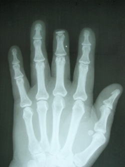
Where is the second finger fractured?
The second finger is transversely fractured through the head of the second metacarpal, with the fracture off-ended and volarly displaced.
Is the fifth finger amputated?
The fifth finger is obliquely amputated through the proximal shaft of the proximal phalanx. The finger is absent, imaged separately showing no further fracture of the phalanges. The MCP joint is normal.
What is the postoperative care of a residual limb?
The postoperative care of the residual limb is handled by medical professionals who regularly change the compression dressings. The goal is to support the rapid absorption of blood and fluid build-up in the tissues around the wound.
What is a prosthesis?
The prosthesis protects the sensitive residual limb from direct contact with objects and the socket is individually fitted to the particular situation. Please note that a prosthesis is an aid, but cannot entirely replace the complex functions of a hand. You must anticipate certain limitations, even with a prosthesis.
How does an amputation affect your hand?
An amputation affects not only the function of your hand, but its appearance as well. Initially, the situation will be unusual for you - you have to learn what you can still do after the amputation and what functions you no longer have. For many of those affected, the initial impact of an amputation is major.
How does losing a finger affect a person?
The loss of a finger or parts of the hand sometimes affects behaviour in public. Some of those affected want to conceal the hand from others. In rare cases, the patient has difficulty accepting his or her own hand after an amputation. If this is the case, the person should seek help and take part in post-amputation therapy as well as occupational ...
Can you use compression dressings on residual limb?
In addition, slight pressure is applied using compression dressings to compress the residual limb as much as possible. If the residual limb is to be fitted with a silicone prosthesis later, it is advisable to use compression aids made of silicone now. It cannot be predicted how long you need until the wound is healed.
Is a finger amputation a minor operation?
Take your time to adjust to your new situation. While the amputation of a finger is a relatively minor amputation, it is still an operation.
Can you get a prosthesis after an amputation?
Being fitted with a prosthesis can be an option that will help you to return to your normal life after an amputation.
What is the technique of index finger ray amputation?
A to D, Technique of index finger ray amputation. After the appropriate skin flaps are raised, dissection proceeds from dorsal to volar. After division of the extensor digitorum communis and extensor indicis proprius, the index finger metacarpal is transected at its flare, distal to the extensor carpi radialis longus insertion. Following division of the lumbrical, dissection proceeds volarly where the neurovascular bundles are identified. The digital nerves are transposed into the interosseous space to prevent the formation of a symptomatic neuroma.
How to do a ray amputation?
The distal ends of the incision are carried over the proximal phalangeal skin to preserve sufficient skin for closure of the commissure between the index and ring fingers. This technical point is important when performing ray amputations of the middle two rays to preserve skin and prevent iatrogenic web contracture. Alternatively, a web-saving incision that avoids placing the scar in the new web space can be used ( Figure 49.22 ).
What is a V-Y flap?
In 1970, Atasoy and associates described a triangular volar “V-Y” advancement flap ( Figure 49.4 ) for reconstruction of the distal pad with preservation of length when bone is exposed. It is indicated for dorsal oblique or transverse distal fingertip amputations beyond the midpart of the nail. The flap is contraindicated in injuries in which there is an oblique amputation with more palmar skin loss than dorsal skin loss and in situations in which there is extensive skin loss as a result of the injury.
What is the most common type of amputation?
An amputation of the fingertip is the most common type of amputation seen in the upper extremity and at the same time provokes the greatest controversy because of the multitude of treatment options. Multiple techniques have been developed to advance local skin or transfer soft tissue to ensure coverage of an area where bone is exposed. The surgeon, in consultation with each patient, must choose the type of coverage that appears to be most appropriate for that individual’s needs and within the technical abilities of the surgeon. Regardless of choice of treatment, the goals of preserving functional length and restoring adequate sensibility remain constant. When one is comparing the various treatment options available or looking for guidance, the injury zone classification systems of Hirase, Allen, and Tamai provide a useful frame of reference ( Figure 49.1 ).
What is an island flap?
A number of elegant and ingeniously conceived island flaps have been described for coverage of fingertip amputations. These consist of flaps raised on their neurovascular pedicle, and advantages include the avoidance of prolonged digital immobilization in an awkward position, single-stage reconstruction permitting early rehabilitation, introduction of an independent blood supply to provide a good soft tissue bed for nerve grafting or repair, satisfactory restoration of a well-padded sensate digital pulp, the potential for a composite tissue transfer, a wide selection of donor sites, and a greater arc of rotation for mobilization of the flap over longer distances. These include homodigital and heterodigital island flaps and can be anterogradely or retrogradely based. However, island flaps are more technically demanding and are associated with complications such as flap failure, joint contracture, and cold intolerance. In addition, they have not proved to be more advantageous than simpler methods of achieving coverage of a fingertip amputation. The use of an island flap may be beneficial in select cases, and the reader is referred to comprehensive review articles to become familiar with the indications and surgical techniques.
How long did Sturman and Duran monitor fingertip amputation?
Sturman and Duran monitored 235 patients with a fingertip amputation for a minimum of 1 year and reported their late outcomes. Of the patients who had split-thickness skin grafts, 70% reported tenderness in the area of the graft, and in 41%, the tenderness was considered to be marked. Cold sensitivity was present in 49% of the patients who had split-thickness skin grafts, including 27 of the 53 who had thumb or index tip amputations. Fifty-nine percent of the latter group avoided the injured digit when the pickup test was administered. Thirty-two percent of them noted diminished touch sensibility. The average two-point discrimination measured was 5 mm, considerably greater than the normal 2 mm.
What tendon is released to the index finger?
Distal release of the FDP tendon to the index finger may lead to a “lumbrical-plus” deformity. As the independent FDP and its lumbrical move proximally, tension increases in the lumbrical tendon and its contribution to the intrinsic extensor of the PIP joint. As active flexion of the digit is attempted, further proximal migration of the lumbrical may put sufficient tension on this lateral band to cause paradoxic extension of the PIP joint. Sectioning the lumbrical tendon allows the superficialis to regain control of the PIP joint and may alleviate the problem. In my experience, this rarely occurs, and sectioning of the lumbrical is unnecessary at the time of the amputation.
