
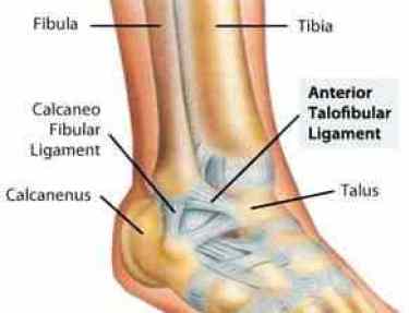
The most common mechanism of injury in ankle sprains is a combination of plantar flexion and inversion. The lateral stabilizing ligaments, which include the anterior talofibular, calcaneofibular and posterior talofibular ligaments, are most often damaged. The anterior talofibular ligament is the most easily injured.
What causes a lateral collateral ligament sprain?
A lateral collateral ligament (LCL) sprain occurs when there is a tear in the ligaments on the outside of the knee. Causes include sports injuries and accidents. Symptoms include pain, swelling ...
Should I Ice a ligament sprain?
Ice: experts recommend applying an ice pack to the injured area for 20 minutes at a time, four to eight times a day. Compression: put pressure on the injured area to decrease swelling. This can mean elastic wraps, air casts, and splints. For more severe ankle sprains, your doctor may recommend a walking boot which keeps air pressure on the ...
Can ligaments heal on their own. posterior cruciate ligament?
Posterior cruciate ligament tears tend to be partial tears with the potential to heal on their own. People who have injured just their posterior cruciate ligaments are usually able to return to sports without knee stability problems.
What are the grades of ligament tears?
- grade 1: (minor sprain) high signal is seen medial (superficial) to the ligament, which looks normal
- grade 2: (severe sprain or partial tear) high signal is seen medial to the ligament, with high signal or partial disruption of the ligament
- grade 3: complete disruption of the ligament
How do you treat a calcaneofibular ligament sprain?
Treatment / Management During the initial inflammatory response, RICE (rest, ice, compression, and elevation) is implemented in the first 4 to 5 days. Immobilization with cast or boots can be applied in the first week to reduce swelling and pain, after which, brace or taping can be provided for a return to activity.
How long does a torn calcaneofibular ligament take to heal?
ligament. Mild tenderness and swelling around the ankle, typically recovers in 5-14 days. Partial tearing of anterior talofibular ligament and some tearing of the calcaneofibular ligament. Moderate tenderness and swelling around the ankle, typically will take 2-3 weeks to recover.
Can you walk on a torn calcaneofibular ligament?
It can become overly stretched in a sprain, partially torn, or even completely torn (ruptured). Small tears of the ATFL will cause pain, tenderness, and swelling, but walking is usually still possible. Larger ATFL tears will cause greater pain, swelling and bruising, and you may have difficulty walking.
What does the calcaneofibular ligament do?
Function. The CFL provides lateral stabilization to the ankle joint and subtalar joint. The ligament allows flexion and extension movements to occur at the ankle and serves as a primary restraint to inversion in neutral or dorsiflexed position at the talocrural joint.
How do you injure your calcaneofibular ligament?
Calcaneofibular ligament injuries typically occur along with an anterior talofibular ligament injury within the scope of a lateral ankle sprain. The typical mechanism is excessive supination of the rearfoot on an externally rotated lower leg, leading to an inversion-internal-rotation type injury 1,2.
Can walking on a sprained ankle make it worse?
Yes. That's the very short answer. According to the National Association of Athletic Trainers, ankle injuries, including sprains, are very often undertreated. Ignoring treatment, including excessive movement of the ankle through unnecessary walking, leads to a greater risk of worsening the injury.
What does a CFL tear feel like?
Symptoms. Swelling and tenderness occur over the injured ligament, distal to the lateral malleolus. Pain is felt during weightbearing and when moving the ankle joint. Effusion of blood causes hematoma and bruising behind and below the lateral malleolus.
Do you need crutches for a sprained ankle?
Your doctor may recommend not putting any weight on the injured area for 48 to 72 hours, so you may need to use crutches. A splint or brace also may be helpful initially. But don't avoid all activity. Even with an ankle sprain, you can usually still exercise other muscles to minimize deconditioning.
How should you sleep with a sprained ankle?
Elevating a sprained ankle reduces the accumulation of fluid in the joint. This can ease swelling, which may also help reduce pain. Try sleeping with the foot and ankle propped up on pillows at a level higher than the heart. When sitting or resting, use pillows or a footrest to keep the foot and ankle elevated.
How do you test for calcaneofibular ligament?
1:012:26The Talar Tilt Test | Lateral Ankle Sprain - YouTubeYouTubeStart of suggested clipEnd of suggested clipIn order to test the calcaneofibular ligament. Bring your patient's foot into the anatomical.MoreIn order to test the calcaneofibular ligament. Bring your patient's foot into the anatomical. Position. So that the ligament is perpendicular to the long axis of the tailless.
What test is used to determine if there is damage to the calcaneofibular ligament?
It is also known as the inversion stress test and it stresses the calcaneo-fibular ligament, Procedure: The patient is positioned in sitting or supine lying with the knee in full extension. The examiner stabilizes the distal leg with one hand while the other hand holds the heel with the ankle in neutral position.
Do ankle ligaments heal on their own?
Nonsurgical Treatment Nearly all isolated low ankle sprains can be treated without surgery. Even a complete ligament tear (Grade 3) will heal without surgical repair if it is immobilized and rehabilitated appropriately.
How do you know if you tore your calcaneofibular ligament?
MRI. The usual appearances of calcaneofibular ligament injury are thickening in case of a sprain or chronic injury, ligament contour irregularities, partial discontinuity, and signal intensity changes in case of partial tear and complete discontinuity and fiber retraction in case of a complete tear 3-5.
What is Grade 2 sprain of the calcaneofibular ligament?
Grade 2 is a partial tear. Grade 3 is a full tear. Often, the level of pain in a grade 3 full tear is less than a partial tear and results in the patient seeking less treatment. A more descriptive grading should include the level of instability and the potential number of ligaments torn.
What is the most commonly torn ankle ligament?
The most common and significant ligament tears include tears in the Anterior TaloFibular Ligament (ATFL), CalcaneoFibular Ligament (CFL), and the large Deltoid ligament complex.
Does a torn ATFL and CFL require surgery?
Clinical Management Of ATFL Rupture The concern would be that they develop chronic instability in the ankle or an anterior ankle impingement long term, which would require surgery. This can occur in patients who either present to physiotherapy late, or are non- compliant with their rehab.
CFL and AFTL
The calcaneofibular ligament injury rarely occurs without also injuring that anterior talofibular ligament. During an ankle sprain, the anterior talofibular ligament comes to tension first due to its anterior location, and then once the AFTL fails, the calcaneofibular ligament becomes injured.
Instability
This form of ankle instability involves talocural joint and the inability to control talar tilt. This form of ankle instability can cause the ankle to become stiff, swollen, painful, and lead to a sensation of the ankle giving out.
Diagnosis
Calcaneofibular ligament orthopedic tests include the talar tilt test, anterior drawer test, and tenderness with palpation of the ankle ligament.
About the Author: Dr. Justin Dean
Dr. Justin Dean is a clinical educator and Chiropractor based in Los Angeles.
What is a calcaneofibular injury?
Calcaneofibular ligament injuries typically occur in conjunction with an anterior talofibular ligament injury within the scope of a lateral ankle sprain and are rarely found isolated. The injuries can comprise either ligament tears, avulsion fractures, or both.
What are the symptoms of calcaneofibular ligament injury?
The usual appearances of calcaneofibular ligament injury are thickening in case of a sprain or chronic injury, ligament contour irregularities, partial discontinuity, and signal intensity changes in case of partial tear and complete discontinuity and fiber retraction in case of a complete tear 3-5.
Which ligament should be assessed for possible deltoid ligament injury?
In the case of combined anterior talofibular and calcaneofibular ligament injury , the deltoid ligament should also be assessed for possible deltoid ligament injury.
Which ligament is the second most common injury?
The calcaneofibular ligament is the second most common ligament injured (after the anterior talofibular ligament) in a very high percentage of lateral ankle sprains. Isolated calcaneofibular ligament injury is rare 1,2.
What is the treatment for a swollen ankle?
Management is nevertheless most often conservative and includes initial treatment with rest, ice, compression and elevation ( RICE) and semi-rigid ankle brace and/or a short-term period of immobilization in more severe cases 6.
Can a lateral ankle sprain be a calcaneofibular injury?
Conversely, an unstable ankle joint after sustaining a lateral ankle sprain will more likely coincide with a calcaneofibular ligament injury. The calcaneofibular ligament can additionally be assessed with the inversion stress test in a dorsiflexed position 1,2.
Can a lateral ankle sprain cause instability?
Clinical signs and symptoms are that of an acute lateral ankle sprain, but in case of a calcaneofibular injury they will be typically more severe and also depending on the grade of injury will result in talocrural joint instability and talar tilt. Conversely, an unstable ankle joint after sustaining a lateral ankle sprain will more likely coincide ...
Presentation
About 6 months prior to the presentation, the patient had the right ankle inversion injury. The patient presented with a complaint of persistent pain in the lateral ankle region.
Case Discussion
The case shows isolated calcaneofibular ligament sprain with a normal anterior talofibular ligament. Ankle inversion is a common injury involving athletes and nonathletes. Injuries involving anterior talofibular ligament with/without calcaneofibular ligament are common. However, an isolated injury to the calcaneofibular ligament is considered rare.
What is the function of the calcaneofibular ligament?
The calcaneofibular ligament’s responsibility is to control inversion. Inversion involves turning the foot on its side, so the bottom of the foot faces the opposite foot. The calcaneofibular ligament connects the talus and calcaneus (heel) bones of the foot.
How thick is the talar ligament?
The ligament is two centimeters long, five millimeters wide, and three millimeters thick. Damage to this ligament occurs when the foot twists too much while the toes point upwards towards the shin. Doctors diagnose damage with a talar tilt test.
What bones are in the ankle?
Calcaneofibular ligament. The ankle bones include the calcaneus, cuboid, external cuneiform, internal cuneiform, middle cuneiform, navicular, and talus. The talus sits at the top, under the fibula and tibia (the bones of the lower leg).
What is the function of ligaments and tendons?
Ligaments and tendons (types of fibrous connective tissue) connect the leg bones with the ankle bones, thus preventing slippage. They also offer stability during movement. Tendons protect the ligaments. When a person is standing, the ligament is slack.
What is a sprain ankle?from ncbi.nlm.nih.gov
Ankle sprains are a common reason for presentation to the emergency department, accounting for approximately 7% to 10% of visits and up to 40% of all sports injuries.[1] The majority of ankle injuries are sports-related and involve the lateral ankle compartment. The lateral ankle ligaments consist of the anterior talofibular ligament (ATFL), the calcaneofibular ligament (CFL), and the posterior talofibular ligament (PTFL). Differentiating between ATFL-superimposed CFL injuries vs. isolated CFL injuries are challenging as clinical exams yield low sensitivities; however, it is commonly accepted that the ATFL is largely involved in the majority of ankle sprains, accounting for two-thirds of lateral ankle injuries.[2] While there is limited literature available for isolated CFL injuries, combined ATFL and CFL involvement are the second most common injury pattern of the lateral ankle.[3] As such, discussions about CFL injuries in the literature are largely embedded in lateral ankle injuries. This article will discuss the shared characteristics of lateral ankle injuries and identify the unique qualities of CFL injuries.
How rare are CFL injuries?from ncbi.nlm.nih.gov
Isolated CFL injuries are rare as reported incidences are commonly classified under lateral ligament injuries. Thirty thousand ankle sprains occur each day, accounting for 25% to 40% of sports injuries. The lateral ligament compartment is involved in 85% of ankle injuries, with a daily incidence of one in 10,000. [1][5][6]
What is the CFL ligament?from ncbi.nlm.nih.gov
The CFL is reinforced by either the lateral talocalcaneal ligament or anterior talocalcaneal ligament, in 35% and 42% of cases, respectively.[7] The CFL resists inversion in both plantarflexion and dorsiflexion and stabilizes the subtalar joint during plantarflexion. While combined inversion and supination is the mechanism of injury to the lateral ankle, an isolated CFL injury occurs from inversion in extreme dorsiflexion. [3][6] The CFL can sustain a loading force of 109 plus or minus 28 N, and while working with ATFL stretch during high-grade ankle sprains, it can withstand a force of 345 N.[1]
How thick is the talar ligament?from healthline.com
The ligament is two centimeters long, five millimeters wide, and three millimeters thick. Damage to this ligament occurs when the foot twists too much while the toes point upwards towards the shin. Doctors diagnose damage with a talar tilt test.
What is the talus?from healthline.com
The talus sits at the top, under the fibula and tibia (the bones of the lower leg). Ligaments and tendons (types of fibrous connective tissue) connect the leg bones with the ankle bones, thus preventing slippage. They also offer stability during movement. Tendons protect the ligaments.
What bones are in the ankle?from healthline.com
Calcaneofibular ligament. The ankle bones include the calcaneus, cuboid, external cuneiform, internal cuneiform, middle cuneiform, navicular, and talus. The talus sits at the top, under the fibula and tibia (the bones of the lower leg).
How long does it take for a CFL to heal?from ncbi.nlm.nih.gov
The progression of healing following initial injury has three distinct phases: inflammatory (1 to 10 days ), proliferative (4 to 8 weeks), and remodeling phase (up to one year).[13] As such, each biological phase offers a unique therapeutic window for intervention.
What is the calcaneofibular ligament?
The calcaneofibular ligament is part of the lateral ankle ligament complex consisting of the ATFL, CFL, and posterior talofibular (PTFL) ligaments. The CFL is typically 6-8 mm thick and runs along a length of 20 mm in an oblique course from the tip of the lateral malleolus posteriorly and inferiorly, attaching to the trochlear eminence of the calcaneus. The CFL is situated inferior to the ATFL with occasional fibers that connect between them. 2 The CFL is also seated deep to the peroneal tendon complex and is near completely covered by its posteromedial portion. 3 The ligament crosses the ankle and subtalar joints, the only ligament that spans 2 separate joints laterally. The ligament is taut in flexion, extension and varus angulation but relaxes during valgus stress to the ankle. The CFL is often seen in close apposition to a smaller ligament that lies medial and anterior, the lateral talocalcaneal ligament (LTCL).
What is lateral ligament injury?
Injury to the lateral ligamentous structures usually occur after suffering an inversion type injury to the ankle. When the ankle is stressed, the vector forces are greatest laterally. If these forces are greater than the tensile strength of the ligament then the lateral ligaments can tear or rupture. Typically these injuries occur with the ankle also in plantar flexion and the ATFL is the first ligament to tear. It is postulated that the CFL could tear in the absence of ATFL if the ankle is inverted in neutral or dorsiflexed position. On examination, patients can present with tenderness to palpation over the CFL, correlating to a 72 percent chance of ligament injury. 10 In addition to pain, edema and ecchymosis are often visualized. Joint instability can be assessed using the anterior drawer test at the ankle which could relate to injury to the ATFL, CFL, or both. Clinically, a 3 grade system is used to classify the injuries. Grade 1 is a minor injury or sprain. Grade 2 and 3 injuries respectively equate to partial or complete tears. Lateral ankle stress test maneuvers have been shown to correlate poorly with the degree of ligament disruption. 11 Therefore, MR imaging is essential in proper diagnosis.
What is the role of the CFL in the ankle?
The CFL provides lateral stabilization to the ankle joint and subtalar joint. The ligament allows flexion and extension movements to occur at the ankle and serves as a primary restraint to inversion in neutral or dorsiflexed position at the talocrural joint. The CFL also serves to resist subtalar joint inversion. This limits talar tilt. In cases of absence of the LTCL, the CFL has a greater stabilizing role to the subtalar joint. 4 The CFL is tense in dorsiflexion and is a secondary stabilizer to the ATFL. 5 The CFL increases in length in dorsiflexion and pronation. 6 Extreme inversion forces to the ankle can cause injury and ultimately rupture to the CFL. It has been shown that the CFL tension is increased mainly in inversion and dorsiflexion while the ATFL acts as the primary restraint in inversion and plantarflexion. 7 The CFL is typically not injured in isolation because the ATFL is the weakest ligament and the great majority of ankle inversion injuries occur with at least some degree of plantar flexion (Figure 4). The least commonly torn stabilizing ankle ligament is the PTFL. An anatomic classification of ankle sprains has been developed. A first degree sprain consists of a partial or complete tear of the ATFL; a second degree sprain consists of partial or complete tearing of the both the ATFL and CFL, and a third degree sprain consists of injury to all of the lateral ligaments, including the PTFL. 8
What happens if you have a CFL injury?
More chronic injury to the CFL can result in scarring/thickening or ligament attenuation/non-visualization without surrounding edema. (Figure 9)
What is the best imaging for ankle ligamentous complex?
MRI is the study of choice in evaluation of the lateral ankle ligamentous complex. A high resolution extremity coil is best suited for visualization of these small structures. The CFL is best imaged when the ankle is placed in plantar flexion (roughly 20 degrees) as this can lessen the magic angle effect 9 which can obscure visualization. The CFL (and the ATFL) appears as a low signal fiber bundle on all imaging sequences in the normal state. The CFL is best seen on coronal and axial views but is usually not visualized in its entirety on one single image. Multiple continuous images are typically needed to trace the course of the ligament. The CFL appears band-like extending in the anteroposterior direction and courses parallel to the lateral calcaneal wall with the foot in plantar flexion (Figure 5a). The CFL on coronal views is more round in shape but occasionally can be imaged in its entirety depending on the obliquity of the slice selection (Figure 5b).
How to treat grade 1 and 2 ankle ligamentous complex?
The ankle is stabilized in a boot for immobilization. Most injuries respond well to this conservative treatment with physical therapy also performed. There is no standard treatment protocol for the rare isolated injuries of the CFL. Surgery is usually considered for younger persons or athletes with grade 3 sprains. Surgery is also considered in older patients whose symptoms are refractory to the conservative treatments.
What is the ligament that supports the lateral ankle?
Roughly 20 percent of all sports-related injuries occur at the lateral ankle. The calcaneofibular ligament is an important lateral stabilizing ligament of the ankle. The main function of the ligament is to provide support to the subtalar joint. This ligament courses from the lateral malleolus to the calcaneus, deep to the peroneal tendons, crossing both the talocrural (ankle) and subtalar joints, making this the longest of the stabilizing ankle ligaments. The ligament is most commonly damaged during inversion injuries to the ankle, and usually there is an associated injury to the anterior talofibular ligament (ATFL). The CFL is rarely torn in isolation and only a few case reports exist. 1
What happens when your ankle is sprained?
A sprain occurs when your ankle is forced to move out of its normal position, which can cause one or more of the ankle's ligaments to stretch, partially tear or tear completely.
Why do ankles sprain?
Poor physical condition. Poor strength or flexibility in the ankles may increase the risk of a sprain when participating in sports.
What is the ligament that holds your ankle together?
Most ankle sprains involve injuries to the three ligaments on the outside of your ankle. Ligaments are tough bands of tissue that stabilize joints and help prevent excessive movement. An ankle sprain occurs when you roll, twist or turn your ankle in an awkward way. This can stretch or tear the ligaments that help hold your ankle bones together.
How to treat a sprained ankle?
Although self-care measures and over-the-counter pain medications may be all you need, a medical evaluation might be necessary to reveal how badly you've sprained your ankle and to determine the appropriate treatment.
What is the purpose of ligaments in ankle?
Ligaments help stabilize joints, preventing excessive movement. A sprained ankle occurs when the ligaments are forced beyond their normal range of motion. Most sprained ankles involve injuries to the ligaments on the outer side of the ankle. Treatment for a sprained ankle depends on the severity of the injury.
What happens if you don't treat a sprained ankle?
Failing to treat a sprained ankle properly, engaging in activities too soon after spraining your ankle or spraining your ankle repeatedly might lead to the following complications: Chronic ankle pain. Chronic ankle joint instability. Arthritis in the ankle joint.
What to do if you have a sprain in your ankle?
Call your doctor if you have pain and swelling in your ankle and you suspect a sprain. Self-care measures may be all you need, but talk to your doctor to discuss whether you should have your ankle evaluated. If signs and symptoms are severe, you may have significant damage to a ligament or a broken bone in your ankle or lower leg.
What is a high ankle sprain?
This, as we talked about earlier, is a syndesmotic injury, or your classic high ankle sprain. This is the injury to the syndesmotic ligaments that connect the fibula to the tibia, and so, when the ankle externally rotates, that's what puts stress on these ligaments.
Why does my ankle hurt after a sprain?
According to Dr. Farber, "Non-healing ankle sprains are typically caused by discreet damage to the interior bones, cartilage and ligaments of the foot or at its junction with the tibia and fibula." For this reason, and because these injuries may also involve entrapped fluid or impinging scar tissue, many reasons for persistent pain after ankle sprains are not apparent on X-ray.
What is the injury of the ankle in basketball?
Look at #50 there as he comes down and rolls his ankle. If you watch a little linger, you get this close-up view. As his foot comes down, you see that rolling mechanism of the ankle. That's a classic eversion ankle sprain injury while playing basketball that we see quite commonly.
How long does it take for an ankle sprain to heal?
Most ankle sprains will heal with standard RICE therapy (rest, ice, compression and elevation) within two to 12 weeks. But for the patients with sprains that do not heal over time with standard therapy, both the cause and next steps for treatment can be unclear.
What kind of brace did the girl in the skis wear?
She was seen by the ski patrol on the hill, told that she just had a sprain. She used a lace-up ankle brace for a couple of weeks but really continues to have a lot of pain, especially along the anterolateral aspect of the ankle, and especially when the ankle is really everted and rolled out to the side.
How to diagnose a fibula?
You can diagnose this with what's called a squeeze test where you squeeze the fibula against the tibia proximally in the leg, just below the knee. That should recreate pain at the ankle. People often complain of pain over the tibia where you put your hand or your thumb. That's not a positive test. It has to be pain that they feel down in the ankle.
Can you see a talus injury on an MRI?
X-rays are often negative. Sometimes, you can see some lucency in the talus that suggests there's an injury there but, in more acute injuries, it's often very hard to see, so MRI or CT scan can be very helpful.
What is the calcaneofibular ligament?from radsource.us
The calcaneofibular ligament is part of the lateral ankle ligament complex consisting of the ATFL, CFL, and posterior talofibular (PTFL) ligaments. The CFL is typically 6-8 mm thick and runs along a length of 20 mm in an oblique course from the tip of the lateral malleolus posteriorly and inferiorly, attaching to the trochlear eminence of the calcaneus. The CFL is situated inferior to the ATFL with occasional fibers that connect between them. 2 The CFL is also seated deep to the peroneal tendon complex and is near completely covered by its posteromedial portion. 3 The ligament crosses the ankle and subtalar joints, the only ligament that spans 2 separate joints laterally. The ligament is taut in flexion, extension and varus angulation but relaxes during valgus stress to the ankle. The CFL is often seen in close apposition to a smaller ligament that lies medial and anterior, the lateral talocalcaneal ligament (LTCL).
What is a sprain ankle?from ncbi.nlm.nih.gov
Ankle sprains are a common reason for presentation to the emergency department, accounting for approximately 7% to 10% of visits and up to 40% of all sports injuries.[1] The majority of ankle injuries are sports-related and involve the lateral ankle compartment. The lateral ankle ligaments consist of the anterior talofibular ligament (ATFL), the calcaneofibular ligament (CFL), and the posterior talofibular ligament (PTFL). Differentiating between ATFL-superimposed CFL injuries vs. isolated CFL injuries are challenging as clinical exams yield low sensitivities; however, it is commonly accepted that the ATFL is largely involved in the majority of ankle sprains, accounting for two-thirds of lateral ankle injuries.[2] While there is limited literature available for isolated CFL injuries, combined ATFL and CFL involvement are the second most common injury pattern of the lateral ankle.[3] As such, discussions about CFL injuries in the literature are largely embedded in lateral ankle injuries. This article will discuss the shared characteristics of lateral ankle injuries and identify the unique qualities of CFL injuries.
What is lateral ligament injury?from radsource.us
Injury to the lateral ligamentous structures usually occur after suffering an inversion type injury to the ankle. When the ankle is stressed, the vector forces are greatest laterally. If these forces are greater than the tensile strength of the ligament then the lateral ligaments can tear or rupture. Typically these injuries occur with the ankle also in plantar flexion and the ATFL is the first ligament to tear. It is postulated that the CFL could tear in the absence of ATFL if the ankle is inverted in neutral or dorsiflexed position. On examination, patients can present with tenderness to palpation over the CFL, correlating to a 72 percent chance of ligament injury. 10 In addition to pain, edema and ecchymosis are often visualized. Joint instability can be assessed using the anterior drawer test at the ankle which could relate to injury to the ATFL, CFL, or both. Clinically, a 3 grade system is used to classify the injuries. Grade 1 is a minor injury or sprain. Grade 2 and 3 injuries respectively equate to partial or complete tears. Lateral ankle stress test maneuvers have been shown to correlate poorly with the degree of ligament disruption. 11 Therefore, MR imaging is essential in proper diagnosis.
What is the CFL ligament?from ncbi.nlm.nih.gov
The CFL is reinforced by either the lateral talocalcaneal ligament or anterior talocalcaneal ligament, in 35% and 42% of cases, respectively.[7] The CFL resists inversion in both plantarflexion and dorsiflexion and stabilizes the subtalar joint during plantarflexion. While combined inversion and supination is the mechanism of injury to the lateral ankle, an isolated CFL injury occurs from inversion in extreme dorsiflexion. [3][6] The CFL can sustain a loading force of 109 plus or minus 28 N, and while working with ATFL stretch during high-grade ankle sprains, it can withstand a force of 345 N.[1]
What is the ligament that supports the lateral ankle?from radsource.us
Roughly 20 percent of all sports-related injuries occur at the lateral ankle. The calcaneofibular ligament is an important lateral stabilizing ligament of the ankle. The main function of the ligament is to provide support to the subtalar joint. This ligament courses from the lateral malleolus to the calcaneus, deep to the peroneal tendons, crossing both the talocrural (ankle) and subtalar joints, making this the longest of the stabilizing ankle ligaments. The ligament is most commonly damaged during inversion injuries to the ankle, and usually there is an associated injury to the anterior talofibular ligament (ATFL). The CFL is rarely torn in isolation and only a few case reports exist. 1
How rare are CFL injuries?from ncbi.nlm.nih.gov
Isolated CFL injuries are rare as reported incidences are commonly classified under lateral ligament injuries. Thirty thousand ankle sprains occur each day, accounting for 25% to 40% of sports injuries. The lateral ligament compartment is involved in 85% of ankle injuries, with a daily incidence of one in 10,000. [1][5][6]
What is the function of ligaments and tendons?from healthline.com
Ligaments and tendons (types of fibrous connective tissue) connect the leg bones with the ankle bones, thus preventing slippage. They also offer stability during movement. Tendons protect the ligaments. When a person is standing, the ligament is slack.
Epidemiology
Clinical Presentation
- Clinical signs and symptoms are that of an acute lateral ankle sprain, but in case of a calcaneofibular injury they will be typically more severe and also depending on the grade of injury will result in talocrural joint instability and talar tilt. Conversely, an unstable ankle joint after sustaining a lateral ankle sprain will more likely coincide with a calcaneofibular ligament injury. T…
Pathology
- Calcaneofibular ligament injuries typically occur along with an anterior talofibular ligament injury within the scope of a lateral ankle sprain. The typical mechanism is excessive supination of the rearfoot on an externally rotated lower leg, leading to an inversion-internal-rotation type injury 1,2. Isolated injury is rare but can occur on supinat...
Radiographic Features
- Plain radiographs may show a lateral malleolar tip or avulsion fracture other findings include lateral malleolar soft tissue swelling. The calcaneofibular ligament will be hypoechoic and thickened or swollen in case of a sprain. Partial tears might show anechoic defects and undulated or irregular ligament fibers. Complete tears might show anechoic defects, ligament stumps displ…
Radiology Report
- In case of a calcaneofibular injury the report should look similar to that of a lateral ankle sprain and should include the description of the following: 1. calcaneofibular ligament injury (sprain, partial tear, complete tear) 2. anterior (and/or posterior) talofibular ligament injury (sprain, partial tear, complete tear) 3. concomitant subtalar joint injury 4. avulsion injuries of the lateral malleolu…
Treatment and Prognosis
- Calcaneofibular ligament injuries are the result of a more serious form or higher grade lateral ankle sprain and the calcaneofibular ligament provides a significant contribution to lateral ankle stability 6,7. Management is nevertheless most often conservative and includes initial treatment with rest, ice, compression and elevation (RICE) and semi-rigid ankle brace and/or a short-term …
Differential Diagnosis
- anterior talofibular ligament injury
- peroneal tendon dislocation/retinaculum injury
- ankle fractures
- osteochondral injury
Practical Points
- Calcaneofibular ligament injuries usually go along with anterior talofibular ligament injuries, which should be looked out for on MRI or ultrasound in the assessment of an ankle injury. In the case of combined anterior talofibular and calcaneofibular ligament injury, the deltoid ligament should also be assessed for possible deltoid ligament injury.