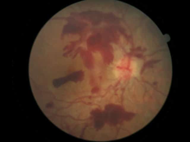
What is the membrane that lines the inside of the eye?
a transparent membrane covering the eyeball and under surface of the eyelid Mucous membrane that lines the eyelid and covers the white portion of the eye. A thin mucous membrane, helps keep the outside of the eye moist, lines the inner surface of the eyelids and folds back over the white of the eye CILIARY BODY
What is the conjunctiva membrane?
The conjunctiva is a thin membrane lining the inside of your eyelids (both upper and lower) and covering the outer portion of the sclera (white part of the eye). It doesn't cover the cornea, which is the clear covering on the front of the eye.
What is the inner surface of the eyelid called?
The part lining the inner surface of the eyelids is called the palpebral or tarsal conjunctiva. The part covering the sclera is called the bulbar conjunctiva.
What is the function of the mucous membrane of the eye?
Mucous membrane that lines the eyelid and covers the white portion of the eye. A thin mucous membrane, helps keep the outside of the eye moist, lines the inner surface of the eyelids and folds back over the white of the eye
What is the membrane that lines the eyelids and bends over the surface of the eyeball?
What is the white part of the eye?
What is the pigmented area at the centre of the retina?
Which layer of the eyeball is light sensitive?
Which part of the retina contains the most acute image formation and colour vision?
What does it mean when your eyes are red?
See 1 more
About this website

What is the membrane that lines the eyelid and covers the white portion of the eye?
Mucous membrane that lines the eyelid and covers the white portion of the eye.
What is the middle layer of the eyeball?
The opaque middle layer of the eyeball a thin highly vascular layer between the sclera and the retina. It supplies blood to the retina and conducts arteries and nerves to other structures in the eye.
What is the hole in the center of the eye that allows light to enter the eyeball?
A hole in the center of the iris of the eye that allows light to enter the eyeball. The diameter of pupil is controlled by the iris in response to the brightness of the light.
Which structure controls the shape of the lens?
Structure surrounding the lens that connects the choroid and iris. It contains ciliary muscles, which control the shape of the lens, and it secretes aqueous humor.
Where is the inner layer of the eye?
Inner layer. It's found at the back of your eye and is a thin layer of tissue made up of light receptors and sensory neurons.
Which part of the cornea contains aqueous humor?
between the back of the cornea and the front of the lens, contains aqueous humor
What is the thin layer of the retina called?
Epiretinal membranes are thin, transparent layers of fibrous tissues that form a film on the inner surface of the retina.
What is the difference between retinal tear and vitreous detachment?
Retinal tear or detachment: A retinal tear is a break in the retina whereas retinal detachment occurs when the retina pulls away from the back of the eye. Injuries: Eye injuries or traumas can cause ERMs.
How does a vitrectomy work?
During a vitrectomy, the surgeon will make tiny cuts in the affected eye and remove the fluid from inside the eye. The surgeon will then hold and gently peel the epiretinal membrane from the retina and replace the fluid in the eye. Finally, the doctor places a pad and shield on the eye to protect it from infection or injury.
Can you develop ERM in one eye?
Having risk factors for ERMs does not guarantee that someone will develop this condition in one eye or both. Also, someone who does not have any risk factors could develop an ERM.
What is the white part of the eye called?
Some people confuse the conjunctiva for the white part of the eye, which is the sclera. The sclera is a tough, opaque, fibrous tissue. This connective tissue helps to maintain the shape of your eyeball, while the conjunctiva is a mucous membrane covering the outer part of your sclera.
What is the capsule that surrounds the eyeball?
Tenon’s capsule is a sheath that surrounds the eyeball and merges with the conjunctiva in the limbal area. This capsule protects the eye and prevents ocular infections from spreading behind the eye.
What Color Is the Conjunctiva?
The conjunctiva is very thin and practically transparent. Except for the blood vessels, any tissue underneath is visible.
What does pterygia look like?
Pinguecula are small, yellowish bumps, while pterygia look like wedge-shaped growths that extend from the conjunctiva to the cornea. Most of these growths can be managed with lubricating drops and the use of sunglasses protection.
What is the area where the conjunctiva meets the cornea?
The area where the conjunctiva meets the cornea is called the limbus. Tenon’s capsule is a sheath that surrounds the eyeball and merges with the conjunctiva in the limbal area. This capsule protects the eye and prevents ocular infections from spreading behind the eye. The part lining the inner surface of the eyelids is called ...
Where are the conjunctival lymphoma?
Conjunctival lymphomas are fleshy, salmon-pink patches that often form in the fornices or bulbar conjunctiva. They are usually painless and can be hidden by the lower or upper eyelid. The ophthalmologist will take a biopsy of the growth and consult an oncologist to determine whether systemic lymphoma is present or if the lymphoma is localized to the eye. Systemic lymphoma requires treatment with immunotherapy or chemotherapy, while localized growths can be treated with external beam radiation therapy.
What are the problems associated with the conjunctiva?
Many conditions can affect the conjunctiva. Some are common and cause mild symptoms, while others are rare and can be vision-threatening or even life-threatening. Several of these conditions include:
What is the membrane that lines the eyelids and bends over the surface of the eyeball?
Thin Transparent mucous membrane that lines the eyelids and bends over the surface of the eyeball.
What is the white part of the eye?
Tough opaque tissue that serves as the eyes protective outer coat - appears as the white of the eye.
What is the pigmented area at the centre of the retina?
Small pigmented area at the centre of the retina - because it is pigmented yellow it absorbs the UV light being recieved through the lens and acts like a natural sunblock.
Which layer of the eyeball is light sensitive?
Innermost layer of eye ball - light sensitive and multi-layered. Attached to the brain by the optic nerve - changes light waves into nerve impulses which the brain then converts into visible images,
Which part of the retina contains the most acute image formation and colour vision?
Central to the retina, contains fovea, the area of most acute image formation and colour vision.
What does it mean when your eyes are red?
Describes the normal red light reflected back from the back of the eye when light is shone on it . An abnormal or absent of it can suggest cataracts or retinoblastoma in children.
