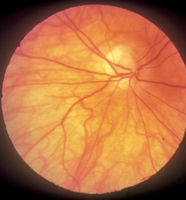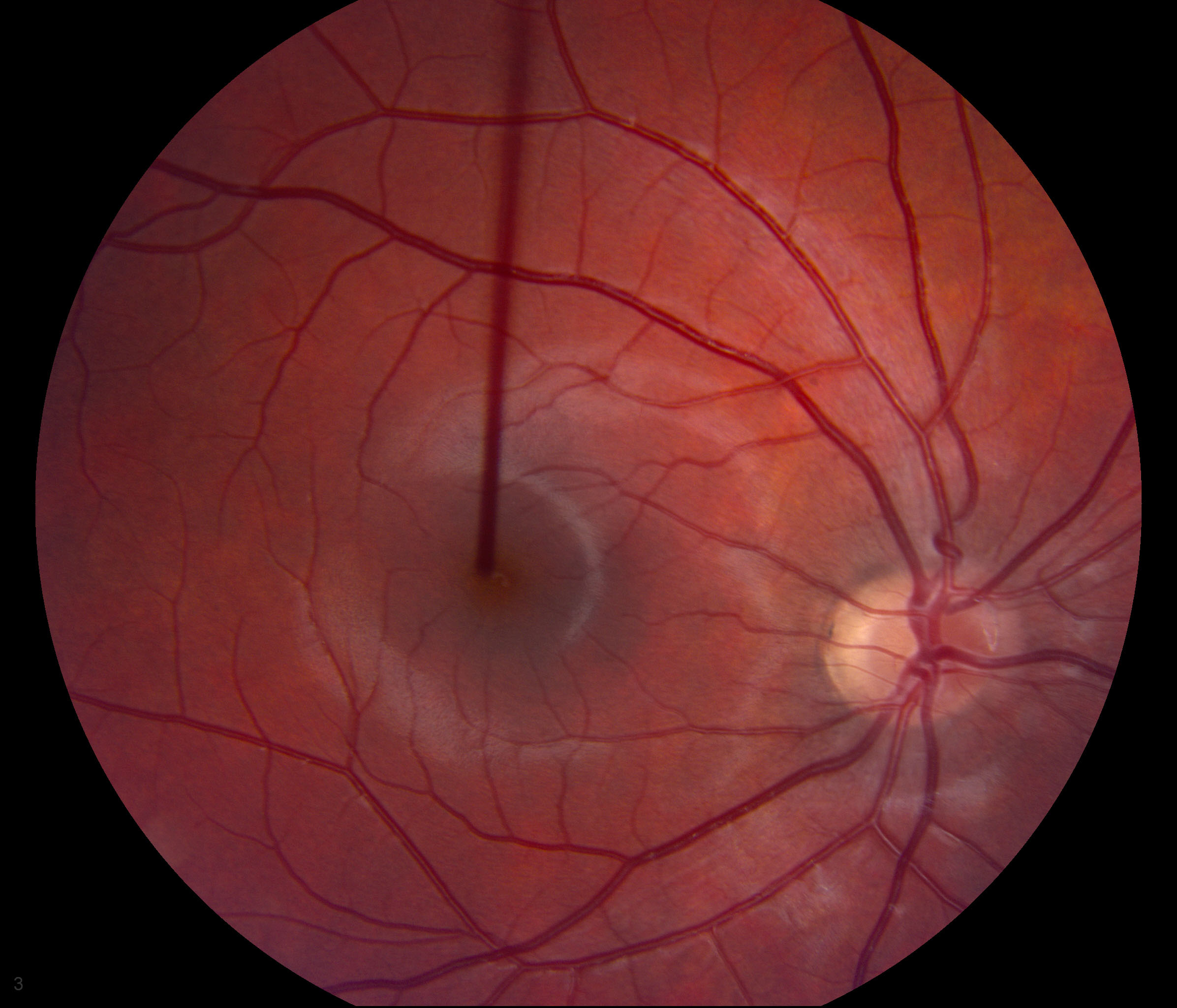
Tilted disc syndrome (Fuch's Coloboma): a congenital abnormality (birth defect) of the eye that causes the optic nerve to enter the retina (this area is called the optic disc) at an abnormal angle. In tilted disc syndrome, the abnormal entry of the optic nerve causes an abnormal position and angle of the optic disc.
What is tilted disc syndrome?
In tilted disc syndrome, the abnormal entry of the optic nerve causes an abnormal position and angle of the optic disc. Tilted disc syndrome is a benign condition that is thought to affect 1% to 2% of people.
How common are tilted discs in the eye?
Tilted discs are fairly common, especially in cases of high myopia. They are usually bilateral (80%). The superotemporal aspect of the optic nerve is typically elevated, which can mimic optic nerve edema. There can be situs inversus of the retinal vessels (increased branching of the retinal vessels nasally).
What is the pathophysiology of tilting of the optic disc?
Disease. It is characterized by inferonasal tilting of the optic disc and most commonly occurs bilaterally. TDS also has an association with high myopia as one fifth of patients with greater than 5 diopters of myopia have tilted discs. Bitemporal superior visual field defects are present in about one-fifth of patients with tilted discs...
What are the sequelae of tilted optic disks?
Visual sequelae described with tilted optic disks include myopia, astigmatism, visual field loss, deficient color vision, and retinal abnormalities. Although the natural course of tilted optic disks is nonprogressive, the anomaly can be mistaken for tumors of the anterior visual pathway, edema of the optic nerve head, or glaucoma.
What causes tilted optic disc?
Tilted optic discs often arise due to acquired changes related to the progression of myopia, known as myopic tilted disc. Because tilted disc syndrome arises from a congenital anomaly, the signs are considered nonprogressive. However, as an acquired condition, myopic tilted disc is often progressive.
How common is a tilted optic nerve?
The occurrence of tilted optic discs appears in about 1 to 2% of the population.
What does tilted disc syndrome look like?
The disc looks oval and lopsided, usually elevated superiorly and depressed inferiorly. There is a crescent commonly inferior to the disc, and hypopigmentation in a wedge shaped area next to the defective portion of the disc.
What is an abnormal optic disc?
Abnormalities of the optic disc may reflect eye disease (such as glaucoma), problems in development (as in various syndromes), or CNS disease (such as increased intracranial pressure). Each optic nerve is composed of about 1.2 million axons deriving from the retinal ganglion cells of one eye.
Can optic nerve damage be caused by stress?
In fact, continuous stress and elevated cortisol levels negatively impact the eye and brain due to autonomous nervous system (sympathetic) imbalance and vascular dysregulation; hence stress may also be one of the major causes of visual system diseases such as glaucoma and optic neuropathy.
Can you go blind from optic nerve damage?
Your optic nerves are vital to your eyesight. Damage to these nerves can lead to temporary or permanent vision loss. Glaucoma is the most common optic nerve disorder. If left untreated, optic nerve damage can lead to blindness.
What causes optic nerves to swell?
Besides MS , optic nerve inflammation can occur with other conditions, including infections or immune diseases, such as lupus. Rarely, another disease called neuromyelitis optica causes inflammation of the optic nerve and spinal cord.
Where is the optic disc?
The optic disc, also known as the optic nerve head, forms a slightly raised spot on the nasal side of the retina. Here, there are no photoreceptors, hence it is known as the blind spot of the eye. The optic disc has a slight cup-shaped depression, called the physiologic cup.
What is a high myopia?
High myopia: A rare inherited type of high-degree nearsightedness is called high myopia. It happens when your child's eyeballs grow longer than they should or the cornea is too steep. High myopia is usually defined as myopia with a refractive error greater than -6.
Is tilted optic disc normal?
Tilted optic disks are a common finding in the general population. An expression of anomalous human development, the tilted disk appears rotated and tilted along its axes. Visual sequelae described with tilted optic disks include myopia, astigmatism, visual field loss, deficient color vision, and retinal abnormalities.
How does the optic disc affect vision?
Although optic nerve drusen do not usually affect vision, peripheral vision loss may occur. It is usually mild and goes unnoticed by the patient. Visual field exams may be performed to monitor for decreased peripheral vision in older children.
What are the disc signs of glaucoma?
Characteristics of a glaucomatous ONHgeneralised/focal enlargement of the cup.disc haemorrhage (within 1 disc diameter of ONH)thinning of neuroretinal rim (usually at superior & inferior poles)asymmetry of cupping between patient's eyes.loss of nerve fibre layer.More items...
What causes asymmetrical optic nerves?
The presence of asymmetry between the optic nerve cup between the two eyes of an individual is considered an early sign of glaucomatous damage42 clinically and is a predictor of future damage in ocular hypertensive patients.
What percentage of people have optic nerve hypoplasia?
The frequency of Corpus Callosum Hypoplasia in the general population is estimated to be 1.8-2.1 per 10,000, and it affects 2.3% of developmentally disabled people.
What is the most common cause of optic nerve damage?
The most common is poor blood flow. This is called ischemic optic neuropathy. The problem most often affects older adults. The optic nerve can also be damaged by shock, toxins, radiation, and trauma....These may include:Brain tumor.Cranial arteritis (sometimes called temporal arteritis)Multiple sclerosis.Stroke.
Is optic nerve hypoplasia rare?
The prevalence of this disease is approximately 1 in 10,000 children, according to the National Organization of Rare Diseases. ONH accounts for approximately 25% of vision loss in infants, according to a report from the Oman Journal of Ophthalmology.
What is tilted disc syndrome?
Dr. Brujic describes tilted disc syndrome as a variant-something that is recorded by an eye care professional, but is not considered a pathology. It occurs when the nerve enters the eye at an oblique angle rather than a perpendicular one.
How many people have tilted optic discs?
The occurrence of tilted optic discs appears in about 1 to 2% of the population.
Is there a difference in macular nerve fiber thickness?
There were some differences in macular nerve fiber layer thickness, but not in retinal nerve fiber layer thickness. The tilted disc group was shown to suffer from a greater degree of myopia than the non-tilted disc group.
Is myopic eye disorder benign?
While it is generally considered to be a benign and congenital—albeit uncorrectable—condition, a recent study in the journal Optometry & Vision Science took a closer look, using optical coherence tomography to compare differences between myopic eyes with and without tilted discs.
Can glaucoma be caused by tilted discs?
"It is one of these situations where if you have a glaucoma suspect with tilted discs, you always wonder if there is an influence from the tilt that is actually creating an altered retinal nerve fiber layer artifact," says Mile Brujic, O.D., who practices in Ohio. "What this shows is that when you compare it to optic nerves in myopes that aren't tilted, there is no difference between the two. That tells us we can look at these tilted discs and, with a higher level of confidence, we can make better treatment decisions when we are doing optical coherence tomography on those individuals."
What is tilted disc syndrome?
Tilted disc syndrome (TDS), also known as Fuch’s Coloboma, is a congenital anomaly that occurs in 1 to 2% of the population. While mostly understood as a nonhereditary process, reports of autosomal dominant inheritance exist. It is characterized by inferonasal tilting of the optic disc and most commonly occurs bilaterally. TDS also has an association with high myopia as one fifth of patients with greater than 5 diopters of myopia have tilted discs. Bitemporal superior visual field defects are present in about one-fifth of patients with tilted discs and other features of TDS include inferior or inferonasal crescent, irregular orientation of retinal vessels (situs inversus), and an ectasia of the lower fundus or inferior staphyloma.
What are the visual field defects in TDS?
Other types of defects in TDS include altitudinal or arcuate defects that may be confused with glaucomatous changes.
What causes TDS?
TDS is thought to be caused by oblique insertion of the optic nerve and retinal vessels due to incomplete closure of the embryonic fissure of the eye. Additionally, there is hypoplasia and thinning of the retinal, choroidal, and scleral layers with focal hypopigmentation and ectasia of the inferonasal posterior wall of the globe. It is unclear whether both the disc anomalies and inferior staphylomas co-exist at birth or whether the inferior staphyloma deepens with time.
What is myopia in physical examination?
Physical Examination. Myopia, tilted optic disc, situs inversus of retinal vessels (a nasal detour of the temporal retinal vessels as they emerge from the disc before turning back temporally), scleral crescent located inferiorly or inferonasally, inferior staphyloma.
What is the name of the study that examined the shape of the eyes and structure of optic nerves in the eyes with?
8. Shinohara K, et al. Analyses of shape of eyes and structure of optic nerves in eyes with tilted disc syndrome by swept-source optical coherence tomography and three-dimensional magnetic resonance imaging. Eye (2013) 27, 1233-1242
When was TDS first described?
While Fuchs made early descriptions of tilted optic discs, the first clear description of TDS was by Rucker in 1944. To date, TDS has mainly been analyzed by ophthalmoscopic examination; however, OCT, CT, and MRI have also been used to characterize the abnormalities in TDS.
Does TDS cause myopia?
It is characterized by inferonasal tilting of the optic disc and most commonly occurs bilaterally. TDS also has an association with high myopia as one fifth of patients with greater than 5 diopters of myopia have tilted discs.
What is a tilted optic disk?
The congenital tilted optic disk (or tilted disk) appears as if the optic nerve enters the eye at an oblique angle while being rotated along its anterior–posterior axis. The anomaly is a relatively common anatomical variant, having been reported in 0.4–3.5% of populations ( Table 1 ). 31, 46, 94, 115, 129, 137 This nearly nine-fold range reflects both the wide phenotypic spectrum of the anomaly and the criteria used to define tilted disk. Most papers published on tilted disks do not objectively define the condition, which could tend to underestimate borderline or equivocal cases. 31, 94 Overlapping features with myopic disks may explain the variation in prevalence in some studies. You et al, 137 for instance, excluded eyes with myopia greater than 8 diopters (D), when other population-based studies did not.129 To avoid confusion with myopic disks, some investigators use inferior or nasal tilting as inclusion criteria. 129
What is the shape of the right tilted disk?
Fig. 4. Right tilted disk with a vertically elliptical shape. The long axis is at the 6–12 o'clock meridian. There is no torsion. The disk is surrounded by an eccentric rim of atrophic retinal pigment epithelium (annular crescent). Macular staphyloma is present.
What causes correctable visual impairment in tilted optic disk?
The most common cause of correctable visual impairment in tilted disk is refractive error. Specifically, patients with tilted optic disks have an increased prevalence of myopia and astigmatism. 129, 137
Why are tilted optic disks included in the differential diagnosis of chiasmal lesions?
Because the visual field of patients with bilateral tilted optic disks may falsely localize to the optic chiasm, tilted disks are included in the differential diagnosis of chiasmal lesions, such as pituitary adenoma.
Why are myopic crescents indistinguishable from tilted disks?
Clinically and histologically, myopic disks with oblique insertions and acquired crescents are indistinguishable from tilted disks because both have absent or severely attenuated RPE, Bruch's membrane, and choroid. Overlapping nomenclature further confuses the issue. Myopic crescents are also referred to as beta peripapillary atrophy,85 a term used to describe the crescents in tilted disk and the peripapillary tissue of glaucomatous or glaucoma-suspect disks. Histopathologically, these areas lack photoreceptors, RPE, and choroid. Juxtapapillary sclera may be “stretched” or attenuated in myopic eyes, but otherwise appears unremarkable.85
How to tell if a disk is tilted?
In clinical practice, the diagnosis of tilted disk is based on ophthalmoscopic appearance. Clinically, tilted disks appear as an exaggerated oval or D-shaped optic nerve head with one hemisphere of the disk more elevated than the contralateral half ( Fig. 2 ). When viewed with an ophthalmoscope, the anomalous optic nerve appears to be entering the eye at an acute angle rather than perpendicular to the scleral canal. The orientation of the tilt is most commonly in the inferonasal direction, with the superotemporal portion of the disk elevated and the inferonasal disk flat ( Fig. 1 ). 53 The direction of the tilt may also be horizontal, vertical, or along an oblique axis ( Fig. 7 ). The elevated portion of the disk margin may appear indistinct or blurred ( Fig. 8 ). As will be discussed later, this blurring of the disk margin may simulate optic disk swelling and, consequently, mimic papilledema when bilateral.138 The elevated margin of the disk is usually adjacent to intact neurosensory retina, pigment epithelium, and choroid, and the depressed portion of the disk is commonly contiguous with thin choroid and attenuated retinal pigment epithelium (RPE).
Why is the disk tilted?
Localized absence of ganglion cells and their failure to make synaptic connection in the lateral geniculate body may result in hypoplastic development of other supportive tissue. Accordingly, the disk becomes tilted because of the imbalance in number of ganglion cells and supportive tissue within the optic nerve.34
What is tilted eye?
Tilted: Most optic nerves enter the eye straight on, like a cable pushed into a styrofoam ball perpendicular to the surface. In some eyes, the optic nerve enters at an angle, leading to a tilted appearance. This occurs most commonly in nearsighted people (myopes).
Is an angled optic nerve pathologic?
Angled nerve head: The ophthalmologist examines the optic nerve head with the ophthalmoscope. This structure is usually flat although it may vary in interior detail. In some people the blood vessels exit the nerve in a tilted manner which gives the whole disk the appearance of tilt. This can also occur in high myopia. By itself, it is not pathologic.
What is tilted disc syndrome?
Tilted disc syndrome (TDS), also known as Fuch’s Coloboma, is a congenital anomaly that occurs in 1 to 2% of the population. While mostly understood as a nonhereditary process, reports of autosomal dominant inheritance exist. It is characterized by inferonasal tilting of the optic disc and most commonly occurs bilaterally. TDS also has an association with high myopia as one fifth of patients with greater than 5 diopters of myopia have tilted discs. Bitemporal superior visual field defects are present in about one-fifth of patients with tilted discs and other features of TDS include inferior or inferonasal crescent, irregular orientation of retinal vessels (situs inversus), and an ectasia of the lower fundus or inferior staphyloma.
What are the visual field defects in TDS?
Other types of defects in TDS include altitudinal or arcuate defects that may be confused with glaucomatous changes.
What causes TDS?
TDS is thought to be caused by oblique insertion of the optic nerve and retinal vessels due to incomplete closure of the embryonic fissure of the eye. Additionally, there is hypoplasia and thinning of the retinal, choroidal, and scleral layers with focal hypopigmentation and ectasia of the inferonasal posterior wall of the globe. It is unclear whether both the disc anomalies and inferior staphylomas co-exist at birth or whether the inferior staphyloma deepens with time.
What is myopia in physical examination?
Physical Examination. Myopia, tilted optic disc, situs inversus of retinal vessels (a nasal detour of the temporal retinal vessels as they emerge from the disc before turning back temporally), scleral crescent located inferiorly or inferonasally, inferior staphyloma.
What is the name of the study that examined the shape of the eyes and structure of optic nerves in the eyes with?
8. Shinohara K, et al. Analyses of shape of eyes and structure of optic nerves in eyes with tilted disc syndrome by swept-source optical coherence tomography and three-dimensional magnetic resonance imaging. Eye (2013) 27, 1233-1242
When was TDS first described?
While Fuchs made early descriptions of tilted optic discs, the first clear description of TDS was by Rucker in 1944. To date, TDS has mainly been analyzed by ophthalmoscopic examination; however, OCT, CT, and MRI have also been used to characterize the abnormalities in TDS.
Does TDS cause myopia?
It is characterized by inferonasal tilting of the optic disc and most commonly occurs bilaterally. TDS also has an association with high myopia as one fifth of patients with greater than 5 diopters of myopia have tilted discs.
Where is the optic disc located?
The optic disc is placed 3 to 4 mm to the nasal side of the fovea. It is a vertical oval, with average dimensions of 1.76mm horizontally by 1.92mm vertically. There is a central depression, of variable size, called the optic cup.
What is the function of the optic disc?
Function. The optic disc or optic nerve head is the point of exit for ganglion cell axons leaving the eye. Because there are no rods or cones overlying the optic disc, it corresponds to a small blind spot in each eye.
How to see blood flow in optic disc?
Blood flow in the retina and choroid in the optic disc region can be revealed non invasively by near-infrared laser Doppler imaging. Laser Doppler imaging can enable mapping of the local arterial resistivity index, and the possibility to perform unambiguous identification of retinal arteries and veins on the basis of their systole - diastole variations, and reveal ocular hemodynamics in human eyes. Furthermore, the Doppler spectrum asymmetry reveals the local direction of blood flow with respect to the optical axis. This directional information is overlaid on standard grayscale blood flow images to depict flow in the central artery and vein.
What imaging method shows blood flow in the optic disc?
Blood flow in the optic disc revealed by holographic laser Doppler imaging.
Why is there no rod in the optic disc?
Because there are no rods or cones overlying the optic disc, it corresponds to a small blind spot in each eye. The ganglion cell axons form the optic nerve after they leave the eye. The optic disc represents the beginning of the optic nerve and is the point where the axons of retinal ganglion cells come together.
What is the terminal portion of the optic nerve?
The terminal portion of the optic nerve and its entrance into the eyeball, in horizontal section. The optic disc or optic nerve head is the point of exit for ganglion cell axons leaving the eye. Because there are no rods or cones overlying the optic disc, it corresponds to a small blind spot in each eye.
What is the difference between a pale and an orange disc?
A pale disc is an optic disc which varies in colour from a pale pink or orange colour to white. A pale disc is an indication of a disease condition.
What is tilted nerve?
On histopathology, there is oblique insertion of the optic nerve, elevation of superotemporal disc, and posterior ectasia inferiorly. Tilted nerves are independently associated with suprasellar tumors, and craniosynostoses ( specifically Crouzon and Apert syndromes).
What is the most common optic disc anomaly?
Optic nerve hypoplasia is the most common optic disc anomaly encountered in ophthalmic practice.
What is optic nerve pit?
The histopathologic description of an optic nerve pit is herniation of dysplastic retina into a collagen pocket with a defect in the lamina cribrosa.
Which aspect of the optic nerve is typically elevated, which can mimic optic nerve edema?
The superotemporal aspect of the optic nerve is typically elevated, which can mimic optic nerve edema.
What is the triad of hypoplasia?
Consists of the triad: ON hypoplasia, absent septum pellucidum/agenesis of corpus callosum, and pituitary dwarfism
Why is the Morning Glory disc anomaly named?
The morning glory disc anomaly named because it closely resembles the morning glory flower. There is also a geyser in Yellowstone called Morning Glory for the same reason.
How many congenital disc anomalies are there?
Because there are a few associated systemic conditions that we need to know as part of our training, there is certainly some possibility that a question or two may address these congenital disc anomalies. Fortunately, although there are many more than will be listed in this article, there are essentially 6 congenital disc anomalies that you should be familiar with before you take the OKAP.
