How long does it take for vitreous hemorrhage to clear?
In many cases, they resolve spontaneously in 2-3 months, although if the bleeding is not reabsorbed, it may require a surgery called vitrectomy. Vitreous hemorrhages may be of different degrees. Severe bleedings may cause a sudden and complete loss of vision, while mild ones cause blurred vision or the appearance of floaters.
What Chamber of the eye contains the vitreous humor?
• The ocular fundus is the largest chamber of the eye and contains vitreous humor, a clear, gelatinous substance, composed mostly of water and encapsulated by a hyaloid membrane. The vitreous humor occupies about 2/3 of the eyes volume and helps maintain the shape of the eye.
What causes blood behind the eye?
Risk factors for subconjunctival hemorrages include:
- Diabetes
- High blood pressure
- Having a "cold" or allergies (that increase coughing and sneezing)
- Wearing contact lenses (increases eye rubbing)
- Use of aspirin or blood thinners
- Aging (over age 50)
- Blood clotting disorders
- Vitamin K deficiency
What does the vitreous gel do for the human eye?
Vitreous gel, also called vitreous humor, is a clear gel which fills most of the interior of the eyeball. One of the main functions of this gel is simply to enable the eyeball to hold its spherical shape, as without the gel the eyeball would collapse. The gel also helps hold the retina in place against the interior wall of the eyeball.
Is vitreous hemorrhage serious?
Vitreous hemorrhage is an urgent situation because it can cause permanent vision loss. You should go to your doctor or emergency room as soon as you notice any symptoms of this condition. An ophthalmologist's first step will be to check for a retinal tear or detachment that can cause permanent vision loss.
What are the causes of vitreous haemorrhage?
The most common causes include proliferative diabetic retinopathy, vitreous detachment with or without retinal breaks, and trauma. Less common causes include vascular occlusive disease, retinal arterial macroaneurysm, hemoglobinopathies, age-related macular degeneration, intraocular tumors, and others.
What are the symptoms of vitreous hemorrhage?
Vitreous haemorrhage symptoms Symptoms range from the sudden appearance of spots or floaters in your vision to a sudden blurring of vision; and in severe cases sudden blindness. Some people find that their vision tends to be worse in the morning, as the blood has settled to the back of their eye during the night.
Can you go blind from vitreous hemorrhage?
A vitreous haemorrhage can be severe and result in legal blindness, or it may be mild and result only in annoying black floaters. The severity of visual loss is related to the density of the haemorrhage and the underlying cause for the bleeding.
What is the treatment for vitreous hemorrhage to the eye?
Normally, no treatment is needed for a vitreous hemorrhage. The blood should clear by itself and your vision will be restored. Unfortunately, this may take up to several months. Your eye doctor will follow up with you and monitor this condition until it goes away.
How do you manage vitreous hemorrhage?
After establishing the aetiology and source of vitreous haemorrhage, management is individually tailored. The management options are observation, laser photocoagulation, cryotherapy and pars plana vitrectomy. The choice depends on several factors.
How common is a vitreous hemorrhage?
Vitreous hemorrhage (VH) is defined as the presence of blood within the vitreous cavity, which is the space lined posteriorly by the retina and anteriorly by the ciliary body, zonular fibers, and posterior lens capsule (Figure 1). Vitreous hemorrhage is not uncommon with an incidence of seven cases per 100,000.
How do you check for vitreous hemorrhage?
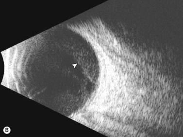
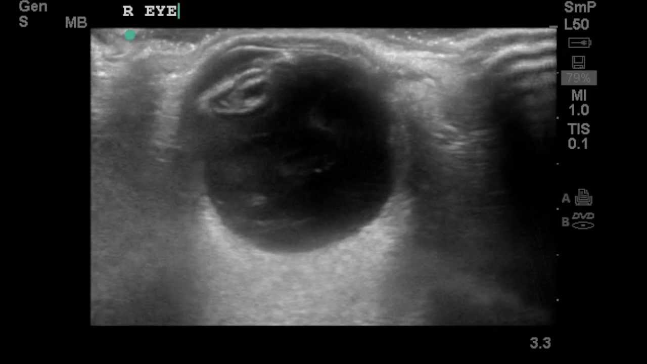
If the vitreous hemorrhage obscures a complete view to the retina, B-scan ultrasonography can detect vitreous hemorrhage, PVD, retinal tears, retinal detachment, tractional membranes, intraocular tumors, and foreign bodies.
What is treatment for vitreous?
An ophthalmologist who is a specialist in retina and vitreous surgery removes the vitreous through a small incision (vitrectomy). The vitreous is replaced with a solution to help your eye maintain its shape. Surgery may not remove all the floaters, and new floaters can develop after surgery.
How do you sleep with a vitreous hemorrhage?
Patients with vitreous hemorrhage should avoid laying down flat in bed, and if possible, should sleep in a reclining chair or with several pillows under their head in bed. Vitrectomy surgery can be considered for non-clearing vitreous hemorrhage. The cause of the hemorrhage might also need treatment.
How long does it take for vitreous blood to clear?
Many cases of Vitreous hemorrhage will have Complete resolution of bleeding, which may occur within 4-6 weeks. This is because of tendency of intraocular structures (Trabecular meshwork) to absorb it.
How do I restore my vitreous?
There is no specific treatment for vitreous degeneration; a vitrectomy laser surgery can be performed to help alleviate any vitreous floaters.
What is the most common cause of retinal hemorrhages?
These are dense, dark red, sharply outlined, and are seen in disorders that affect the pre-venular deep capillary layer. Common causes for such hemorrhages include diabetic retinopathy, retinal vein occlusions, ocular ischemic syndrome, sickle cell retinopathy, and juxta foveal telangiectasia.
How common is a vitreous hemorrhage?
Vitreous hemorrhage (VH) is defined as the presence of blood within the vitreous cavity, which is the space lined posteriorly by the retina and anteriorly by the ciliary body, zonular fibers, and posterior lens capsule (Figure 1). Vitreous hemorrhage is not uncommon with an incidence of seven cases per 100,000.
How do you prevent vitreous hemorrhage?
Prevention of vitreous haemorrhage involves preventing the underlying causes. This includes careful and regular management of diabetic eye disease (which tends to be worse in less well-controlled diabetes) and high blood pressure, and giving up smoking.
What can damage the vitreous humor?
With aging, the vitreous humor undergoes vitreous degeneration, acquiring a thinner liquid consistency. This can lead to vitreous floaters, or small disruptions in the visual field such as spots.
What is the proliferative process of vitreoretinopathy?
Proliferative vitreoretinopathy. After vitreous hemorrhage, proliferative vitreoretinopathy can occur. It is thought that macrophages and chemotactic factors induce fibrovascular proliferation, which can lead to scarring and subsequent retinal detachment. Ghost cell glaucoma.
Why is hemosiderosis bulbi a serious complication?
Hemosiderosis bulbi is a serious complication thought to be caused by iron toxicity as hemoglobin is broken down. Since hemolysis occurs slowly, the iron-binding capacity of proteins in the vitreous usually outpaces the slow rate of hemolysis, thereby avoiding hemosiderosis bulbi.
What is the leading cause of vitreous hemorrhage in people younger than 40?
Blunt or perforating trauma can injure intact vessels directly and is the leading cause of vitreous hemorrhage in people younger than 40.
How many cases of vitreous hemorrhage per 100,000?
Vitreous hemorrhage has an incidence of seven cases per 100,000, which makes it one of the most common causes of acutely or subacutely decreased vision. Although the diagnosis of vitreous hemorrhage is generally straightforward, management is dictated by uncovering the underlying etiology.
What are the symptoms of vitreous hemorrhage?
Signs and Symptoms. The symptoms of vitreous hemorrhage are varied but usually include painless unilateral floaters and/or visual loss. Early or mild hemorrhage may be described as floaters, cobwebs, haze, shadows or a red hue. More significant hemorrhage limits visual acuity and visual fields or can cause scotomas.
How fast does blood clear from vitreous hemorrhage?
The blood is typically cleared from within the vitreous hemorrhage at a rate of approximately 1 percent per day. Blood outside the formed vitreous resolves more quickly. Vitreous hemorrhage is cleared more quickly in syneretic and vitrectomized eyes, and more slowly in younger eyes with well-formed vitreous.
What are the three main categories of vitreous hemorrhage?
The mechanisms of vitreous hemorrhage fall into three main categories: abnormal vessels that are prone to bleeding, normal vessels that rupture under stress, or extension of blood from an adjacent source. (See “Mechanisms of Vitreous Hemorrhage.”)
What causes a closed globe injury?
Closed globe injury from blunt trauma: Compression of the globe in an anterior-posterior direction causes the equator of the globe to bulge in a coronal plane. Especially in a young patient with formed vitreous and strong adherence of the vitreous to the retina, this bulging in the coronal plane causes inward-directed traction exerted by the vitreous on the retina. The fact that the vitreous base, an area of especially strong vitreous attachment, is near the equator contributes to the tractional force the vitreous exerts on the retina in this area. This inward-directed tractional force on the retina by the vitreous can cause a retinal dialysis, but it can also result in retinal tears and vitreous hemorrhage as retinal vessels may be ruptured. Blunt trauma may also rupture blood vessels associated with the iris and ciliary body, causing hyphema and spillover hemorrhage into vitreous.
What are the risk factors for vitreous hemorrhage?
Risk Factors. The population at risk for vitreous hemorrhage will have the demographic and clinical characteristics according to its causes. For example, poorly controlled diabetics with end-organ damage such as proliferative diabetic retinopathy are at high risk.
Why do my eyes bleed?
The neovascular vessels grow into the vitreous and are fragile. Normal eye movements, acute PVD, and fibrovascular contraction of the neovascularization can all cause these vessels to bleed. Retinal macroaneurysms and acutely occluded retinal venules in retinal vein occlusion may rupture, causing vitreous hemorrhage.
What is vitreous hemorrhage?
Vitreous Hemorrhage is a relatively common cause of acute vision loss, having an incidence of approximately 7 cases per 100,000, 4.8 per 10000 in Taiwan, and may vary according to population characteristic, geography, and other factors. It is therefore frequently encountered by ophthalmologists and Emergency Room professionals alike due to its often rapid onset which causes painless, but substantial vision loss. Although the diagnosis of vitreous hemorrhage is often straightforward to make on funduscopic examination or ultrasonography, further investigation may be required to determine the underlying etiology.
What causes retinal venules to rupture?
Rather, increased intracranial pressure causes increased pressure in retinal venules, causing them to rupture. Sub-internal limiting membrane (sub-ILM) hemorrhage is noted and intraoperatively a break may be noted in the ILM through which the blood is thought to reach the vitreus gel from the sub-ILM cavity.
What causes blood to expand into the vitreous cavity?
Extravasation of blood into the vitreous cavity is generally caused by two basic mechanisms: Rupture of normal vessels through mechanical force: Closed globe injury from blunt trauma: Compression of the globe in an anterior-posterior direction causes the equator of the globe to bulge in a coronal plane.
When to use anti-VEGF?
Many surgeons use pre-operative anti-VEGF agents 1 to 7 days before pars plana vitrectomy for vitreous hemorrhage in diabetics , as regression of neovascular membranes reduces intra- and post-operative bleeding and dissection of tissue may become easier. Several small studies support this belief, although other small studies refute it . There is concern, however, that these patients frequently fail their pre-anesthesia testing and that their surgery may be cancelled after the anti-VEGF agent has been given, potentially exacerbating tractional retinal detachment. For this reason, many surgeons wait until the patient is medically cleared for surgery before giving the anti-VEGF agent.
What is vitreous haemorrhage?
Vitreous haemorrhage occurs when blood leaks into the vitreous humour inside the eye. The leaked blood most commonly comes from blood vessels at the back of the eye. This is more likely to happen if the blood vessels have been damaged (eg, by trauma) or are particularly fragile (because of eye disease related to diabetes).
How is vitreous haemorrhage prevented?
Prevention of vitreous haemorrhage involves preventing the underlying causes. This includes careful and regular management of diabetic eye disease (which tends to be worse in less well-controlled diabetes) and high blood pressure, and giving up smoking.
Why do you see a specialist on the same day?
If you experience a vitreous haemorrhage then you will usually be seen by a specialist on the same day. This is because sudden loss of vision is considered an eye emergency. The aim is to ensure accurate diagnosis and to avoid permanent loss of vision which could occur if there is a retinal detachment behind the bleeding.
What is the name of the substance that helps the eye keep its shape?
Vitreous Haemorrhage. Vitreous haemorrhage is bleeding into the jelly-like filling of the back part of your eye. This substance is the vitreous humour. It helps the eye keep its shape and is normally clear, allowing light from outside the eye to pass through it to reach the retina.
How many people have vitreous haemorrhage?
Vitreous haemorrhage affects about 7 per 100,000 people each year. This makes it one of the most common causes of sudden deterioration in vision. It most often affects only one eye.
How long does it take for a retinal tear to clear?
This means laser treatment to bleeding vessels and any other abnormal vessels, and repair to any tears in the retina. After this it is a matter of waiting for the blood to slowly clear. This can take several weeks.
When is a vitrectomy performed?
Vitrectomy is also sometimes performed if the blood in the vitreous is clearing very slowly and vision remains impaired. Waiting is commonly the chosen option once the bleeding has stopped.
What is posterior vitreous detachment?
Posterior vitreous detachment (PVD) – A PVD can occur as part of the natural aging process of the eye. Over time, the posterior vitreous undergoes changes such as shrinking (vitreous syneresis). A hemorrhage can happen when the vitreous suddenly separates from the blood vessels that surround it (4-12% of cases) or when it suddenly pulls away from and tears the retina (11-44% of cases).
What is the most common cause of vitreous hemorrhage in adults?
These traumatic injuries make up the majority of all cases of vitreous hemorrhages in people ages 40 years and under. Proliferative diabetic retinopathy (PDR) – PDR is a common cause of vitreous hemorrhages in adults with diabetes, accounting for 31-54% of cases.
How does vitrectomy work?
A vitrectomy corrects vitreous hemorrhages by removing a part of or the entire vitreous. If the hemorrhage is mild, surgery may not be necessary. If the hemorrhage is severe, immediate surgery may be necessary as it may indicate an urgent situation such as a retinal detachment.
What is vitreous hemorrhage?
Vitreous hemorrhage. Contact an eye doctor immediately if you're experiencing a rapid onset of blurriness or loss of vision. A vitreous hemorrhage can quickly escalate into a very serious situation. A vitreous hemorrhage occurs when blood from ruptured blood vessels leaks into the vitreous humor, the clear gel-like fluid of the eyeball.
How many cases of vitreous hemorrhage in the US?
A vitreous hemorrhage occurs when blood from ruptured blood vessels leaks into the vitreous humor, the clear gel-like fluid of the eyeball. It has an incidence of 7 cases per 100,000 people each year in the U.S. In the eyeball, vitreous humor (or just “vitreous”) is located in the vitreous chamber, the cavity between the lens (at the front) ...
Why is it important to detect vitreous hemorrhage early?
Early detection by an eye doctor is imperative as a vitreous hemorrhage can worsen and cause serious issues.
What causes a vitreous to rupture?
Trauma or eye injury – Trauma to the eye from a direct impact by a blunt object or a puncture by a sharp object can cause blood vessels around the vitreous to be stressed, rupture and then leak into the vitreous. These traumatic injuries make up the majority of all cases of vitreous hemorrhages in people ages 40 years and under.
What happens when the vitreous detachment is near the retina?
As one gets older, pockets of fluid can develop in the vitreous. When these pockets develop near the back of the eye, the vitreous can pull away from the retina and possibly tear it. Posterior vitreous detachment accounts for 3.7–11.7% of vitreous hemorrhage cases.
What is the term for the extravasation of blood into the vitreous humor of the eye?
Specialty. Ophthalmology. Vitreous hemorrhage is the extravasation, or leakage, of blood into the areas in and around the vitreous humor of the eye. The vitreous humor is the clear gel that fills the space between the lens and the retina of the eye. A variety of conditions can result in blood leaking into the vitreous humor, ...
How to diagnose a vitreous hemorrhage?
Diagnosis. Vitreous hemorrhage is diagnosed by identifying symptoms, examining the eye, and performing tests to identify the cause. Some common tests include: Examination of the eye with a microscope. Pupil dilation and examination.
How to fix retinal tear?
The goal of the treatment is to fix the cause of the hemorrhage as quickly as possible. Retinal tears are closed by laser treatment or cryotherapy, and detached retinas are reattached surgically.
What causes a bleed in the back of the eye?
Trauma. Some injuries can cause blood vessels in the back of the eye to bleed. Trauma is the leading cause of vitreous hemorrhage in young people, and accounts for 12–18.8% of cases in adults.
How to treat a hemorrhage in the eye?
In most cases, the patient is advised to rest with the head elevated 30–45°, and sometimes to put patches over the eyes to limit movement prior to treatment in order to allow the blood to settle. The patient is also advised to avoid taking medications that cause blood thinning (such as aspirin or similar medications).
What causes a blood vessel to break in the back of the eye?
Diabetic retinopathy. The most common cause found in adults is diabetic retinopathy. Abnormal blood vessels can form in the back of the eye of a person with diabetes. These new blood vessels are weaker and prone to breaking and causing hemorrhage.
What is the prevalence of vitreous hemorrhage?
The number of new cases of vitreous hemorrhage tends to coincide with the frequency of causative disease. Additionally, this measurement will also depend on the particular study population, mean age of the patients, and geographical region the study is conducted in.
What is the most common cause of vitreous hemorrhage in young people?
This number is about six percent in London and 19.1 percent in Sweden. The most common cause of vitreous hemorrhage in young people is trauma.
What causes a cloudy eyeball?
A vitreous hemorrhage may result directly from retinal tears or neovascularization (bleeding from newly formed blood vessel). This leads to blood leaking inside the eyeball, often resulting in the clouding of vitreous humor and vision impairment. This may vary from having a few “floaters,” blurred sight, or seeing only dark with a possibly reddish tinge.
How many cases of vitreous hemorrhage per 100,000?
It is estimated that vitreous hemorrhage has an incidence of seven cases per 100,000, making it one of the most common causes of acute and subacute decreases in vision.
What can be detected with an ultrasound?
Ultrasounds can detect many causes of vitreous hemorrhage, including posterior vitreous detachment, retinal tears and detachments, tumors, and foreign bodies. Other possible tests include a computed tomography (CT) scan in cases of penetrating injury and an angiogram to show the blood vessels in the back of the eye.
What is vitreous humor?
Vitreous humor is a clear gel-like substance that fills the space between the lens and the retina of the eyeball. Nearly 99 percent of this fluid consists of water, with the remaining one percent made of collagen and hyaluronic acid, giving it its gel-like consistency. The space in which vitreous humor resides represents 80 percent of the eye, ...
What is the procedure to remove the vitreous?
If there are excessive amounts of blood obscuring the ability to find the source of bleeding and preventing treatment, your doctor may suggest a vitrectomy, which involves the removal of the entire vitreous to get a better view of the back of the eye.
What is a vitreous hemorrhage?
Vitreous hemorrhage has variable symptoms depending on the amount of bleeding: vision of spots that are suspended in vision ( floaters ), blurred vision or complete and sudden loss of vision.
Why do diabetics bleed?
Patients with diabetic retinopathy or central retinal vein occlusion experience a lack of oxygen in the retina, which stimulates the creation of abnormal vessels known as neovessels, that may break and cause these bleedings .
What causes a floater in the eye?
Vitreous hemorrhages may be of different degrees. Severe bleedings may cause a sudden and complete loss of vision, while mild ones cause blurred vision or the appearance of floaters. Eye injuries, surgical interventions and vascular disorders are its most common causes.
What is the main symptom of a vitreous hemorrhage?
The main symptom in a patient suffering a vitreous hemorrhage is a sudden loss of vision , but it all depends on the severity of hemorrhage.
Why can't light go through vitreous humor?
When there’s blood within the vitreous humor, this substance loses its transparency, reason why light cannot go through it and the patient experiences vision loss.
How long does it take for a bleed to reappear?
A wait-and-see approach is usually adopted, as mildest bleedings are usually spontaneously reabsorbed in a 2 or 3-month period.
Why is it important to go to the ophthalmologist?
It is important to go to the ophthalmologist and undergo an examination in order to check there is no other complication associated, such as retinal detachment or glaucoma.
What causes vitreous hemorrhage?
The most common causes include proliferative diabetic retinopathy, vitreous detachment with or without retinal breaks, and trauma.
Can vitrectomy be done spontaneously?
Occasionally, hemorrhage does not resolve spontaneously and vitrectomy surgery is necessary and beneficial.

Vitreous Hemorrhage Symptoms
- Vitreous hemorrhage can appear in many different ways. Symptoms can include unilateral floaters and/or vision loss. Some people with a mild case experience certain symptoms earlier on. These signs include:2 1. Floaters 2. Haze 3. Cobwebs 4. A red hue 5. Shadows The symptoms may be worse in the morning due to blood pooling in your eye while lying do...
Causes
- A vitreous hemorrhage can have several causes. A few common causes are:34 1. A broken abnormal blood vessel (caused by conditions like diabetic retinopathy) 2. A detached or torn retina 3. An eye injury 4. Bleeding elsewhere in the eye that leaks into the vitreous 5. Bleeding in the brain 6. Posterior vitreous detachment(which happens when the vitreous gel gets more liqui…
Diagnosis
- A healthcare provider specializing in conditions of the eyes, such as an ophthalmologist, will perform an eye exam. They may also ask about your medical history and whether you've had any recent accidents or injuries. Your healthcare provider may perform additional testing to help diagnose a vitreous hemorrhage, including:3 1. Blood tests: To pinpoint the cause of bleeding 2. …
Treatment
- Although the bleeding may go away in a couple of months, your ophthalmologist may want you to return for occasional follow-ups to monitor bleeding.1 Your provider may recommend using a few pillows to elevate your head when you sleep to minimize blood pooling in your affected eye. They may recommend avoiding heavy lifting and strenuous activity as well. You may also be advised t…
Prevention
- You can't always prevent vitreous hemorrhage. However, there are a few things you can do to decrease your risk:3 1. Keep your blood sugarunder control if you have diabetes. 2. See an eye doctor regularly for eye exams, especially if you have diabetesor sickle cell disease. 3. Wear protective eyewear when playing sports, working with sharp tools, or shooting firearms.
When to See A Healthcare Provider
- If you have been diagnosed with a vitreous hemorrhage, watch for any changing symptoms in the eye. Contact your provider if the symptoms are not improving. Seek immediate medical attention if you have new vision loss or see flashes of light.5
Summary
- A vitreous hemorrhage is bleeding occurring in the fluid of the eye. Some causes of vitreous hemorrhage are injury and diabetic retinopathy. Treatment will vary depending on the cause and severity of the vitreous hemorrhage. Regular eye exams and the use of protective eyewear can reduce your chances of developing a vitreous hemorrhage.
A Word from Verywell
- Vision changes or loss from a vitreous hemorrhage can cause fear. Always check with your healthcare provider if you have changes to your vision, have been diagnosed with a vitreous hemorrhage, and have changing symptoms in your affected eye. Diagnosis can help determine the exact cause of vitreous hemorrhage. That health condition can also be treated to prevent furthe…
Epidemiology
Structure
Pathophysiology
Clinical significance
Causes
Symptoms
- The symptoms of vitreous hemorrhage are varied but usually include painless unilateral floaters and/or visual loss. Early or mild hemorrhage may be described as floaters, cobwebs, haze, shadows or a red hue. More significant hemorrhage limits visual acuity and visual fields or can cause scotomas. Patients often say vision is worse in the morning as...
Diagnosis
Prognosis
Treatment
Research
Contraindications
Miscellaneous