What is the function of the alar fascia?
The alar fascia is a subdivision of the prevertebral fascia and belongs to the cervical fascia. The alar fascia bridges between the transverse processes of cervical vertebrae and separates the danger space from the retropharyngeal space.
Where does the alar fascia attach to the skull?
The alar fascia bridges between the transverse processes of cervical vertebrae and separates the danger space from the retropharyngeal space. Recent findings show that the fascia alar seems not to be attached to the base of skull but origin from the level of the first cervical vertebra (C1).
Is gray's description of the spaces in the alar fascia correct?
So, it is clear that Gray's description concerning the spaces, especially the ‘danger space’ and retropharyngeal space, is confusing in terms of limitations and connections. In contrast, Hafferl lists the fascia intercarotica but does not mention the alar fascia (Hafferl, 1969 ).
What is the superficial palmar fascia?
The superficial palmar fascia is a fanlike palmar fascial extension into which the palmaris longus inserts. The palmar cutaneous branch of the median nerve (PCN) lies between the flexor carpi radialis and palmaris longus tendons in the distal forearm, but its branches may be found up to 6 mm ulnar to the thenar crease in the palm ( Fig. 12-4 ).

Where is the alar fascia?
The alar fascia is a layer of fascia, sometimes described as part of the prevertebral fascia, and sometimes as in front of it. Section of the neck at about the level of the sixth cervical vertebra.
What is the alar fascia in the neck?
The alar fascia is a thin fibroareolar membrane separating the (anterior) true retropharyngeal space from the (posterior) danger space. It is the ventral component of the deep layer of the deep cervical fascia.
What does alar fascia surround?
The muscular layer surrounds the infrahyoid muscles. The visceral layer has also been described under different names, such as the pretracheal or buccopharyngeal fascia. The deep layer of deep cervical fascia is formed by the prevertebral and alar fasciae (Vieira et al.
What is in the Alar space?
The danger space or alar space, is a region of the neck. The common name originates from the risk that an infection in this space can spread directly to the thorax, and, due to being a space continuous on the left and right, can furthermore allow infection to spread easily to either side.
How many fascias are in the neck?
three fascial layersIt consists of three fascial layers (or sheaths), which are: The investing layer of deep cervical fascia. Pretracheal layer of deep cervical fascia. The prevertebral layer of deep cervical fascia.
What is fascia in the body?
What is fascia? Fascia is a thin casing of connective tissue that surrounds and holds every organ, blood vessel, bone, nerve fiber and muscle in place. The tissue does more than provide internal structure; fascia has nerves that make it almost as sensitive as skin. When stressed, it tightens up.
What are the 3 layers of fascia?
IntroductionClassification System.Superficial Fascia.Visceral Fascia.Parietal Fascia.
What is fascia in the head?
Head fascia covers the skull and merges and entwines into the neck. When we accidentally pull our neck, most of the time the fascia has been 'jerked' out its original shape resulting in pain. Fascia in the head becomes very tight when we are fatigued, dehydrated or stressed, leading to headaches.
What is the difference between deep fascia and superficial fascia?
The superficial fascia (i.e. tela subcutanea, hypodermis, subcutaneous tissue) is used to describe the connective that separates the skin from the underlying muscle tissue. The deep fascia is a dense, organized, connective tissue located deep to the skin and subcutaneous tissue.
Is there fascia in the neck?
The structures found in the neck are surrounded by a layer of subcutaneous tissue called the superficial fascia, while there are also layers of deep cervical fascia which distribute the structures in the neck into different compartments.
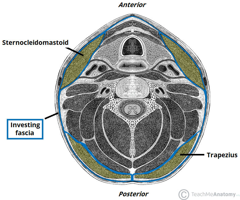
What is the investing fascia?
Deep fascia (or investing fascia) is a fascia, a layer of dense connective tissue that can surround individual muscles and groups of muscles to separate into fascial compartments. This fibrous connective tissue interpenetrates and surrounds the muscles, bones, nerves, and blood vessels of the body.
Where is the danger space?
neckThe danger space is a deep compartment of the head and neck located behind the true retropharyngeal space, extending from the skull base to the mediastinum.
What is neck fascia?
The deep cervical fascia (or fascia colli in older texts) lies under cover of the platysma, and invests the muscles of the neck; it also forms sheaths for the carotid vessels, and for the structures situated in front of the vertebral column. Its attachment to the hyoid bone prevents the formation of a dewlap.
What is the investing fascia?
Deep fascia (or investing fascia) is a fascia, a layer of dense connective tissue that can surround individual muscles and groups of muscles to separate into fascial compartments. This fibrous connective tissue interpenetrates and surrounds the muscles, bones, nerves, and blood vessels of the body.
What's in carotid sheath?
The carotid sheath is an important landmark in head and neck anatomy and contains several vital neurovascular structures, including the carotid artery, jugular vein, vagus nerve, and sympathetic plexus. It extends upwards from the arch of the aorta and terminates at the skull base.
What is the retropharyngeal space?
The retropharyngeal space (RPS) is an anatomical region that spans from the base of the skull to the mediastinum. Its location is anterior to the prevertebral muscles and posterior to the pharynx and esophagus.
1 Definition
The alar fascia is a subdivision of the prevertebral fascia and belongs to the cervical fascia.
2 Anatomy
The alar fascia bridges between the transverse processes of cervical vertebrae and separates the danger space from the retropharyngeal space. Recent findings show that the fascia alar seems not to be attached to the base of skull but origin from the level of the first cervical vertebra (C1).
3 Clinical relevance
Infections can spread via the danger space from the neck to the mediastinum.
What is the alar fascia?
Alar fascia. The alar fascia is a thin fibroareolar membrane separating the (anterior) true retropharyngeal space from the (posterior) danger space. It is the ventral component of the deep layer of the deep cervical fascia. Notably, in the well patient, the alar fascia is not usually visible on cross-sectional imaging, ...
Which vertebral level is the alar fascia fused with?
Inferiorly, there is progressive fusion anteriorly with the visceral ( buccopharyngeal) fascia between the T2 and T4 vertebral levels. Laterally, the alar fascia fuses with the lateral wall of each carotid sheath. Additionally, some fibers are contributed by the medial raphe extensions of the posterior middle pharyngeal constrictor 2.
Which level of the carotid sheath fused anteriorly with the prevertebral fascia?
superior: level of C1, where it may fuse posteriorly with the prevertebral fascia 1 or transition to loose areolar tissue 2. inferior: level of T2, where it fuses anteriorly with visceral fascia. lateral: carotid sheaths bilaterally.
Where does the prevertebral fascia connect to the skull base?
Superiorly, the prevertebral fascia extends until the level just above the C1 anterior tubercle, where it either fuses with prevertebral fascia posteriorly 1, or narrowed to a median raphe before transitioning to loose areolar tissue 2. It does not connect to the skull base as a distinct layer.
Does the alar fascia attach to the skull base?
Although the alar fascia has a well defined attachment in the midline at the level of C1, it does not attach to the skull base laterally, which is a potential point of entry into the danger space posteriorly 4.
Is alar fascia visible on CT?
Alar fascia is not usually resolved on imaging; however, it may be visible on contrast-enhanced CT as a thin membrane in the retropharyngeal space in patients with extensive regional edema e.g. post-radiation therapy 2. retropharyngeal abscess.
What is the role of the palmar fascia?
Palmar fascia serves several purposes including support of the skin, tendon, and bones. In Dupuytren disease, myofibroblasts are abundant and have increased collagen production. Three histological stages have been described: proliferative (isolated nodules), involutional (nodules with cords), and residual (stable end-stage disease) [18]. The proliferative phase is characterized by a hypercellular state with an increase in the number of disorganized myofibroblasts and collagen strands. During the involutional stage, there are fewer myofibroblasts; alignment of these cells along lines of flexor and extensor stresses forms dense “tension lines.” Finally, during the residual phase, the myofibroblasts are replaced by firm collagen deposits with formation of linear collagen bundle cords similar to mature scar tissue. The rate of progression of the three stages is variable among patients, and the disease tends to affect the dominant hand more often.
Why is the palmar fascia incised?
The skin flaps are elevated and palmar fascia is incised to fully expose the underlying nerves and vessels.
Where is the superficial transverse palmar ligament located?
The superficial transverse palmar ligament is identified at the distal palmar crease level and lies deep to the pretendinous band. This ligament is not involved in Dupuytren's contracture.
Which part of the palmar fascia is abnormal?
Surgical Anatomy. The palmar fascia, which in Dupu ytren disease is abnormal, is composed of thenar, hypothenar, and palmar aponeuroses; palmar is involved most often (Fig. 84.4 ). Normal anatomy has bands of aponeurosis. These bands, when diseased, become the pathologic cords of Dupuytren disease.
What happens during the residual phase of myofibroblasts?
Finally, during the residual phase, the myofibroblasts are replaced by firm collagen deposits with formation of linear collagen bundle cords similar to mature scar tissue. The rate of progression of the three stages is variable among patients, and the disease tends to affect the dominant hand more often.
Which MR shows the dominant focus of plantar fibromatosis?
Sagittal STIR MR shows the dominant focus of plantar fibromatosis to have homogeneous ↓ SI. The lentiform shape that blends with the plantar fascia is typical. Involvement of the plantar fascia lateral band is less common than the medial band.
Can you remove diseased fasciae with a limited fasciectomy?
In a limited fasciectomy, all the diseased fasciae are excised, while normal fasciae are left in place. Complete removal of diseased fasciae is impossible, but this procedure is the most widely used technique for Dupuytren's contracture because of its predictable outcome and low complication rate.
Structure
It is bounded at the top by the skull base, at the front by the alar fascia and behind by the prevertebral fascia. It comes to an end at the level of the diaphragm.
Clinical significance
On CT or MRI it is only visible when distended by fluid or pus, below the level of T1-T6, as the retropharyngeal space ends at this level, allowing distinction between the two entities.
What is the fascia alaris?
The fascia alaris is a thick wall of dense connective tissue which extends between the carotid sheath and the fascia prevertebralis more or less in a sagittal plane. It extends cranial to the skull base and caudal to the level of C6 or C7. The fascia is also a guide for the inferior root of the ansa cervicalis profunda (Figures 1 and 8 ). The inferior root can pass the internal jugular vein medially or laterally; the fascia alaris may be developed more medially (Figure 1) or laterally (Figure 8 ).
What is the fascia cervicalis?
The fascia cervicalis media covers and envelops the infrahyoid muscles, which are also called the musculi detractores larynges, or ‘strap muscles’ in ENT surgery. It has a strong and aponeurotic character, mainly in the cranial and medial areas. As the fascia is limited to the lateral border of the omohyoid muscles, it is only visible in the medial corner of the lateral cervical triangle. This is the area in which the omoclavicular triangle is formed and the external jugular vein must pass through the fascia on its way to its termination at the confluence of the internal jugular and subclavian veins, called ‘venous confluence/angle of Pirogoff'. Its caudal insertion is at the dorsal aspect of either the manubrium of the sternum or the clavicle (Figures 1 and 3 ).