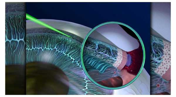
Why is argon laser used in eye surgery?
Macular degeneration is sometimes treated with an argon or krypton laser. In this treatment, the laser is used to destroy abnormal blood vessels so that haemorrhage or scarring will not damage central vision.
What is the use of the laser in ophthalmology?
It stands for Light Amplification by Stimulated Emission of Radiation. Laser has particular use in ophthalmology because the eye can be used as an optical device. The transparency of the front part of the eye, the cornea, allows light such as LASER to reach almost all the tissues of the eye.
Which laser is widely used in ophthalmology?
Current ophthalmic lasers Of these instruments, the diode laser is the laser most frequently used by the veterinary ophthalmologist; its tissue damage is caused by photocoagulation (Fig. 2.15).
What is argon laser Retinopexy?
Argon laser retinopexy is a safe and reliable technique for the treatment of symptomatic flap tears and other retinal breaks that are at a high risk of developing retinal detachment.Apr 16, 2004
Which laser is used for retina?
Frequency-doubled Nd-YAG Laser (532 nm) It is commonly used nowadays in treatment of many retinal conditions (proliferative diabetic retinopathy, diabetic macular edema, vein occlusions etc.).Aug 7, 2021
What does an argon laser look like?
The argon laser contains argon gas and produces a visible blue-green beam with wavelengths of 488 nm and 514 nm, which are absorbed selectively by hemoglobin, melanin, and other pigments that lie under the retina.
Is argon laser painful?
Occasional discomfort may be felt but treatment is usually pain-free. Sometimes a local anaesthetic injection around the eye may be required. Will eye drops be needed? Eye drops will need to be instilled by the nurse into one or both eyes 30 minutes before your laser treatment.
What is laser photocoagulation used for?
Laser photocoagulation is a type of laser surgery for the eyes. It is done to treat age-related macular degeneration (AMD). AMD is a condition that can lead to loss of vision. The retina is the layer of cells in the back of your eye that converts light into electrical signals.
What is trabeculoplasty?
Trabeculoplasty is a laser treatment for an eye condition called glaucoma. Glaucoma is a long-standing condition that damages the optic nerve due to increased eye pressure. Trabeculoplasty helps to open the drainage area of the eye and reduce the eye pressure.
What happens during argon laser treatment?
This procedure does not need any hospitalization and can be carried out by an eye specialist called as an ophthalmologist.
Is there any side effects or risk in this treatment?
The eye drops used before the procedure may blur your vision for three to four hours even after treatment.
What wavelength is the argon laser?
The argon laser contains argon gas and produces a visible blue-green beam with wavelengths of 488 nm and 514 nm , which are absorbed selectively by hemoglobin, melanin, and other pigments that lie under the retina. The beam is readily transmitted down a fiberoptic bundle, allowing endobronchial surgery (Fig. 40-8).
How long do argon lasers last?
However, they have relatively long lifetimes, often operating for several thousand hours before requiring gas tube replacement. Both argon- and krypton-ion lasers emit several monochromatic wavelengths simultaneously, which can be isolated using a Littrow prism inserted between the laser mirrors flanking the gas tube.
What is the wavelength of a laser used for DR?
Argon laser. Argon laser is a common laser used for treating DR. The main spectral peaks of the argon laser are 488 nm (blue) and 514 nm (green) i.e. relatively shorter wavelengths. Modern argon lasers remove blue wavelength light, allowing photocoagulation to be achieved at 514 nm which reduces light scatter as well as preventing absorption ...
What lasers are used for flow cytometry?
Argon- and krypton-ion lasers make excellent laser sources for flow cytometry. They produce high-quality Gaussian TEM 00 beams with low noise and good power stability, particularly in the higher-power water-cooled units. They are large lasers and produced copious amounts of waste heat, requiring a complex cooling system. However, they have relatively long lifetimes, often operating for several thousand hours before requiring gas tube replacement. Both argon- and krypton-ion lasers emit several monochromatic wavelengths simultaneously, which can be isolated using a Littrow prism inserted between the laser mirrors flanking the gas tube. Argon-ion lasers emit powerful lines at 488 and 514.5 nm, allowing initial excitation of fluorescein and rhodamine (and later emission of phycobiliproteins and their tandem dyes) ( Shapiro, 2003 ). Argon-ion lasers also produce a relatively strong series of lines in the ultraviolet, particularly at 351 and 364 nm ( Fig. 1). This allows the use of UV-excited dyes such as DAPI, which had first been used in mercury arc lamp systems, and also allowed the use of UV physiological probes such as the calcium indicator indo-1. Water-cooled argon lasers also produce a relatively strong line at 457 nm; this wavelength permits such applications as chromosome sorting using the DNA dyes chromomycin A3 and mithramycin, and simultaneous excitation of green, yellow, and cyan fluorescent protein ( Gray and Cram, 1990; Van den Engh et al., 1984 ). Argon-ion lasers have formed the excitation core of most commercial flow cytometers in the past 25 years.
What is an argon beam coagulator?
The argon beam coagulator (ABC) is an electrocoagulation system that should not be confused with the argon laser. No eyewear is required. The instrument achieves hemostasis by using inert gas as a medium to conduct radiofrequency energy (Fig. 77-4 ). The gas is emitted as a constant flow at room temperature from a handpiece and nozzle, which blows away blood and debris to optimize visualization. The first large clinical series utilizing the ABC for splenic salvage was published in 1991. 1 This report concluded that most spleens with superficial lacerations are easily salvaged with standard topical maneuvers and that the ABC offers a technical advantage in patients with deep parenchymal injuries. In the ensuing decade, the ABC achieved wide acceptance in the management of both spleen and other solid organ injuries.
What is the purpose of the Ar ion laser?
The laser beam, which may be focused on the retina, is used to prevent retinal detachment in addition to other medical and surgical applications. For all these applications, a few watts to tens of watts of laser output are necessary.
Is a laser trabeculoplasty effective for XFS?
Laser Therapy. Argon laser trabeculoplasty (ALT) is particularly effective, at least early on, in eyes with XFS. The baseline IOP is usually higher in XFG than in eyes with POAG undergoing ALT and thus the initial drop in IOP is greater in XFG.
How does an argon laser work?
The various energy level of argon ion is shown in the figure. When electric discharge passes through the tube, the Argon atoms are pumped to energy level E3 by two-step of collision with electrons. In the first step , neutral argon atoms are ionized and raised to energy level E1 ...
What is the argon laser made of?
The Argon ion Laser consists of a long and narrow discharge tube made of beryllium oxide filled with organ gas having two Windows at its ends inclined at Brewster’s angle.
What is an ion laser?
An ion laser is a gas laser in which ionized gas is used as the active or lasing medium. The electrical pumping method is used to achieve the population inversion in an ion laser. The pumping process is a two-step process in ion Laser. In the first step, the electron in the discharge tube makes a collision with neutral atoms ...
What is the active medium of a Krypton ion laser?
In the Krypton ion Iron laser, the active or lasing medium is krypton ions. The population inversion is achieved through the electrical pumping method. The working of krypton ion Laser is similar to that of Argon ion Laser. However, the output krypton iron laser has spectral lines in the visible region of the spectrum.
Why do argon ions migrate?
Due to the high current density, the Argon ions migrate continuously toward the cathode and electrons toward the anode. Ions having low mobility tend to accumulate at the cathode, get neutralized, and diffuse slowly back into the discharge tube.
What is the energy level of argon?
In the first step, neutral argon atoms are ionized and raised to energy level E1 (known as the ground state of argon ions). In the second step, the ion in the ground state is excited to energy level E3.
Which laser has a spectral line?
However, the output krypton iron laser has spectral lines in the visible region of the spectrum. On the other hand, the output of the argon-ion laser has a spectral line in the visible region, ultraviolet region, and near-visible region of the spectrum.
Retinal fluorescence and disease diagnosis
Thanks to a combination of scanning laser ophthalmoscopy and computer processing, scientists can study naturally occurring retinal fluorescence, which could serve as an early-warning marker of glaucoma, macular degeneration and other eyesight-stealing diseases.
Improving retinal imaging
Scientists are constantly trying to improve SLO technology with adaptive optics and other techniques to increase resolution and image finer details in the structure of the retina.
Laser eye surgery
Lasers are essential surgical tools for the 21st-century ophthalmologist. With these devices, surgeons can treat a vision-robbing disease such as glaucoma, macular degeneration and diabetic retinopathy. They can also make the patient’s existing vision crisp and clear.
The latest on LASIK
Laser-assisted in situ keratomileusis, or LASIK, is a popular elective surgery with people whose natural vision is worse than 20/20. The procedure for reshaping the cornea to reduce refractive error is now routine in the United States, but researchers have continued to refine the technology incrementally.
Fixing damaged corneas
Sometimes ophthalmic surgeons need to repair corneas damaged by burns, acids, accidents or other trauma. Conventionally, they use sutures to attach transparent slices of sterilized human amniotic membrane—roughly 30 µm thick—to the damaged corneal tissue. However, inflammation and scarring are frequent side effects of the surgery.
Looking forward
Many research teams around the world are vigorously pursuing higher-resolution retinal imaging using AO, optical coherence tomography (OCT) and lasers with exotic optics. Improved surgical techniques with lasers could result in faster healing and less scarring.
