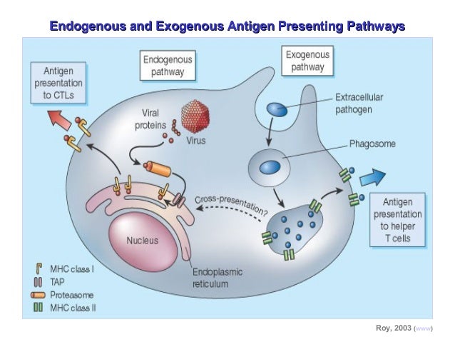
What are extracellular antigens? Extracellular antigens can bind to professional antigen presenting cells (APCs An antigen-presenting cell (APC) or accessory cell is a cell that displays antigen complexed with major histocompatibility complexes (MHCs) on their surfaces; this process is known as antigen presentation. T cells may recognize these complexes using their T cell receptors (TCRs). T…Antigen-presenting cell
What are the different types of antigen presenting cells?
There are three types of antigen-presenting cells in your body: macrophages, dendritic cells and B cells. One of their jobs is to act like a detective, showing the other cells of the immune system suspects they think are invading your body.
How are antigens recognized on the cell surface?
Antigen–MHC complexes are recognized on the cell surface by the TCR of CD4 + T cells, which then activate other immune effector cells. B cells presenting antigen using MHC class II molecules are induced to secrete antigen-specific antibody by interaction with activated CD4 + Th2 cells of the same specificity.
What is the meaning of extracellular?
Definition of extracellular : situated or occurring outside a cell or the cells of the body extracellular digestion extracellular enzymes : situated or occurring outside a cell or the cells of the body extracellular digestion extracellular enzymes
How are extracellular antigens transported by exogenous pathway?
In the exogenous pathway, extracellular antigens are internalized by APCs and degraded to peptides within endosomes. These peptides are bound to MHC class II and transported to the APC surface for recognition by CD4 ++ T cells (usually Th).

What is intracellular antigens?
Intracellular Antibodies. Intracellular antibodies, which may also be called intrabodies, are antibodies that are produced in the cell, and bind an antigen within the same cell.
How are extracellular antigens processed?
Secreted DC proteases act extracellularly to process intact proteins into antigenic peptides. Peptides produced by such activity are efficiently loaded onto cell surface class II MHC molecules.
What are the three types of APCs?
The main types of professional APCs are dendritic cells (DC), macrophages, and B cells.
What is an example of an antigen?
Antigens are known to trigger the immune system. They can be endogenous, exogenous, or autoantigens. Examples of antigens are pollen, bacteria, parasitic worms, and viruses.
What is an example of an exogenous antigen?
Exogenous antigens include particles considered foreign within the organism. For example, allergens (such as pollen), proteins from transplanted tissues and organs, and parts of microorganisms (such as coat, capsule, cell wall, flagella, fimbria, or toxin of bacteria, viruses, etc.)
What are the types of antigen processing pathways?
Antigens are internalized by several pathways, including phagocytosis, macropinocytosis, and endocytosis, and eventually traffic to a mature or late endosomal compartment where they are processed and loaded onto MHC-II molecules.
What cells are APC?
Antigen-presenting cells (APCs) are a heterogeneous group of immune cells that mediate the cellular immune response by processing and presenting antigens for recognition by certain lymphocytes such as T cells. Classical APCs include dendritic cells, macrophages, Langerhans cells and B cells.
What is the difference between MHC and APC?
An antigen-presenting cell (APC) or accessory cell is a cell that displays antigen bound by major histocompatibility complex (MHC) proteins on its surface; this process is known as antigen presentation. T cells may recognize these complexes using their T cell receptors (TCRs).
What is a cell surface antigen Why is it important?
Cell markers, also known as cell surface antigens, serve as monograms to help identify and classify cells.
What are 4 types of antigens?
There are different types of antigens on the basis of origin:Exogenous Antigens. Exogenous antigens are the external antigens that enter the body from outside, e.g. inhalation, injection, etc. ... Endogenous Antigens. ... Autoantigens. ... Tumour Antigens. ... Native Antigens. ... Immunogen. ... Hapten.
What are three antigens examples?
An antigen may be a substance from the environment, such as chemicals, bacteria, viruses, or pollen. An antigen may also form inside the body.
What is the difference between an antibody and an antigen?
Antigen vs antibody An antigen is a foreign substance that enters your body. This can include bacteria, viruses, fungi, allergens, venom and other various toxins. An antibody is a protein produced by your immune system to attack and fight off these antigens.
How antigen is processed and presented on the surface of MHC?
The usual process of antigen presentation through the MHC I molecule is based on an interaction between the T-cell receptor and a peptide bound to the MHC class I molecule. There is also an interaction between the CD8+ molecule on the surface of the T cell and non-peptide binding regions on the MHC class I molecule.
How does body recognize foreign antigen?
In contrast to the immunoglobulins, which interact with pathogens and their toxic products in the extracellular spaces of the body, T cells only recognize foreign antigens that are displayed on the surfaces of the body's own cells.
How do antibodies recognize and inactivate foreign antigens?
Antibodies recognize foreign invading microorganisms by specifically binding to a pathogen's proteins or antigens, facilitating their neutralization and destruction.
How are antigens made?
Any portion of bacteria or viruses, such as surface protein, coat, capsule, toxins, and cell wall, can serve as antigens. Moreover, a combination of lipid or nucleic acid with proteins or polysaccharides can form more complex antigens, such as lipopolysaccharides.
Which cells take up antigens?
Antigen presentation takes place very rapidly upon entry of antigen into lymphoid tissues. Presumably macrophages and Langerhans-dendritic cells take up the antigen and are responsible for the early recruitment and activation of CD4 T cells. B cells that have surface immunoglobulin molecules with specificity for the antigen also participate by binding the antigen, processing it and presenting it to the CD4 T cells.
What is the role of antigen presenting cells?
Antigen presenting cells (macrophages) fulfil a role of protein digestion and presentation in an appropriate form for ‘helper’ T cells. Whereas antibody can recognize antigens present in whole bacteria, virus, or parasites, the cytotoxic T cell is incapable of this, but will identify changes in the cell membrane or MHC antigens of infected cells.
What are the roles of intestinal epithelial cells in presentation of antigens?
Current dogma suggests that the M cells are the predominant antigen-sampling cells of the gut, which pass internalized antigen to the macrophage cells of the Peyer’s patches , and the latter serves as the major antigen-presenting cells (AP Cs) which inititate T- and B-cell priming in response to oral immunization. However, an alternative pathway of antigen uptake and presentation provided by the absorptive epithelial cells of the intestinal mucosa has been suggested by Bromander et al. (1993). These investigators analyzed the effect of CT exposure on alloantigen presentation by cultured intestinal epithelial cells. The crypt, small intestinal epithelial cell line, IEC-17, derived from the duodenum of Sprague–Dawley rats was cultured with IFNγ to induce optimal expression of MHC II and in the presence or absence of CT ( Quaroni and Isselbacher, 1981 ). After 48 hr, the IEC-17 cells were washed, treated with mitomycin C, and incubated with histoincompatible spleen cells. Alloantigen-stimulated CD4 + T-cell proliferation was then determined by 3 H-thymidine incorporation. CT enhanced the antigen presentation of IEC-17 cells and this enhancement was not due to an upregulation of MHC II expression, but rather a dose-dependent increase in IL-1 and IL-6 secretion by IEC-17 cells. The data suggest that the potent adjuvanticity of CT may be attributed to its ability to enhance the co-stimulating ability of APCs such as intestinal epithelial cells of the mucosal immune system. However, it was noted that intestinal epithelial cells have only been found to process and present antigens in vitro and that whether these cells are capable of functioning as APCs in vivo remains to be examined. Two limitations of the above study are the relevance of using a crypt cell line (IEC-17) rather than a non-crypt intestinal cell line, such as Henle-407 cells, and the nature of the antigen that is presented. Specifically, alloantigen presentation is an atypical form of antigen presentation which does not require presentation in the context of self-MHC and therefore, the enhanced “presentation” observed in this study might not accurately reflect the conventional antigen-processing and -presenting capability of intestinal epithelial cells in vitro.
What is the presentation of antigens?
Antigen presentation is the expression of antigen molecules on the surface of a macrophage or other antigen-presenting cell in association with MHC class II molecules when the antigen is being presented to a CD4+ helper T cell or in association with MHC class I molecules when presentation is to CD8 + cytotoxic T cells. For appropriate presentation, it is essential that peptides bind securely to the MHC class II molecules, since those that do not bind or are bound only weakly are not presented and fail to elicit an immune response. Following interaction of the presented antigen and MHC class II molecules with the CD4 + helper T cell receptor, the CD4 + lymphocyte is activated, IL-2 is released, and IL-2 receptors are expressed on the CD4 + lymphocyte surface. The IL-2 produced by the activated cell stimulates its own receptors, as well as those of mononuclear phagocytes, increasing their microbicidal activity. IL-2 also stimulates B cells to synthesize antibody. Whereas B cells may recognize a protein antigen in its native state, T cells only recognize the peptides, that result from antigen processing, in the context of major histocompatibility complex molecules.
What is the role of MHC in the immune system?
Antigen processing and presentation through the major histocompatibility complex (MHC) is a critical step in initiating effective cell-mediated adaptive immune response against pathogens. Downregulation of the cell surface MHC class I molecules is a key viral immune evasion strategy. KSHV encodes two zinc finger membrane proteins MIR1 and MIR2 (also called K3 and K5) which are E3 ubiquitin ligases for modulation by ubiquitylation of MHC I molecules on the infected cell surfaces ( Coscoy and Ganem, 2000, 2001 ). The ubiquitylated MHC I molecules then undergoes endocytosis and is degraded in the lysosome ( Coscoy et al., 2001 ). Interestingly, it has also been demonstrated that MIR1 is able to act as an E3 ubiquitin ligase on ubiquitylation of lysineless molecules ( Cadwell and Coscoy, 2005, 2008; Coscoy and Ganem, 2003 ). Additionally, MIR2 dowregulates ICAM-1 and B7-2 which are ligands for NK cell-mediated cytotoxicity receptors ( Ishido et al., 2000 ). The second strategy for KSHV to evade the adaptive immune system is to encoding several viral chemokines (vCCL). At least three vCCLs have been identified and known to inhibit Th1 cell-mediated immune responses by binding with cellular chemokine receptors on Th1 helper cells which result in blocking signal transduction of G-proteins ( Means et al., 2002; Moore and Chang, 2003 ). Additionally, KSHV also encodes its own versions of the chemokine receptors like vGPCR ( Sodhi et al., 2004a ). vGPCR is a relative of cellular IL-8 receptors CXCR1 and CXCR2 ( Sodhi et al., 2004a ). Similar to K1, vGPCR is also constitutively active and induces an array of proinflammatory cytokines and growth factors such as IL-1β, IL-6, IL-8, TNF-α, VEGF, and bFGF through AP-1, NF-kB, and HIF1 pathways ( Montaner et al., 2004; Schwarz and Murphy, 2001; Sodhi et al., 2000 ). However, vGPCR activates several downstream kinases including Lyn, JNK, Akt, and p38 not only by constitutive activation but also by certain induction of chemokines like IL-8 ( Montaner et al., 2001; Sodhi et al., 2000, 2004b ).
What is the mechanism by which the antigenic environment is sampled and information imparted to the effector arms of?
Antigen presentation is the mechanism by which the antigenic environment is sampled and information imparted to the effector arms of the adaptive immune system, B and T lymphocytes. Depending on the precise context, antigen presentation can result in either activation or tolerization of lymphocytes, respective examples being the response to a pathogen challenge or tolerance to self-antigen. An antigen is subjected to either endogenous or exogenous processing and the resulting peptides are expressed on the surface of the antigen presenting cell (APC) bound to either major histocompatibility complex (MHC) class I or II molecules (see Chapter 8 ). The type of MHC molecule involved in the presentation not only reflects the source of the antigen, but also has the primary role in determining the ensuing immune response to it.
What is the role of T cells in cellular immunity?
A crucial difference between the T cell and the B cell is in the nature of the antigen which it ‘recognizes’. Whereas the B cell and its secreted product, antibody, recognizes whole and extracellular antigens, the T lymphocyte is incapable of such recognition and can only ‘see’ small linear peptides of digested protein presented as part of a cell membrane and in direct association with the membrane major histocompatibility (MHC) antigens. Antigen presenting cells (macrophages) fulfil a role of protein digestion and presentation in an appropriate form for ‘helper’ T cells. Whereas antibody can recognize antigens present in whole bacteria, virus, or parasites, the cytotoxic T cell is incapable of this, but will identify changes in the cell membrane or MHC antigens of infected cells.
What does "extracellular" mean?
Definition of extracellular. : situated or occurring outside a cell or the cells of the body extracellular digestion extracellular enzymes. Keep scrolling for more.
What does "extracellular" mean in medical terms?
Medical Definition of extracellular. : situated or occurring outside a cell or the cells of the body extracellular digestion extracellular enzymes.
What are the genes that encode MHC class I?
Human MHC class I molecules are encoded by a series of genes – HLA-A, HLA-B and HLA-C (HLA stands for ‘Human Leukocyte Antigen’ , which is the human equivalent of MHC molecules found in most vertebrates). These genes are highly polymorphic, which means that each individual has his/her own HLA allele set. The consequences of these polymorphisms are differential susceptibilities to infection and autoimmune diseases that may result from the high diversity of peptides that can bind to MHC class I in different individuals. Also, MHC class I polymorphisms make it virtually impossible to have a perfect tissue match between donor and recipient, and thus are responsible for graft rejection.
How are peptides derived from ER?
Prior to entering the ER, peptides are derived from the degradation of proteins, which can be of viral- or self origin. Degradation of proteins is mediated by cytosolic- and nuclear proteasomes, and the resulting peptides are translocated into the ER by means of TAP.
What are MHC class II molecules?
MHC class II molecules are expressed by APCs, such as dendritic cells (DC), macrophages and B cells (and, under IFNγ stimuli, by mesenchymal stromal cells, fibroblasts and endothelial cells, as well as by epithelial cells and enteric glial cells). MHC class II molecules bind to peptides that are derived from proteins degraded in the endocytic pathway. MHC class II complexes consists of α- and β-chains that are assembled in the ER and are stabilised by invariant chain (Ii). The complex of MHC class II and Ii is transported through the Golgi into a compartment which is termed the MHC class II compartment (MIIC). Due to acidic pH, proteases cathepsin S and cathepsin L are activated and digest Ii, leaving a residual class II-associated Ii peptide (CLIP) in the peptide-binding groove of the MHC class II. Later, the CLIP is exchanged for an antigenic peptide derived from a protein degraded in the endosomal pathway. This process requires the chaperone HLA-DM, and, in the case of B cells, the HLA-DO molecule. MHC class II molecules loaded with foreign peptide are then transported to the cell membrane to present their cargo to CD4+ T cells. Thereafter, the process of antigen presentation by means of MHC class II molecules basically follows the same pattern as for MHC class I presentation.
What happens to MHC class I molecules?
MHC class I complexes at the cell surface may dissociate as time passes and the heavy chain can be internalised. When MHC class I molecules are internalised into the endosome, they enter the MHC class-II presentation pathway. Some of the MHC class I molecules can be recycled and present endosomal peptides as a part of a process which is called cross-presentation.
What is the process of presenting endosomal peptides?
Some of the MHC class I molecules can be recycled and present endosomal peptides as a part of a process which is called cross-presentation. The usual process of antigen presentation through the MHC I molecule is based on an interaction between the T-cell receptor and a peptide bound to the MHC class I molecule.
What are the three genes that make up the MHC class II?
MHC class II polymorphism. Like the MHC class I heavy chain, human MHC class II molecules are encoded by three polymorphic genes: HLA-DR, HLA-DQ and HLA-DP. Different MHC class II alleles can be used as genetic markers for several autoimmune diseases, possibly owing to the peptides that they present.
What are the key elements of adaptive immunity?
In order to be capable of engaging the key elements of adaptive immunity (specificity, memory, diversity, self/nonself discrimination ), antigens have to be processed and presented to immune cells. Antigen presentation is mediated by MHC class I molecules, and the class II molecules found on the surface of antigen-presenting cells (APCs) ...
How do extracellular and intracellular bacteria differ?
Immune Responses to Extracellular and Intracellular Bacteria Can Differ. Infection by extracellular bacteria induces production of humoral antibodies, which are ordinarily secreted by plasma cells in regional lymph nodes and the submucosa of the respiratory and gastrointestinal tracts.
Why are antibodies important in pathogenic bacteria?
Extracellular bacteria can be pathogenic because they induce a localized inflammatory response or because they produce toxins. The toxins, endotoxin or exotoxin, can be cytotoxic ...
What is the toxin produced by bacteria?
An excellent example of this is the toxin produced by diphtheria, which exerts a toxic effect on the cell by blocking protein synthesis. Endotoxins, such as lipopolysaccharides (LPS), are generally components of bacterial cell walls, while exotoxins, such as diphtheria toxin, are secreted by the bacteria.
Where are MHC class II antigens found?
MHC Class II antigens are found on antigen-presenting cells such as B cells, macrophages, monocytes, and dendritic cells. MHC Class I molecules are displayed on many more cells types, as long at the cell has a nucleus
Which class of lymphocytes responds to MHC class II/antigen complexes?
The class of lymphocytes called Blank 1 of 1 T cells respond to MHC class II/antigen complexes; they activate B cells and effector T cells. helper. A (n) Blank 1 of 1-mediated immune response is effective against extracellular antigens, such as bacteria, viruses, and toxins when they are outside the cell.. antibody.
What is an APC cell?
An APC is a cell that phagocytizes an antigen and displays fragments of it on its surface for recognition by other cells of the immune system; chiefly dendritic cells, macrophages and B cells.
Extracellular Matrix Definition

Extracellular Matrix Function
- Living tissue can be thought of as a dynamic meshwork of cells and liquid. Despite their close proximity to each other, the cells of a tissue are not simply tightly wound together. Instead, they are spaced out with the help of the extracellular meshwork. The matrix will act as a kind of filler that lies between the otherwise tightly packed cells in a tissue. Furthermore, not only is the matri…
Extracellular Matrix Components
- The extracellular matrix is mostly made up of a few key ingredients: water, fibrous proteins, and proteoglycans. The main fibrous proteins that build the extracellular matrix are collagens, elastins, and laminins. These are all relatively sturdy protein macromolecules. Their sturdiness lends the extracellular matrix its buffering and force-resisting properties that can withstand environmenta…
Quiz
- 1. Which of the following is not a fibrous protein type mentioned? A. Elastin B. Proteoglycan C. Collagen D.Laminin 2. Identify the distinction between fibrous protein and proteoglycans, per the article: A. Fibrous protein is more capable of handling aqueous environments B. Proteoglycans serve more of a filler role in the spaces between the cells in a tissue C. Fibrous proteins resist ag…