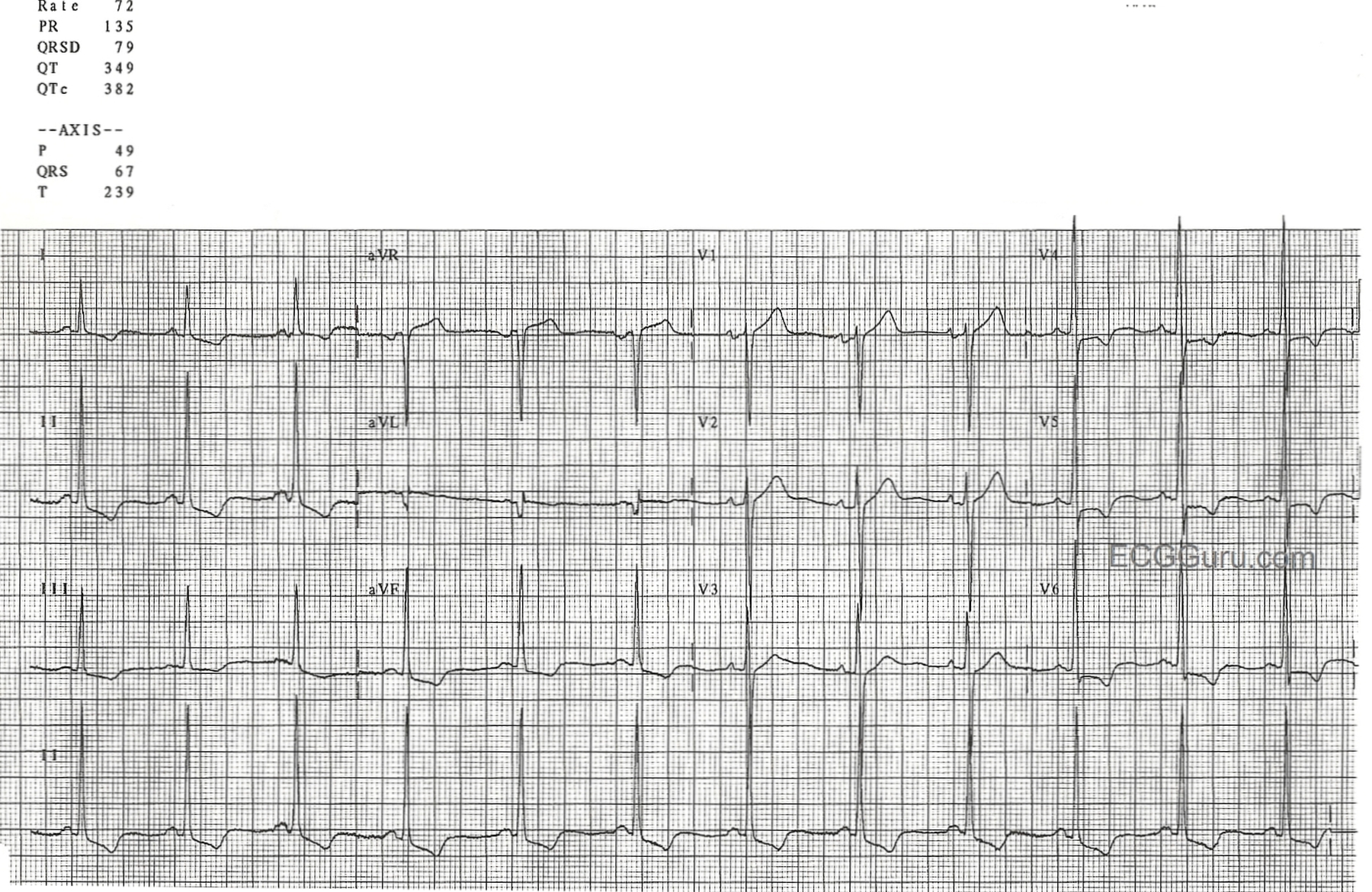
S Wave. The S wave is the first downward deflection of the QRS complex that occurs after the R wave. However, a S wave may not be present in all ECG leads in a given patient. In the normal ECG, there is a large S wave in V1 that progressively becomes smaller, to the point that almost no S wave is present in V6.
What does each wave of an ECG represent?
What does each wave of an ECG represent? There are three main components to an ECG: the P wave, which represents the depolarization of the atria; the QRS complex, which represents the depolarization of the ventricles; and the T wave, which represents the repolarization of the ventricles. Click to see full answer.
What is an S wave?
S waves are a type of transverse wave. What this means is that the oscillations of an S wave’s particles are perpendicular to the wave propagation’s direction. The main restoring force is because of the shear stress. Therefore, the propagation of S waves cannot take place in liquids with very low or zero viscosity.
What is the normal speed of an ECG?
A standard ECG is recorded at 25mm/sec and with a frequency cut off of no lower than 150Hz in adults, and 250Hz in children. On the standard ECG paper, with standard calibration, the squares represent: The standard calibration signal will look like this: This will be present at the beginning or end of all four rows of the trace, and shows:
What wave comes after the T wave of an ECG?
U wave on ECG occurs after the T wave and is usually seen in the mid precordial leads. In hypokalemia, T wave becomes flattened and U wave becomes prominent (or apparently so because of near absence of T waves). Important conditions associated with U waves are systemic hypertension, aortic and mitral regurgitation and coronary artery disease [1].

What does the S wave on an ECG represent?
the S wave signifies the final depolarization of the ventricles, at the base of the heart.
What does a large S wave indicate?
A prominent S-wave in lead I is typically present in cases of congenital heart disease, valvular heart disease, and cor pulmonale that cause right ventricular enlargement and fibrosis.
What is abnormal S wave?
36. An S wave is often absent in leads V5 and V6. An S wave of less than 0.3 mV in lead V1 is considered abnormally small. If the amplitude of the entire QRS complex is less than 1.0 mV in each of the precordial leads, the voltage is considered abnormally low.
What happens at the S wave?
An S wave, or shear wave, is a seismic body wave that shakes the ground back and forth perpendicular to the direction the wave is moving.
What causes a large S wave on ECG?
It is concluded that a prominent S wave in lead I alone or together with lead V6 in ECGs of middle-aged and elderly patients suggests the presence of a disease affecting the pulmonary criculation or the left ventricle of the heart.
What causes deep S wave?
Left ventricular hypertrophy causes increased R-wave amplitudes in V4–V6 and deeper S-waves in V1–V3.
What is the most common ECG abnormality in adults?
The most common ECG abnormalities were T-wave abnormalities. Average heart rate corrected QT interval was longer in women than men, similar in whites and blacks and increased with age, whereas the average heart rate was higher in women than men and in blacks than whites and decreased with age.
Where is the S wave in ECG?
The S wave is the first downward deflection of the QRS complex that occurs after the R wave. However, a S wave may not be present in all ECG leads in a given patient. In the normal ECG, there is a large S wave in V1 that progressively becomes smaller, to the point that almost no S wave is present in V6.
Should I worry about abnormal ECG?
An abnormal ECG can mean many things. Sometimes an ECG abnormality is a normal variation of a heart's rhythm, which does not affect your health. Other times, an abnormal ECG can signal a medical emergency, such as a myocardial infarction /heart attack or a dangerous arrhythmia.
Why do S waves cause more damage?
S waves are more dangerous than P waves because they have greater amplitude and produce vertical and horizontal motion of the ground surface.
What is difference between P and S waves?
P waves can travel through liquid and solids and gases, while S waves only travel through solids. Scientists use this information to help them determine the structure of Earth. For example, if an earthquake occurs on one side of Earth, seismometers around the globe can measure the resulting S and P waves.
Can you feel S waves?
When an earthquake occurs, it makes seismic waves, which cause the shaking we feel. Seismic waves are essentially just the jiggling of the ground in response to the force put on the ground by the earthquake, similar to the way the jello in a bowl responds to a tap to the side of the bowl.
Which lead has the largest S wave?
lead V2The S wave is deepest in the right precordial leads, usually in lead V2. The S wave amplitude decreases as the left precordium is approached.
How would you describe the behavior of an S wave?
S Waves—secondary body waves that shear, or cut the rock they travel through sideways at right angles to the direction of motion; cannot travel through liquid; produce vertical and horizontal motion in the ground surface.
Which statements best explains the characteristics of S waves?
The correct answer is option C. S waves, or secondary waves, can only travel solid rock, that is the crust and mantle.
What are the characteristics of S waves?
S-wave Motion S Wave—secondary body waves that oscillate the ground perpendicular to the direction of wave travel. They travel about 1.7 times slower than P waves. Because liquids will not sustain shear stresses, S waves will not travel through liquids like water, molten rock, or the Earth's outer core.
Is the presence of the S wave clinically significant?
The presence or absence of the S wave does not bear major clinical significance. Rarely is the morphology of the S wave discussed.
Is there a S wave in V1?
In the normal ECG, there is a large S wave in V1 that progressively becomes smaller, to the point that almost no S wave is present in V6. A large slurred S wave is seen in leads I and V6 in the setting of a right bundle branch block. The presence or absence of the S wave does not bear major clinical significance.
Is the S wave present in lead I?
Rarely is the morphology of the S wave discussed. In the setting of a pulmonary embolism, a large S wave may be present in lead I — part of the S1Q3T3 pattern seen in this disease state.
What is the P wave in ECG?
ECG interpretation starts with assessment of the P-wave and PR interval. The P-wave is generated by depolarization (activation, contraction) of the atria. The PR interval is the interval between the start of the P-wave and the start of the QRS complex. The PR interval determines whether impulse transmission from atria to ventricles is normal. The isoelectric (flat) line between the end of the P-wave and the start of the QRS complex is called the PR segment. The PR segment is the baseline (also referred to as reference line or isoelectric line) of the ECG curve. Thus, when measuring the amplitude of a wave on the ECG, the PR segment is the baseline. Refer to Figure 1.
What is the T wave?
The T-wave reflects the rapid repolarization (recovery) of the myocardium and T-wave changes occur in numerous conditions. T-wave changes are frequently misunderstood. The transition from the ST segment to the T-wave should be smooth. The normal T-wave is somewhat asymmetric, with a steeper downward slope.
What is the QT interval?
QT duration reflects the total duration of ventricular depolarization (activation) and repolarization (recovery). It is measured from the start of the QRS complex to the end of the T-wave. The QT interval increases at slower heart rates and vice versa (i.e it decreases at higher heart rates). Therefore, to judge whether the QT interval is normal it is necessary to take the heart rate into account. The heart rate adjusted QT interval is the corrected QT interval, or simply the QTc interval. A long QTc interval causes electrical instability in the ventricles and this may cause lethal ventricular arrhythmias.
What is the heart rate adjusted QT interval?
The heart rate adjusted QT interval is the corrected QT interval, or simply the QTc interval. A long QTc interval causes electrical instability in the ventricles and this may cause lethal ventricular arrhythmias.
Which ventricle is larger, the QRS or the adipose?
Because the left ventricle is usually considerably larger than the right ventricle, the QRS complex is actually a reflection of the electrical potentials generated by the left ventricle.
Is ventricular depolarization a QRS complex?
In other words, if ventricular depolarization only generates a Q-wave and an R-wave, that complex may still be referred to as a QRS complex. However, one may also be more explicit and refer to such a complex as a QR complex.
What is an ECG machine?
ECG (electrocardiogram) in veterinary medicine is a recorder of the electrical activity of the heart at rest.
What is the purpose of a veterinary ECG?
Uses of the veterinary ECG: To monitor the depth of anesthesia, pain, and stress. To observe post-surgical patients. To monitor an animal with a chronic condition . It is useful when arrhythmia is suspected.
What is the S-T interval?
S-T-interval: after QRS in the time when the ventricle is depolarized. It can be used to diagnose ischemia or hypoxia.
Is ECG machine important for veterinary?
There is no doubt that ECG is an important monitoring tool during and after surgery in a veterinary clinic.
How does an ECG work?
This electrical activity is transmitted throughout the body and can be picked up on the skin. This is the principle behind the ECG. An ECG machine records this activity via electrodes on the skin and displays it graphically. An ECG involves attaching 10 electrical cables to ...
What is the ECG chapter?
Chapter 3Conquering the ECG. Besides the stethoscope, the electrocardiogram (ECG) is the oldest and most enduring tool of the cardiologist. A basic knowledge of the ECG will enhance the understanding of cardiology (not to mention this book). Electrocardiography. At every beat, the heart is depolarized to trigger its contraction.
How to become confident in reading ECG?
A normal ECG tracing is provided in Figure 6. The only way to become confident at reading ECGs is to practice. It is important to be methodical – every ECG reading should start with an assessment of the rate, rhythm, and axis. This approach always reveals something about an ECG, regardless of how unusual it is.
Which direction do depolarization and repolarization deflections occur?
depolarization and repolarization deflections occur in opposite directions
Where does the wave of depolarization travel?
The wave of depolarization then proceeds rapidly to the bundle of His where it splits into two pathways and travels along the right and left bundle branches. The impulse travels the length of the bundles along the interventricular septum to the base of the heart, where the bundles divide into the Purkinje system. From here, the wave of depolarization is distributed to the ventricular walls and initiates ventricular contraction.
Where does depolarization take place in the heart?
The route that the depolarization wave takes across the heart is outlined in Figure 3. The sinoatrial node (SAN) is the heart's pacemaker. From the SAN, the wave of depolarization spreads across the atria to the atrioventricular node (AVN). The impulse is delayed briefly at the AVN and atrial contraction is completed.
Which wave reflects depolarization of the main mass of the ventricles?
the R wave reflects depolarization of the main mass of the ventricles –hence it is the largest wave

The Normal ECG (EKG) Waves, Intervals, Durations and Rhythm
Overview of The Normal Electrocardiogram
- Figure 1. The classical ECG curve with its most common waveforms. Important intervals and points of measurement are depicted. ECG interpretation requires knowledge of these waves and intervals.
The P-Wave, PR Interval and PR Segment
- ECG interpretation starts with assessment of the P-wave and PR interval. The P-wave is generated by depolarization (activation, contraction) of the atria. The PR interval is the interval between the start of the P-wave and the start of the QRS complex. The PR interval determines whether impulse transmission from atria to ventricles is normal. The isoelectric (flat) line between the end of the …
The QRS Complex
- The QRS complex reflects the depolarization (activation, contraction) of the ventricles. Although it may not always include a Q-wave, R-wave and S-wave, it is still referred to as a QRS complex. In other words, if ventricular depolarization only generates a Q-wave and an R-wave, that complex may still be referred to as a QRS complex. However, one may also be more explicit and refer to s…