[edit on Wikidata] The anterior white commissure (ventral white commissure) is a bundle of nerve fibers which cross the midline of the spinal cord just anterior (in front of) to the gray commissure (Rexed lamina X).
What is the anterior commissure?
The anterior white commissure ( ventral white commissure) is a bundle of nerve fibers which cross the midline of the spinal cord just anterior (in front of) to the gray commissure ( Rexed lamina X).
What is the anterior white commissure of the spine?
The anterior or ventral white commissure is a collection of nerve fibers that cross the midline of the spinal cord and transmit information from or to the contralateral side of the brain. It is present throughout the length of the spinal cord and lies behind the anterior median fissure.
What is the white commissure in a mouse?
· The anterior white commissure is a rectangularly-shaped collection of nerve fibers located directly anterior to the central canal and gray commissure, posterior to the anterior …
Does the anterior commissure mediate interhemispheric transfer of visual information?
anterior commissure (redirected from Anterior white commissure) Also found in: Encyclopedia . commissure [ kom´ĭ-shūr] 1. a site of union of corresponding parts, such as the angle of the …

What is the anterior commissure?
The anterior commissure (also labeled ac) is a large bundle of crossing fibers, which connects the olfactory bulb and parts of the cerebrum to the same areas on the opposite side.
Is the anterior commissure gray or white?
The anterior white commissure (ventral white commissure) is a bundle of nerve fibers which cross the midline of the spinal cord just anterior (in front of) to the gray commissure (Rexed lamina X).
What is anterior white column?
Acronym: The term anterior white column refers to the large bundle of fibers located between the anterior median fissure of the spinal cord and the anterolateral sulcus of the spinal cord.
What is dorsal white commissure?
The white commissure (commissura alba) is a bridge of white substance which connects the ventral columns over the dorsal end of the ventral median fissure and constitutes a conducting path from one side to the other. From: The Mouse Nervous System, 2012.
What does the anterior gray commissure do?
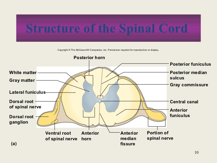
The grey commissure is a thin strip of grey matter that surrounds the central canal of the spinal cord and, along with the anterior white commissure, connects the two halves of the cord. It comprises lamina X in the Rexed classification. Spinal cord. (Grey commissure is #3, near center.)
Where is the anterior commissure located in the brain?
third ventricleThe anterior commissure is located in the anterior wall of the third ventricle at the upper end of the lamina terminalis. It runs across the midline in front of the anterior columns of the fornix, above the basal forebrain and beneath the medial and ventral aspect of the anterior limb of the internal capsule.
What does white matter do in the brain?
In the most general sense, the gray matter of the brain facilitates information processing, and the white matter facilitates information transfer; both are critical for efficient operation of the neural networks responsible for a specific mental domain.
What is the function of the white columns?
White Columns Ascending tracts of nervous system fibers in these columns carry sensory information up to the brain, whereas descending tracts carry motor commands from the brain. Looking at the spinal cord longitudinally, the columns extend along its length as continuous bands of white matter.
What does white matter in the spinal cord do?
The white matter of your brain and spinal cord is composed of bundles of axons. These axons are coated with myelin, a mixture of proteins and lipids, that helps conduct nerve signals and protect the axons. White matter's job is to conduct, process, and send nerve signals up and down the spinal cord.
What is posterior commissure?
The posterior commissure (PC) is a transversely-oriented commissural white matter tract that connects the two cerebral hemispheres along the midline. It is a very important anatomical landmark which is thought to play a role in the visual system, however its functions are still largely unknown.
What is the anterior median fissure of the spinal cord?
The anterior median fissure provides a groove in which the anterior spinal artery sits. From here, it provides the anterior part of the spinal cord. It is sourced from the segmental medullary arteries and the segmental spinal arteries which are sourced from the intercostal arteries.
What commissure means?
Definition of commissure 1 : a point or line of union or junction especially between two anatomical parts (such as adjacent heart valves) 2 : a connecting band of nerve tissue in the brain or spinal cord.
What is the white commissure?
The white commissure (commissura alba) is a bridge of white substance which connects the ventral columns over the dorsal end of the ventral median fissure and constitutes a conducting path from one side to the other.
What is commissural myelotomy?
An alternative procedure called commissural myelotomy is directed at interrupting the axons of spinothalamic tract neurons of both sides as they cross the midline ( Armour, D., 1927; Mansuy, L. et al., 1944; 1976; see Gybels, J. M. and Sweet, W. H., 1989). A lesion is made near the midline of the spinal cord and extending rostrocaudally for as many segments as needed to interrupt the appropriate projections. The lesion should be sufficiently deep to ensure that the crossing spinothalamic axons in the anterior white commissure are cut. Of course, the crossing axons of a number of tracts that accompany the spinothalamic tract, such as spinoreticular axons, are also interrupted, as are many axons in the posterior funciculi. According to the analysis of Dargent, M. et al. (1963), this procedure is apparently particularly effective for vaginal and visceral pain. Interestingly, commissural myelotomy often relieves clinical pain even if on testing there is no hypalgesia. Furthermore, pain and temperature sensation may be lost over a much greater extent of the body than could be predicted from the location and dimensions of the commissural myelotomy ( Gybels, J. M. and Sweet, W. H., 1989 ). Based on evidence reviewed in the following, it now seems likely that the effect of a commissural myelotomy depends at least in part on the interruption of a visceral pain pathway that ascends in the posterior funiculi.
Where do CST axons travel?
Crossed axons in the lateral CST, intermixed with axons of the rubrospinal tract, travel in the lateral funiculus. These CST axons terminate directly and indirectly mainly on LMNs associated with distal musculature, especially for skilled hand and finger movements. The uncrossed anterior CST axons decussate predominantly in the anterior white commissure and terminate directly and indirectly mainly on LMNs that supply medial musculature. A small number of anterior CST axons terminate ipsilateral to the cortical cells of origin. An isolated lesion in the CST in the medullary pyramids results in weakness of contralateral fine, dexterous hand and finger movements. All other lesions involving the CST at other levels (internal capsule, cerebral peduncle, pons), where these descending fibers are intermixed with other descending motor systems, produce contralateral spastic hemiplegia with hypertonus, hyperreflexia, and plantar extensor responses as long-term consequences. Lesions in the lateral CST produce similar symptoms ipsilateral to the damaged lateral funiculus below the level of the lesion.
How is the white matter on both sides of the spinal cord divided?
At the base of the anterior median fissure the white matter on both sides of the cord is continuous via a slender lamina of decussating nerve fibers forming the anterior white commissure.
What is syringomyelia?
Syringomyelia is commonly seen in patients with Chiari malformations in the posterior fossa but may also be a consequence of trauma to the spinal cord, tumors, and infections, The symptoms are highly variable but most frequently include loss of pain and temperature sensations, extremity weakness, and unsteady gait.
Where is a lesion made?
A lesion is made near the midline of the spinal cord and extending rostrocaudally for as many segments as needed to interrupt the appropriate projections. The lesion should be sufficiently deep to ensure that the crossing spinothalamic axons in the anterior white commissure are cut.
Are pyramids gray or white matter?
Roughly 75% of motor fibres housed within the pyramids cross diagonally and posteriorly, and continue down the spinal column as the lateral corticospinal tracts. At this level, the central portion of the medulla contains gray matter, while the outer portions consist of white matter.
What is Lissauer tract?
Answer: Lissauer's tract is a white matter tract in the spinal cord that projects up or down across one or two spinal segments. Somatosensory information arising from the skin enters into the spinal cord via the dorsal horn. Some of those fibers ascend or descend locally, across one or two spinal segments.
What is the dorsal horn?
Medical Definition of dorsal horn : a longitudinal subdivision of gray matter in the dorsal part of each lateral half of the spinal cord that receives terminals from some afferent fibers of the dorsal roots of the spinal nerves. — called also dorsal column, posterior column, posterior gray column, posterior horn.
What does the anterior Spinothalamic tract do?
The anterior spinothalamic tract, also known as the ventral spinothalamic fasciculus, is an ascending pathway located anteriorly within the spinal cord, primarily responsible for transmitting coarse touch and pressure.
What is the function of the anterior white column?
The white matter of the spinal cord is subdivided into dorsal (or posterior), lateral, and ventral (or anterior) columns, each of which contains axon tracts related to specific functions. The dorsal columns carry ascending sensory information from somatic mechanoreceptors (Figure 1.11B).
What is Decussation?
Definition of decussation. 1 : the action of crossing (as of nerve fibers) especially in the form of an X. 2 : a crossed tract of nerve fibers passing between centers on opposite sides of the nervous system.
Where is the substantia Gelatinosa located?
Definition. Substantia gelatinosa is a collection of cells in the gray area (dorsal horns) of the spinal cord. Found at all levels of the cord, it receives direct input from the dorsal (sensory) nerve roots, especially those fibers from pain and thermoreceptors.
What is anterior commissure?
anterior commissure the band of fibers connecting the parts of the two cerebral hemispheres.
What is the middle commissure?
middle commissure a band of gray matter joining the optic thalami; it develops as a secondary adhesion and may be absent.
Which ligamentous attachment of the vocal cords is a common site of extension of squamous cell carcinoma
The anterior ligamentous attachment of the true vocal cords to the thyroid cartilage, which, because it lacks an internal perichondrium, is a common site of extension of squamous cell carcinomas from the anterior commissure.
Which structures are similar in form to the human perihippocampal area?
Evidence is provided for the fact that the limbic structures the fifth temporal gyrus, the amygdala, the hippocampus, the anterior white commissure, the ventral striatium and the perihippocampal area, are similar in form in the two primates, the human and the spider monkey.
What is the anterior commissure?
(left, third from bottom.) The anterior commissure (also known as the precommissure) is a white matter tract (a bundle of axons) connecting the two temporal lobes of the cerebral hemispheres across the midline, and placed in front of the columns of the fornix.
Where is the anterior commissure located?
The anterior commissure (also known as the precommissure) is a white matter tract (a bundle of axons) connecting the two temporal lobes of the cerebral hemispheres across the midline, and placed in front of the columns of the fornix. In most existing mammals, the great majority of fibers connecting the two hemispheres travel through ...
Which lobes connect with anterior commissure?
The anterior commissure also serves to connect the two amygdalae .
Which part of the neocortex carries the fibers of the neocortex?
The anterior commissure serves as the primary mode of interhemispheric communication in marsupials, and which carries all the commissural fibers arising from the neocortex (also known as the neopallium), whereas in placental mammals the anterior commissure carries only some of these fibers).
How many normal controls show the anterior commissure?
Averaged tracking results of ten normal controls showing the Anterior Commissure. Image from Winter and Franz (2014)
Which hemispheres are the corpus callosum found in?
The corpus callosum allows for communication between the two hemispheres and is found only in placental mammals (the eutherians), while it is absent in monotremes and marsupials, as well as other vertebrates such as birds, reptiles, amphibians and fish. The anterior commissure serves as the primary mode of interhemispheric communication in ...
Is anterior commissure larger in women or men?
We also measured the anterior commissure in the same blocks of tissue used for the present hypothalamic study (data not shown) and were unable to replicate a report [by Allen and Gorski] that its cross-sectional area is larger in women than in men.
What is the anterior commissure?
The anterior commissure (also labeled ac) is a large bundle of crossing fibers, which connects the olfactory bulb and parts of the cerebrum to the same areas on the opposite side.
What part of the neocortices is the anterior commissure?
It anteriorly connects two-thirds of the right and left temporal neocortices and the posterior part of the orbital aspect of the frontal lobes. The anterior commissure mediates interhemispheric transfer of visual information, including the visual recall of dreams, and auditory and olfactory information (see Swaab, 2003 ).
Which hemispheres are connected by the anterior commissure?
The anterior commissure, a fiber tract connecting the left and right cerebral hemispheres, has also been studied in relationship to sex and sexual orientation. Nonheterosexual men have been reported to have larger anterior commissures than heterosexual men (Allen and Gorski, 1992 ).