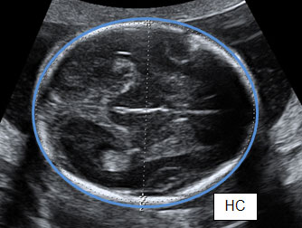
C-Scan refers to the image produced when the data collected from an ultrasonic inspection is plotted on a plan view of the component. The true definition according to BS EN 1330-4:2000 is the 'Image of the results of an ultrasonic examination showing a cross-section of the test object parallel to the scanning surface.'
What is C-SCAN ultrasonic testing?
There can be many formats for collecting and displaying the ultrasonic test data such as A-scan, B-scan, and C-scan. While other mechanisms present the most basic 2D grid plot of the ultrasonic wave, C-scan ultrasonic testing displays the data alongside the depth ( time-of-flight) or amplitude of the wave.
What is an ultrasound scan?
What is an ultrasound? An ultrasound scan is a medical test that uses high-frequency sound waves to capture live images from the inside of your body. It’s also known as sonography.
What is a C-scan used for?
C-scan uses a projection of the ultrasonic data onto a plan view of the component being tested to create an image. This mapping technique is usually utilized for corrosion or thickness mapping, as well as visualization of cracks and inclusions in the component.
What is the difference between CT scan vs ultrasound?
Both CT scan and ultrasound can be used to detect acute abdominal conditions. But if we look at CT scan Vs. Ultrasound abdomen, ultrasound can provide more information about the abdomen, while CT scan may miss fewer cases than ultrasound. CT scan and ultrasound can also be used to detect cancer.

What is B and C scan in ultrasonic testing?
B- scan: If the A- scan - concept is combined with movement of the probe along the surface, a B- scan is the result. It depicts the acoustical side projection of the object, Fig. 4.39. C- scan: Via C- scan are echo amplitudes recorded in relation to probe position.
What type of defects is C scan best used for?
Overall, the C-scan data gives the best indication of lateral defect size and defect depth, even for the sample with the complex barely visible impact damage.
What is the difference between a phased array C scan and a conventional C scan?
A C-scan from a phased array system is very similar to the one from the conventional probe seen above. With phased array systems however, the probe is typically moved physically along one axis while the beam electronically scans along the other according to the focal law sequence.
What is C-scan report?
C-Scan refers to the image produced when the data collected from an ultrasonic inspection is plotted on a plan view of the component. The true definition according to BS EN 1330-4:2000 is the 'Image of the results of an ultrasonic examination showing a cross-section of the test object parallel to the scanning surface.
What is the difference between A-scan and B scan?
There are two main types of ultrasound used in ophthalmologic practice currently, A-Scan and B-scan. In A-scan, or time-amplitude scan, sound waves are generated at 8 MHz and converted into spikes that correspond with tissue interface zones. In B-scan, or brightness amplitude scan, sound waves are generated at 10 MHz.
How echoes of a-scan will be displayed?
a A-Scan or Amplitude mode display The received or the reflected echo signals from the specimen is given to the Y-Plate and time base is connected to the X-Plate of the CRO, so that they are displayed as vertical spikes along horizontal base line as shown in the figure 1.10.
What is the most common type of data display used for the ultrasonic testing of welds?
U. T. ASNT Lvl ITermDefinitionThe most common type of data display used for Ultrasonic examination of weld is ______________.An A-scan displayThe display that plot signal amplitude versus time as called ____________.An A-scan display139 more rows
Which type of data presentation in ultrasonic testing displays a profile or cross sectional view of a test specimen?
B-scan presentations display a profile view (cross-sectional) of a test specimen. Only the reflector depth in the cross-section and the linear dimensions can be determined. A limitation to this display technique is that reflectors may be masked by larger reflectors near the surface.
What does a CT scan do vs MRI?
CT scans take a fast series of X-ray pictures, which are put together to create images of the area that was scanned. An MRI uses strong magnetic fields to take pictures of the inside of the body. CT scans are usually the first choice for imaging. MRIs are useful for certain diseases that a CT scan cannot detect.
What is the difference between a CT scan and a CAT scan?
CAT scan: What's the difference? A CT scan and a CAT scan are the same thing. CT stands for computerized tomography and CAT stands for computerized axial tomography. The original name for this scan was an EMI scan, named after the company that created the technology.
What is C look in operating system?
C-LOOK is an enhanced version of both SCAN as well as LOOK disk scheduling algorithms. This algorithm also uses the idea of wrapping the tracks as a circular cylinder as C-SCAN algorithm but the seek time is better than C-SCAN algorithm.
What is maximum amplitude technique?
The maximum amplitude technique uses a measure of the probe movement between the maximised signals from flaw extremities to size flaws. The 6 dB and 20 dB drop techniques use the reduction in the signal amplitude from the flaw as the probe passes over the edge of the flaw as an indicator of flaw dimensions.
CT Scan vs. Ultrasound: What Is the Difference Between CT Scan and Ultrasound?
CT Scan vs. Ultrasound: There are various types of tools used to treat and diagnose illness. Ultrasound and CT scan are two imaging techniques wide...
What is CT Scan?
CT scan is a computed tomography scan in which a patient has to go through a scanning system that uses x-ray to allow doctors to inside the patient...
What is Ultrasound?
Ultrasound is basically medical sonography that uses high-frequency sound waves to develop images. Ultrasounds are mainly used to see the fetus's d...
How Does Ultrasonography Differ From Radiology and CT Scan?
Ultrasonography can not provide an image of a bony structure. While radiology and CT scan are specific tools for that.
Which Is More Accurate, CT scan or Ultrasound?
CT scan can provide accurate screening for certain diseases like tumors, internal injuries, and other abnormalities inside the body.
Is A CT Scan More Accurate Than An Ultrasound?
CT scan can provide accurate results when detecting diseases like tumors, but when it comes to ultrasound Vs. CT scan abdomen, ultrasound can show...
Which Is Better, CT Scan or Ultrasound?
Both play an essential role in detecting the diseases; CT scans are better for detecting tumors and other internal abnormalities, while ultrasound...
What Is the Difference Between an Ultrasound and A Sonogram?
An ultrasound is a tool that uses sound waves to provide a picture of the inside body, while a sonogram Is a picture given by ultrasound.
What is an A scan?
The A-scan, as shown in Figure below, is the frequently used UT display type by the UT personnel. A-Scan displays the response along the path of the sound beam for a given position of the probe. It also displays the amplitude of the signal coming from the discontinuity as a function of time on the screen. The ‘x’ axis (right) represents the time of flight and indicates the depth of a discontinuity or back wall (thickness). The ‘y’ axis indicates the amplitude of reflected signals (echoes) and can be used to estimate the size of a discontinuity compared to a known reference reflector.
What is the B scan display?
The B-scan display as shown in above figure, right hand side, displays a cross sectional view of the object under test by scanning the probe along one axis. The horizontal axis (left) relates to the position of the probe as it moves along the surface of the object and provides information as to the lateral location of the discontinuity. Echo amplitude is usually indicated by the color or gray scale intensity of echo indications.
What is the A scan method used for?
In ultrasonic flaw detector, A-scan method is used to detect the position and size of the flaws.
Is the transducer stationary in A scan?
This combines the features of both A-Scan as well as B-Scan. In this the transducer is held stationary as in A-scan and echoes appear as dots in the B-scan.
What is a C scan?
Amplitude C-scan is generally used to monitor the behaviour of a specific section of the part. Monitoring the backwall (BW) amplitude can give information on the presence of small mid-wall porosities (BW drops). On the other hand, monitoring a specific composite section can help to spot weak bonding (BW peaks) from the adhesive process between two composite layers.
What is a depth C scan?
Depth C-scan is generally used to monitor the thickness of a part by giving information on the remaining wall thickness or by precisely positioning an anomaly like a disbond, a large porosity or a flaw within the material.
What is an ultrasonic flaw detector?
Ultrasonic flaw detectors record complete A-scan information and can use it to create a two-dimensional C-scan map. Using a gate within the A-scan, each pixel of a C-scan map depicts the depth (time-of-flight) or amplitude of the echoes crossing that gate. Using vivid colour palettes to represent % or mm values, it is then possible to rapidly analyse the complete volumetric integrity of the part.
Can UTmap be used with a T scan?
To improve data alignment precision, UTmap also offers the unique possibility to import 2D CAD drawings of the part (or a simple picture) in the T-scan workspace. The C-scan strips can then be precisely applied with opacity option on the CAD overlay to create even more comprehensive inspection reports.
Why is ultrasound important?
Ultrasound modes determine how an image will be displayed to the radiologist. This is important because it affects what information can be inferred from the screen. The efficiency of your ultrasound tests relies on how effective your equipment is.
What are the different types of ultrasound?
There are five different types of ultrasound modes: 1. A-Mode Ultrasound. A-mode ultrasound is the simplest. The image is shown on the screen in one-dimension. A single transducer scans the body. Using an X and Y access, the collected information is then plotted on the screen as a function of depth. A-mode, or amplitude mode, is ideal ...
What is M mode ultrasound?
M-mode ultrasound works similarly to a stop-motion video . This type takes a collection of A-mode or B-mode ultrasound images and uses them, in effect, to create a video. M-mode ultrasound, or “movement mode”, allows doctors to see the amplitude of movements.
What is the difference between A-mode and B-mode ultrasound?
2. B-Mode Ultrasound. B-mode ultrasound uses linear array transducers to simultaneously scan a plane through the body. These echoes are converted by the machine into a 2D image.
What is an ultrasound scan?
An ultrasound scan is a medical test that uses high-frequency sound waves to capture live images from the inside of your body. It’s also known as sonography. The technology is similar to that used by sonar and radar, which help the military detect planes and ships. An ultrasound allows your doctor to see problems with organs, vessels, ...
Why do doctors order ultrasounds?
Your doctor may order an ultrasound if you’re having pain, swelling, or other symptoms that require an internal view of your organs. An ultrasound can provide a view of the: An ultrasound is also a helpful way to guide surgeons’ movements during certain medical procedures, such as biopsies.
Why do ultrasound transducers rub on skin?
This prevents friction so they can rub the ultrasound transducer on your skin. The transducer has a similar appearance to a microphone. The jelly also helps transmit the sound waves. The transducer sends high-frequency sound waves through your body. The waves echo as they hit a dense object, such as an organ or bone.
What do you do before an ultrasound?
An ultrasound technician, called a sonographer, will apply a special lubricating jelly to your skin.
How do sound waves echo?
Those echoes are then reflected back into a computer. The sound waves are at too high of a pitch for the human ear to hear. They form a picture that can be interpreted by the doctor.
What to tell your doctor before an ultrasound?
Be sure to tell your doctor about any prescription drugs, over-the-counter medications, or herbal supplements that you take before the exam. It’s important to follow your doctor’s instructions and ask any questions you may have before the procedure. An ultrasound carries minimal risks.
What to do if you have abnormalities on an ultrasound?
Should anything abnormal turn up on the ultrasound, you may need to undergo other diagnostic techniques, such as a CT scan, MRI, or a biopsy sample of tissue depending on the area examined. If your doctor is able to make a diagnosis of your condition based on your ultrasound, they may begin your treatment immediately.
What is an A scan?
The A-scan presentation#N#Presentation (Ultrasound) - The method used to show ultrasonic information. This may include A-scans, B-scans or C-scans, displayed on various types of recorders or cathode ray tube instruments.#N#displays the amount of received ultrasonic#N#Ultrasonic - A term referring to acoustic vibration frequencies greater than about 20,000 hertz.#N#energy as a function of time. The relative amount of received energy is plotted along the vertical axis and the elapsed time (which may be related to the sound#N#Sound - Mechanical vibrations transmitted in an elastic gas, liquid, or solid.#N#energy travel time within the material) is displayed along the horizontal axis. Most instruments with an A-scan display#N#A-Scan Display - A data presentation method in which signal amplitude is plotted along the y-axis versus time on the x-axis. The horizontal distance between any two signals represents the material distance between the two conditions causing the signals. In a linear system, the vertical excursion is proportional to the amplitude of the signal.#N#allow the signal to be displayed in its natural radio frequency#N#Frequency - The number of waves that pass a given point in a specified unit of time.#N#form (RF), as a fully rectified RF signal, or as either the positive or negative half of the RF signal. In the A-scan presentation#N#Presentation (Ultrasound) - The method used to show ultrasonic information. This may include A-scans, B-scans or C-scans, displayed on various types of recorders or cathode ray tube instruments.#N#, relative discontinuity#N#Discontinuity - a break in the continuity of a medium or material.#N#size can be estimated by comparing the signal amplitude#N#Amplitude - (1) The maximum absolute value obtained by the disturbance of a wave or any quantity that varies periodically. (2) The vertical height of a received signal on an A-scan. It is measured from peak to peak for an RF presentation or from base to peak for a video presentation.#N#obtained from an unknown reflector to that from a known reflector. Reflector depth can be determined by the position of the signal on the horizontal sweep.
What are the three formats of ultrasonic data?
Ultrasonic data can be collected and displayed in a number of different formats. The three most common formats are know in the NDT world as A-scan, B-scan and C-scan presentations. Each presentation mode provides a different way of looking at and evaluating the region of material being inspected. Modern computerized ultrasonic scanning systems can display data in all three presentation forms simultaneously.