Key Points
- The absorbed elements that pass through the mucosa are picked up from the blood vessels of the submucosa.
- In the gastrointestinal tract, the submucosa is the layer of dense, irregular connective tissue or loose connective tissue that supports the mucosa, as well as joins the mucosa to the bulk of underlying smooth muscle (fibers that run circularly within a layer of longitudinal muscle).
What is the main function of submucosa?
The submucosa, a dense network of connective tissue, blood vessels, lymphatics, neurons, and esophageal glands, primarily functions as a secretory layer.
What is the composition of submucosa?
Histologically, the wall of the alimentary canal shows four distinct layers (from the lumen moving out): mucosa, submucosa, muscularis externa, and either a serous membrane or an adventitia. In the gastrointestinal tract and the respiratory tract the submucosa contains the submucosal glands that secrete mucus.
Which are three features of the mucosa and submucosa?
However, three features of the mucosa and submucosa are unique. These features, which increase the absorptive surface area of the small intestine more than 600-fold, include circular folds, villi, and microvilli (Figure 2).
What is a submucosa?
The layer of tissue under the mucosa (inner lining of some organs and body cavities that makes mucus).
What is the layer of submucosa?
The submucosa, located between the outermost layer of the mucosa and the muscularis externa, is made of connective tissue and several different cell types that include fibroblasts, lymphocytes, eosinophils, macrophages, plasma cells, and mast cells.
Where is submucosa present?
The submucosa is found in all the buccal cavity regions except the attached gingiva and the hard palate covered by masticatory mucosa, where the submucosa layer is absent, and the lamina propria is directly attached to the underlying bone, forming a mucoperiosteum.
What is the structure and function of submucosa?
The stomach and intestines have a thin simple columnar epithelial layer for secretion and absorption. The submucosa is a thick layer of loose connective tissue that surrounds the mucosa. This layer also contains blood vessels, lymphatic vessels, and nerves. Glands may be embedded in this layer.
What is the difference between mucosa and submucosa?
Mucosa -- innermost layer (closest to the lumen), the soft, squishy lining of the tract, consisting of epithelium, lamina propria, and muscularis mucosae. Submucosa -- connective tissue supporting (outside, deep to) the mucosa.
What is the function of the submucosa in the small intestine?
It is responsible for gut movement, or peristalsis. It usually has two distinct layers of smooth muscle: circular and longitudinal. The submucosa is the layer of dense, irregular connective tissue or loose connective tissue that supports the mucosa, as well as joins the mucosa to the bulk of underlying smooth muscle.
What are the subdivisions of the submucosa?
The submucosa has subdivisions of connective tissue, lymph nodules, and nerve fibers. Its major functions are nutrition and protection.
What is the serosa layer composed of?
Adventia layer (or serosa) Contains blood vessels, lymphatics and nerves.
What does the submucosa of the esophagus contain?
Submucosa. The submucosa contains connective tissue as well as lymphocytes, plasma cells, nerve cells (Meissner's plexus), vascular network (Heller plexus), and mucous glands. The esophageal glands are small racemose glands (which have acini arranged like grapes on a stem) of mucous type.
What is the submucosa?
The submucosa (or tela submucosa) is a thin layer of tissue in various organs of the gastrointestinal, respiratory, and genitourinary tracts. It is the layer of dense irregular connective tissue that supports the mucosa ( mucous membrane) and joins it to the muscular layer, the bulk of overlying smooth muscle ...
Why is the submucosa important?
Identification of the submucosa plays an important role in diagnostic and therapeutic endoscopy, where special fibre-optic cameras are used to perform procedures on the gastrointestinal tract. Abnormalities of the submucosa, such as gastrointestinal stromal tumors, usually show integrity of the mucosal surface.
What is a small intestinal submucosa?
Small intestinal submucosa (SIS) is submucosal tissue in the small intestines of vertebrates. SIS is harvested (typically from pigs) for transplanted structural material in several clinical applications, typically biologic meshes. They have low immunogenicity. Some uses under investigation include a scaffold for intervertebral disc regeneration.
Why do you inject dye into the submucosa?
An injection of dye, saline, or epinephrine into the submucosa is imperative in the safe removal of certain polyps . Endoscopic mucosal resection involves removal of the mucosal layer, and in order to be done safely, a submucosal injection of dye is performed to ensure integrity at the beginning of the procedure.
What are the layers of the alimentary canal?
Histologically, the wall of the alimentary canal shows four distinct layers (from the lumen moving out): mucosa, submucosa, muscularis externa, and a either a serous membrane or an adventitia . In the gastrointestinal tract and the respiratory tract the submucosa contains the submuco sal glands that secrete mucus .
What is the submucosa of the stomach?
The submucosa lies under the mucosa and consists of fibrous connective tissue, separating the mucosa from the next layer, the muscularis externa. Layers of stomach lining: Stomach. The serosa is labeled at far right, and is colored yellow.
Which connective tissue is the submucosa?
In the gastrointestinal tract, the submucosa is the layer of dense, irregular connective tissue or loose connective tissue that supports the mucosa, as well as joins the mucosa to the bulk of underlying smooth muscle (fibers that run circularly within a layer of longitudinal muscle).
What vessels run through the mucosa?
Blood vessels, lymphatic vessels , and nerves (all supplying the mucosa) will run through here. Tiny parasympathetic ganglia are scattered around forming the submucosal plexus (or Meissner’s plexus) where preganglionic parasympathetic neurons create synapses with the postganglionic nerve fibers that supply the muscularis mucosae.
What are the layers of the GI tract?
The GI tract is composed of four layers. Each layer has different tissues and functions. From the inside out they are called: 1 Mucosa 2 Submucosa 3 Muscularis 4 Serosa
What is the structure of the gut wall?
General structure of the gut wall: The general structure of the gut wall is illustrated. The submucosa consists of a dense irregular layer of connective tissue with large blood vessels, lymphatics, and nerves that branch into the mucosa and muscularis externa.
Which type of neuron is found in the submucosal plexus?
Tiny parasympathetic ganglia are scattered around to form the submucosal plexus (or Meissner’s plexus) where preganglionic parasympathetic neurons create synapses with postganglionic nerve fibers that supply the muscularis mucosae.
How many layers of muscle are there in the stomach?
The muscularis in the stomach differs from that of other GI organs in that it has three layers of muscle instead of two. Under these muscle layers is the adventitia—layers of connective tissue that are continuous with the omenta.
What is the small intestinal submucosa?
Small intestinal submucosa (SIS) is a resorbable, acellular bio-scaffold composed of extra-cellular matrix (ECM) proteins derived from the jejunum of pigs. SIS has characteristic of an ideal tissue engineered biomaterial and can act as a bioscaffold for remodeling of many body tissues including skin, body wall, musculoskeletal structure, urinary bladder, blood vessels, and supports new blood vessel growth [8,11–13]. SIS consists of three distinct layers of the mammalian small intestine: the lamina propria and muscularis mucosae of the intestinal mucosa, and the tunica submucosa ( Fig. 2.2) [1]. The tunica submucosa is the layer of connective tissue arranged immediately under the mucosa layer of the intestine and is a 100–200 µm thick interstitial ECM: it makes up the bulk of the SIS biopolymer scaffold. SIS induces site-specific remodeling of both organs and the tissue depending on the site of implantation [27]. SIS stimulates host cells to proliferate and differentiate into site-specific connective tissue structures, and this replaces the SIS material within 90 days [36]. SIS’s ability to induce tissue remodeling is associated with angiogenesis, cell migration and differentiation and deposition of ECM [36].
What is the submucosal plexus?
1 ). It consists of connective tissue, blood and lymphatic vessels, and the submucosal plexus (plexus of Meissner). The submucosa may also contain scattered migratory cells, of which mast cells are frequently the predominating cell type.
What is a small intestinal biopolymer scaffold?
Small intestinal submucosa biopolymer scaffolds are made up from the 100 to 200 µm thick interstitial ECM from the tunica submucosa layer. SIS scaffolds must undergo chemical processing to decrease cellular content before implantation. SIS is a thin translucent tissue that consists primarily of type I collagen and GAGs, such as chondroitin sulfate, heprain sulfate, and hyaluronan. It also contains growth factors such as TGF-β and VEGF which encourage growth and growth and differentiation of cells, while simultaneously providing integrity and strength to the scaffold throughout the healing process.144 These scaffolds are mechanically strong, and their inherent porosity encourages a reconstructive healing process by allowing cell infiltration. Mechanical studies have shown that porcine SIS scaffolds stretch considerably more than canine infraspinatus tendons before reaching maximum material properties and have linear moduli an order of magnitude lower than tendon. 142 Additionally it has been reported that these scaffolds have a mean load to failure around 32–38 N. 145
Is the submucosa vascular or vascular?
The submucosa (SM) is highly vascular and the double muscle layer (M) can be seen.

Overview
The submucosa (or tela submucosa) is a thin layer of tissue in various organs of the gastrointestinal, respiratory, and genitourinary tracts. It is the layer of dense irregular connective tissue that supports the mucosa (mucous membrane) and joins it to the muscular layer, the bulk of overlying smooth muscle (fibers running circularly within layer of longitudinal muscle).
Structure
Blood vessels, lymphatic vessels, and nerves (all supplying the mucosa) will run through here. In the intestinal wall, tiny parasympathetic ganglia are scattered around forming the submucous plexus (or "Meissner's plexus") where preganglionic parasympathetic neurons synapse with postganglionic nerve fibers that supply the muscularis mucosae. Histologically, the wall of the alimentary canal shows four distinct layers (from the lumen moving out): mucosa, submucosa, …
Clinical significance
Identification of the submucosa plays an important role in diagnostic and therapeutic endoscopy, where special fibre-optic cameras are used to perform procedures on the gastrointestinal tract. Abnormalities of the submucosa, such as gastrointestinal stromal tumors, usually show integrity of the mucosal surface.
The submucosa is also identified in endoscopic ultrasound to identify the depth of tumours and t…
Discovery
A scientific article published in March 2018 proposed a revision of the anatomical definition of the submucosa. They first saw a non compact tissue which should be submucosa using a technology called endomicroscopy. They hypothesised that the submucosa was not compact as it was previously seen on histological analysis but form a reticular pattern. To confirm their findings, they performed fixed samples of bile duct into a freezing media in order to conserve the shape o…
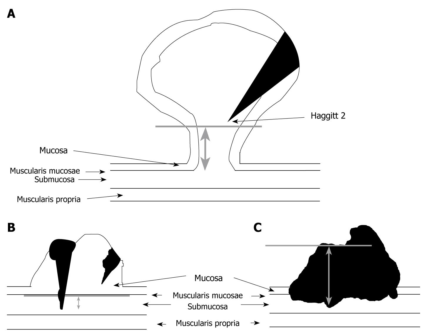
Additional images
• Stomach.
• Section of the human esophagus. Moderately magnified.
• Vertical section of bladder wall.
• General structure of the gut wall showing the submucosa.