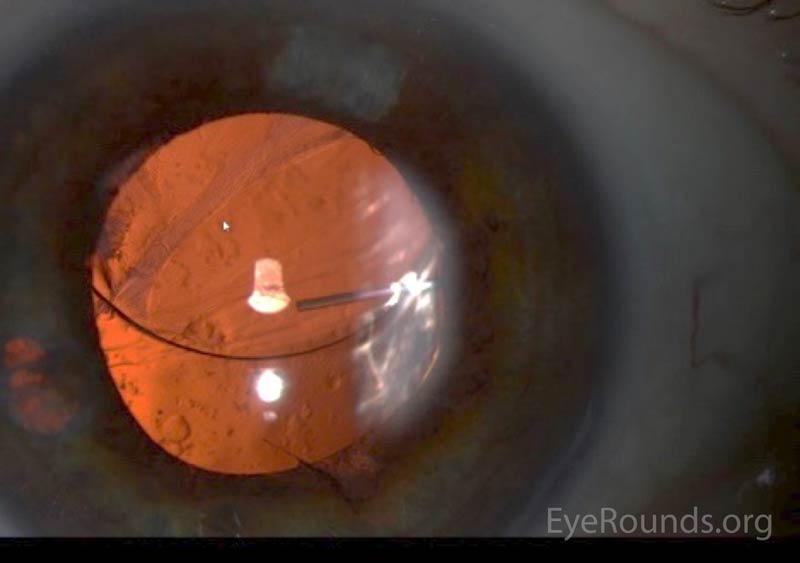
Ciliary flush. Ciliary flush is usually present in eyes with corneal inflammation, iridocyclitis or acute glaucoma, though not simple conjunctivitis. A ciliary flush is a ring of red or violet spreading out from around the cornea of the eye.
What is ciliary flush in eye?
Ciliary flush is usually present in eyes with corneal inflammation, iridocyclitis or acute glaucoma, though not simple conjunctivitis. A ciliary flush is a ring of red or violet spreading out from around the cornea of the eye. The cornea is required to be transparent to transmit light to the retina.
What is the file size for ciliary flush?
Ciliary flush File Size: 90 KB Related: red eye, ciliary flush Cornea/External Disease View Full Image Image License and Citation Guidelines Add to My Bookmarks View Mark Complete Remove Comments Ciliary flush.
Can uveitis cause a ciliary flush?
Ciliary flush occurs with uveitis Overview of Uveitis Uveitis is defined as inflammation of the uveal tract—the iris, ciliary body, and choroid. However, the retina and fluid within the anterior chamber and vitreous are often involved as well.... read more ) but not with uncomplicated conjunctivitis.
What is the meaning of ciliary?
pertaining to or resembling cilia; used particularly in reference to certain eye structures, as the ciliary body or muscle. ciliary body the thickened part of the vascular tunic of the eye, connecting choroid and iris, made up of the ciliary muscle and the ciliary processes.

What is ciliary injection?
Ciliary injection involves branches of the anterior ciliary arteries and indicates inflammation of the cornea, iris, or ciliary body. Conjunctival injection mainly affects the posterior conjunctival blood vessels.
How can you tell the difference between conjunctivitis and scleritis?
Infectious conjunctivitis is usually associated with a discharge and allergic conjunctivitis with itching and chemosis. Scleritis is anatomically deeper and has a distinct appearance. Malignant conjunctival lesions also tend to have a specific appearance and are rarely mistaken for episcleritis.
What is hyperemia of the eye?
Hyperemia, or redness alone in clinical terms, is only a sign of a problem, and may be associated with a broad group of ocular diseases or, possibly, be part of a response to allergic inflammation or irritation.
What is conjunctival injection?
Conjunctival injection or hyperemia is a nonspecific response with enlargement of conjunctival vessels induced by various diseases. Conjunctival injection is an important diagnostic clue for infection or inflammation and can be utilized for the monitoring of the disease progression and response to treatment.
What autoimmune disease causes scleritis?
Causes. Scleritis is often linked to autoimmune diseases. These diseases occur when the body's immune system attacks and destroys healthy body tissue by mistake. Rheumatoid arthritis and systemic lupus erythematosus are examples of autoimmune diseases.
Can an optometrist diagnose scleritis?
One in 10 cases of Scleritis takes the form known as Posterior Scleritis, which affects the sclera of the back part of the eye, so that the front of the eye may appear normal and the optometrist will need to use special instruments to help to make the diagnosis.
What are the signs and symptoms of hyperemia?
Some of the symptoms of passive hyperemia are:Trouble breathing.Pain in the chest.Coughing.Wheezing.Swelling in the limbs.Nausea.Pain.Itchiness.
What is an example of hyperemia?
Active hyperemia is a physiological response to something happening in the body. It is an acute form of hyperemia. For example, there is more blood in the digestive system after a meal, more blood in the muscles after exercise, and more blood in the face when a person blushes.
What is hyperemia caused by?
Active hyperemia is caused by an increased flow of blood into your organs. It usually happens when organs need more blood than usual. Your blood vessels widen to increase the supply of blood flowing in.
Why do people need eye injections?
Intraocular injections are typically needed to treat many serious retinal conditions that can lead to vision loss. Because of this, they should be performed in a timely manner to help patients preserve their existing vision and keep future vision loss to a minimum.
What does injected conjunctiva look like?
Injected conjunctiva is bloodshot eyes. The eyes appear red because of the dilation of blood vessels in the conjunctiva. 2 Bloodshot eyes can be caused by dry air, sun exposure, dust, foreign body, allergies, infection, or trauma.
What type of bacteria causes conjunctivitis?
Bacterial ConjunctivitisInfection of the eye caused by certain bacteria.Can be caused by Staphylococcus aureus, Streptococcus pneumoniae, Haemophilus influenzae, Moraxella catarrhalis, or, less commonly, Chlamydia trachomatis and Neisseria gonorrhoeae.More items...
How can you tell the difference between scleritis and uveitis?
To differentiate uveitis from episcleritis and scleritis, instill a topical cycloplegic (e.g., 0.25% scopolamine) to see if the pain subsides. The more significant the pain, the more likely you are dealing with uveitis.
What does scleritis look like?
Scleritis means that the sclera is inflamed. The inflammation is what makes the white of the eye look red, or sometimes purple. Pain from scleritis is usually severe and is worse at night.
How can you tell the difference between blepharitis and conjunctivitis?
The difference between the two is which part of the eye is affected. Blepharitis involves the inflammation of the eyelids while pink eye affects the conjunctiva....Blepharitis can have similar symptoms to pink eye, such as:Swelling of the conjunctiva.Watery eyes.Eyes feeling itchy or irritated.Crusty eyelids and lashes.
What are the symptoms of scleritis?
What Are Symptoms of Scleritis?pain.tenderness of the eye.redness and swelling of the white part of the eye.blurred vision.tearing.extreme sensitivity to light.
Ciliary Flush
Ciliary flush, Ciliary injection, Ciliary body congestion, ciliary flush, ciliary injection, Ciliary flush (disorder)
Ciliary injection ( C0271108 )
Ciliary flush, Ciliary injection, Ciliary body congestion, ciliary flush, ciliary injection, Ciliary flush (disorder)
Visual acuity
A reduction in visual acuity in a 'red eye' is indicative of serious ocular disease, such as keratitis, iridocyclitis, and glaucoma, and never occurs in simple conjunctivitis without accompanying corneal involvement.
Ciliary flush
Ciliary flush is usually present in eyes with corneal inflammation, iridocyclitis or acute glaucoma, though not simple conjunctivitis. A ciliary flush is a ring of red or violet spreading out from around the cornea of the eye.
Corneal abnormalities
The cornea is required to be transparent to transmit light to the retina. Because of injury, infection or inflammation, an area of opacity may develop which can be seen with a penlight or slit lamp. In rare instances, this opacity is congenital. In some, there is a family history of corneal growth disorders which may be progressive with age.
Pupillary abnormalities
In an eye with iridocyclitis, (inflammation of both the iris and ciliary body), the involved pupil will be smaller than the uninvolved, due to reflex muscle spasm of the sphincter muscle of the iris . Generally, conjunctivitis does not affect the pupils.
Abnormal intraocular pressure
Intraocular pressure should be measured as part of the routine eye examination . It is usually only elevated by iridocyclitis or acute-closure glaucoma, but not by relatively benign conditions. In iritis and traumatic perforating ocular injuries, the intraocular pressure is usually low.
Severe pain
Those with conjunctivitis may report mild irritation or scratchiness, but never extreme pain, which is an indicator of more serious disease such as keratitis, corneal ulceration, iridocyclitis, or acute glaucoma .
Differential diagnosis
Of the many causes, conjunctivitis is the most common. Others include: Usually nonurgent
Symptoms and Signs of Conjunctivitis
Any source of inflammation can cause lacrimation or discharge and diffuse conjunctival vascular dilation. Discharge may cause the eyes to crust overnight. Thick discharge may blur vision, but once discharge is cleared, visual acuity should be unaffected.
Diagnosis of Conjunctivitis
Usually, diagnosis of conjunctivitis is made by history and examination (see table Differentiating Features in Acute Conjunctivitis Differentiating Features in Acute Conjunctivitis Conjunctival inflammation typically results from infection, allergy, or irritation.
Treatment of Conjunctivitis
Most infectious conjunctivitis is highly contagious and spreads by droplets, fomites, and hand-to-eye inoculation. To avoid transmitting infection, physicians must
Key Points
Conjunctivitis typically results from infection, allergy, or irritation.
Drugs Mentioned In This Article
Pediatric orbital tumors most commonly include dermoid tumors, capillary hemangiomas, and lymphangiomas. When a pediatric patient has a capillary hemangioma on the upper eyelid that is affecting vision, which of the following is the most appropriate treatment?
Merck and the Merck Manuals
Merck & Co., Inc., Kenilworth, NJ, USA is a global healthcare leader working to help the world be well. From developing new therapies that treat and prevent disease to helping people in need, we are committed to improving health and well-being around the world. The Merck Manual was first published in 1899 as a service to the community.
