Fat necrosis is a form of necrosis characterized by the action upon fat by digestive enzymes. In fat necrosis the enzyme lipase releases fatty acids from triglycerides. The fatty acids then complex with calcium to form soaps. These soaps appear as white chalky deposits. It is usually associated with trauma of the pancreas or acute pancreatitis.
What is fat necrosis of the liver?
Fat necrosis. Fat necrosis is a form of necrosis characterized by the action upon fat by digestive enzymes. In fat necrosis the enzyme lipase releases fatty acids from triglycerides. The fatty acids then complex with calcium to form soaps. These soaps appear as white chalky deposits.
What is the role of lipase in fat necrosis?
In fat necrosis the enzyme lipase releases fatty acids from triglycerides. The fatty acids then complex with calcium to form soaps.
Should I be worried about fat necrosis?
Although fat necrosis is benign, a doctor may recommend regular screening to ensure they have correctly identified a lump as fat necrosis, because it can be difficult to distinguish oil cysts from other types of lumps. Fat necrosis happens when inflammation affects the supply of blood and oxygen to body fat, causing fat cells to die.
What causes fat necrosis of the breast?
Causes of fat necrosis include trauma or injury to the fat tissue or a result of scar tissue formed during a surgical procedure such as a breast augmentation. Oftentimes, the lumps resulting from fat necrosis will go away on their own. If there is fluid build-up in the lump, the fluid can be removed using a syringe.
What is non enzymatic fat necrosis?
Known as a benign, non-supportive inflammatory process, breast fat necrosis (FN) occurs due to iatrogenic breast trauma. Fat necrosis is an inflammatory, sterile process which has roots in fat aseptic saponification. In this regard, blood and tissue lipase contribute to this disorder.
What does fat necrosis mean?
Listen to pronunciation. (… neh-KROH-sis) A benign condition in which fat tissue in the breast or other organs is damaged by injury, surgery, or radiation therapy.
Should I worry about fat necrosis?
Fat necrosis commonly develops after breast surgery, radiation, or other traumatic injuries to the breast. While the lumps can cause some worry initially, they are harmless in terms of your overall health and don't increase your risk of developing breast cancer.
How do you get rid of fat necrosis?
To remove the fat necrosis, a surgeon will cut out the damaged or dead tissue. Before the operation, they'll provide you with a local or general anesthetic. You may end up with a small scar, but this typically fades as time passes. Liposuction is another option for removing the fat necrosis.
Does fat necrosis need to be removed?
As long as doctors are sure of the diagnosis, fat necrosis and oil cysts usually don't need to be treated. Sometimes fat necrosis goes away on its own. If a needle biopsy is done to remove the fluid in an oil cyst, it can also serve as treatment.
How long does it take for fat necrosis to go away?
Fat necrosis is harmless so you will not usually need any treatment or follow-up. In most cases the body will break it down over time. This could take a few months. It's important to go back to your GP if the lump gets bigger or you notice any other changes to your breasts.
Should I massage fat necrosis?
“Firm lumps resulting from fatty necrosis often resolve on their own or can be directly massaged to break them down,” Dr.
Does fat necrosis grow?
If it doesn't, you may need to have it surgically removed. One option for removal is liposuction and another is a lumpectomy. Can breast fat necrosis grow? Yes, it can grow in different parts of the breast.
How long does necrosis take to develop?
Soft tissue necrosis usually begins with breakdown of damaged mucosa, resulting in a small ulcer. Most soft tissue necroses will occur within 2 years after radiation therapy. Occurrence after 2 years is generally preceded by mucosal trauma.
What are the symptoms of fat necrosis?
Fat necrosis in the breast may occur without symptoms. If symptoms appear, a person will usually notice lumps under the skin. These are often, but not always, around the areola, the part that surrounds the nipple. Fat necrosis and oil cysts are not usually painful.
Should fat necrosis be biopsied?
Fat necrosis can be diagnosed clinically or radiographically in the majority of cases, without the need for biopsy.
How common is necrosis after fat transfer?
According to a study conducted by surgeons from the Cleveland Clinic Multidisciplinary Medical Center of 171 patients who had fat transfer breast reconstruction post-breast cancer surgery, a little over 10 percent experienced fat necrosis an average of 3.4 months after fat transfer.
What causes fat necrosis?
Fat necrosis happens when inflammation affects the supply of blood and oxygen to body fat, causing fat cells to die. It can happen after an injury, surgery, or radiation treatment. Fat necrosis commonly affects the breasts, where it can lead to lumps, skin changes, and oil cysts. These changes are not cancerous.
Does fat necrosis grow?
If it doesn't, you may need to have it surgically removed. One option for removal is liposuction and another is a lumpectomy. Can breast fat necrosis grow? Yes, it can grow in different parts of the breast.
What happens during necrosis?
Necrosis (from Ancient Greek νέκρωσις (nékrōsis) 'death') is a form of cell injury which results in the premature death of cells in living tissue by autolysis. Necrosis is caused by factors external to the cell or tissue, such as infection, or trauma which result in the unregulated digestion of cell components.
Does massage help fat necrosis?
Various types of massage techniques can help to reduce the size, improve the feel of the fat necrosis, and improve esthetics. It's treated like scar tissue which requires firm pressure and moving the tissue in multiple directions.
What is fat necrosis?
Fat necrosis refers to the destruction and death of fat cells in the body, specifically fat cells in the breasts. Learn about fat necrosis, including the symptoms and treatments of this condition. Create an account.
Where does fat necrosis occur?
Fat necrosis refers to the destruction and death of fat cells. Fat necrosis often occurs in breast tissue, occurring in both men and women. However, fat necrosis can occur anywhere in the body that contains fat tissue. Fat necrosis often occurs in the fat tissue of the breasts. Fat necrosis is usually benign, meaning it is harmless.
Why does fat necrosis go away?
Causes of fat necrosis include trauma or injury to the fat tissue or a result of scar tissue formed during a surgical procedure such as a breast augmentation. Oftentimes, the lumps resulting from fat necrosis will go away on their own. If there is fluid build-up in the lump, the fluid can be removed using a syringe.
How to remove fluid from necrotic fat?
Fluid that may form around necrotic fat tissue can be removed using syringe.
Can you take pain medication for fat necrosis?
Additionally, a person can take an over-the-counter pain medication to treat any pain associated with fat necrosis.
Does fat necrosis cause breast cancer?
It is also usually painless and does not increase a person's risk of getting breast cancer later in life. It can occur in men and women of any age but more frequently in older women with larger breasts. Usually, fat necrosis feels like a lump under the skin, sometimes occurring with redness on the skin around the lump.
What is fat necrosis?
Fat necrosis occurs after trauma, foreign body reaction, or a response to a breast malignancy or radiotherapy, especially if tumor necrosis is present. Fat necrosis can radiologically, grossly, and histologically (especially at frozen section) mimic malignancy. The FNA of fat necrosis consists of fat; amorphous debris (degenerating fat); inflammatory cells including neutrophils, plasma cells, and lymphocytes; and numerous lipid-laden macrophages (lipophages). The lipophages have abundant vacuolated cytoplasm (Fig.25.13 ). Multinucleated macrophages and spindle-shaped fibroblastic cells can also be present. The rare hibernoma of the breast should be considered in the differential diagnosis when finely and coarsely vacuolated cells are encountered. Myospherulosis, a possible sequela of fat necrosis, has been reported in FNA material. 145 Spherules measuring 4–7μm each and arranged individually or in sac-like structures are diagnostic.
What is the predominant finding in the later stage of fat necrosis?
In the later stage, collagenous scar is the predominant finding, with seemingly granular histiocytes surrounding oil cysts of varying size. These “oil cysts” contain the free lipid material released by lipocyte necrosis. 73 The greatest clinical importance of fat necrosis is in its mimicry of carcinoma, as noted earlier. There is no known association with carcinoma or carcinoma risk.
What causes fat necrosis in cattle?
Traumatic fat necrosis is typically the result of blunt trauma or chronic pressure on adipose tissue against bony prominences, such as the subcutaneous adipose tissue compressed against the sternum in recumbent cattle. Ischemia is thought to contribute to the cell injury. Inflammation and saponification are inconspicuous in this form of fat necrosis.
What is the term for a rat's fat necrosis?
A generalized form of fat necrosis termed steatitis has also been described in rat adipose tissue. It develops in association with vitamin E or antioxidant deficiency that follows excess dietary polyunsaturated fatty acids of the type found in fish or linseed oils. 81 It is characterized by the presence of widely distributed small yellow foci in fat which are composed of clusters of macrophages containing small lipid vacuoles and lipofuscin pigment.
What is FN in breast cancer?
Fat necrosis (FN) can mimic carcinoma both clinically and mammographically and is commonly seen in patients who have had a previous surgical biopsy or other trauma to the breast. FN is also encountered in male patients.155
What is yellow fat disease?
Nutritional fat necrosis, also known as steatitis or yellow fat disease, is usually the result of feeding a diet high in unsaturated fatty acids and low in vitamin E or other antioxidants, setting the stage for ROS production and lipid peroxidation. Yellow fat disease is often seen in carnivores, such as cats or mink, on a fish-based diet. Affected adipose tissue is firm, nodular, and yellow-brown.
Can biopsy be performed for fat necrosis?
If there is reliable history of previous trauma, observation for a finite period of time may be an option. Without resolution, biopsy must be performed for definitive diagnosis. If the only finding is a lipid‐filled cyst with fibrous capsule calcification typical for fat necrosis, aspiration or continued observation can be considered. If there are also other findings such as a spiculated lesion or indeterminate microcalcifications, the possibility of fat necrosis and an adjacent malignancy must be entertained.
What causes fat necrosis?
The term “necrosis” means the cells have died. Potential causes of fat necrosis include blunt trauma, surgeries, or radiation to a particular area of the body.
Where does fat necrosis occur?
While fat necrosis can occur anywhere on the body where there is fatty tissue, the most common location for it to appear is the breast.
How to diagnose fat necrosis?
Fat necrosis may be diagnosed using an MRI machine. If a person feels a lump that is suspected of being fat necrosis, a doctor will usually recommend an imaging scan . This will identify if the lump could be cancerous or due to another underlying cause.
What is a lump in breast tissue?
However, sometimes the fat cells die, and they release their oily contents. As a result, a lump can form. Doctors call this lump an oil cyst.
What is the term for the cells that have died from fatty tissue?
Treatment. Outlook. Fat necrosis is a condition that occurs when a person experiences an injury to an area of fatty tissue. This can result in the fat being replaced with the oily contents of fat cells. The term “necrosis” means the cells have died.
What imaging tools can be used to identify fat necrosis?
Examples of the imaging tools a doctor may use include: X-ray: X-rays, such as mammography, can be used to visualize areas of fat necrosis.
Is fat necrosis scary?
As a result, the appearance of fat necrosis can be very frightening for a woman who is unfamiliar with fat necrosis.
What Is Fat Necrosis?
To put it in layman's terms, fat necrosis is a collection of dead or damaged fat cells that have hardened and manifested as firm bumps. When the body has restricted oxygen flow as a result of surgery, radiation, or trauma, fat cells can die and release their contents to form a small body of liquified matter that hardens over time.
What Causes Fat Necrosis
The roots of post-op side effects and complications often aren’t black and white, and this is no exception. “Fat necrosis can be caused by the lack of integration of transplanted fat into a specific area when specific areas of fat do not receive an adequate amount of blood supply,” Dr. Buford explains.
How To Prevent & Treat Fat Necrosis
The primary recommendations for preventing fat necrosis are to not smoke or be around those who do before or after surgery, as this restricts blood flow. For those who are post-BBL, follow your surgeon’s instructions for sitting and standing to a T.
The Takeaway
For those who have had autologous fat transfer or breast reconstruction, fat necrosis is a risk. But it is one that is manageable and, in some cases, preventable. Arming yourself with the right information ahead of time will go a long way towards ensuring a smooth recovery and satisfactory outcome.
What is the mechanism of intracellular accumulation?
The inability to digest a complex substrate is a possible mechanism for intracellular accumulations.
What type of receptor produces cytokines after a microbe is recognized in circulation?
Toll-like receptors produce cytokines after a microbe is recognized in circulation.
What causes liquefactive necrosis?
Liquefactive necrosis can be associated from bacterial, viruses, parasites or fungal infections. Unlike coagulative necrosis, liquefactive necrosis forms a viscous liquid mass as the dead cells are being digested. The micro-organisms can release enzymes to degrade cells and initiate an immune and inflammatory response. Cellular dissolution and digestion of dying cells may also release further enzymes, which speeds up the liquefying process. The micro-organisms stimulate the leukocyte to home-in on the necrotic area and release powerful hydrolytic enzymes (such as lysozymes) which causes local damage and cells to be lysed, causing a fluid phase. The enzymes responsible for liquefaction are derived from either bacterial hydrolytic enzymes or lysosomal hydrolytic enzymes. These are proteases (collagenases, elastases), DNases and lysosomal enzymes.
What is the origin of the term "necrosis"?
Introduction. Necrosis comes from the Greek origin nekrōsis meaning “death” and later moved to modern Latin to necrosis. Necrosis can be described as a pathological process of cell death which could have been resulted from infections, hypoxia, trauma or toxins. Unlike apoptosis, necrosis is uncontrolled and release lots ...
How does necrosis start?
Necrosis can start from a process called “oncosis”. Oncosis comes from the Greek origin ónkos, meaning swelling. Oncosis occurs when the mitochondria within a cell are damaged beyond recovery by toxins or hypoxia. ATP is thus not being made, which dysregulate the ionic concentration within the cell as the ionic pumps are no longer functioning. Sodium moves into the cell and water follow, making the cell explode. The content that has been released will attract immune cells which will initiate inflammation and release reactive oxygen species (ROS) and enzymes such as proteases. The surrounding tissues may be damaged, and therefore organs may fail to work. In a way, necrosis alerts the immune system to clean through phagocytosis and start local inflammation. However, if the collateral damage is more significant than the process of healing, necrosis will thus increase in size, killing more healthy cells and decrease surrounding body function, which may cause organ failure. Decomposing tissue will increase in number, and micro-organisms may start to replicate and dominate as the immune system struggle to contain the necrosis. Surgery is thus utilised to remove the necrotic tissue by a procedure called debridement and let the healthy tissue take over and heal. Depending on the severity, time, type and extent of the necrosis, the tissues may never heal back to its original function and integrity.
What happens to cells during coagulative necrosis?
Cells that undergo coagulative necrosis can become dry, hard, and white. What is interesting is that gel-like appearance occurs in dead tissues, but the architecture of the cells is maintained for at least some days. Coagulation occurs as the proteins are degraded and denatured, and an opaque film starts to form.
Where does coagulative necrosis occur?
Coagulative necrosis generally occurs due to an infarct (lack of blood flow from an obstruction causing ischaemia) and can occur in all the cells of the body except the brain. The heart, kidney, adrenal glands or spleen are good examples of coagulative necrosis.
How many patterns of necrosis are there?
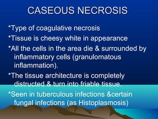
Depending on where (such as which organ) and what type of damage occurred in the body, necrosis will have a specific morphological pattern. There are six distinct patterns that are identifiable, and by identifying the pattern, an underlying cause could be identified. Coagulative.
Why is my pancreas chalky white?
An insoluble salt is created and gives the appearance of a chalky-white area. Infections, viruses, trauma, ischaemia and toxins could be responsible for the pancreas to be damaged and release its enzymes. Breast tissues can also have fat necrosis to which is triggered from trauma.
