What is excitation-contraction coupling?
Excitation-contraction coupling (ECC) is a physiological process that links excitation of muscles by the nervous system to their mechanical contraction. In skeletal muscle, ECC is initiated with an action potential, generated by the somatic nervous system, which causes a depolarisation of the muscle fibre membrane (sarcolemma).
Which receptors are involved in excitation-contraction coupling in skeletal muscle?
Ríos E, Brum G. Involvement of dihydropyridine receptors in excitation-contraction coupling in skeletal muscle. Nature. 1987;325:717–720.
What is excitation in muscle contraction?
Excitation refers to the generation of an action potential within the muscle cell. An action potential is an electrical event, meaning that it involves a change of charge. The electric signal that leads to muscle cell contraction begins as a nerve impulse and is relayed to the muscles via motor neurons.
Is there an allosteric model of excitation-contraction coupling in skeletal muscle?
An Allosteric model of the molecular interactions of excitation-contraction coupling in skeletal muscle. J Gen Physiol. 1993;102:449–481. [PMC free article][PubMed] [Google Scholar]
What is the process of excitation contraction coupling?
Excitation-contraction (E-C) coupling is the process by which the action potential of the motor neuron leads to the synchronous contraction of the myofibrils, of which there may be between hundreds to thousands within a given muscle fiber.
Why does excitation contraction coupling occur?
Excitation contraction coupling is triggered by a nerve impulse delivered by a motor neuron. This becomes an action potential in the membrane of the muscle cell. The membrane voltage change caused by the action potential triggers ECC.
How does the excitation contraction coupling process in muscle fiber?
Excitation-contraction coupling is the mechanism that links plasma membrane stimulation with cross-bridge force production. The muscle receives a neural signal and converts that signal into mechanical force after synapsing at the neuromuscular junction.
What is ATP in excitation-contraction coupling?
ATP provides the energy in our muscles to generate force, through its use by myosin ATPases, and helps to terminate contraction by pumping Ca(2+) back into the sarcoplasmic reticulum, achieved by Ca(2+) ATPase.
What is always the first step in excitation-contraction coupling?
Thus, the excitation-contraction coupling process begins with signaling from the nervous system at the neuromuscular junction (Figure 10.3. 1) and ends with calcium release for muscle contraction.
What happens during excitation contraction coupling quizlet?
Excitation refers to the propagation of action potentials along the axon of a motor neuron. Excitation-contraction coupling is a series of events that occur after the events of the neuromuscular junction have transpired.
How does skeletal muscle contraction?
Skeletal muscle contraction begins first at the neuromuscular junction, which is the synapse between a motoneuron and a muscle fiber. Propagation of action potentials to the motoneuron and subsequent depolarization results in the opening of voltage-gated calcium (Ca2+) channels of the presynaptic membrane.
Where does excitation-contraction coupling occur?
First coined by Alexander Sandow in 1952, the term excitation–contraction coupling (ECC) describes the rapid communication between electrical events occurring in the plasma membrane of skeletal muscle fibres and Ca2+ release from the SR, which leads to contraction.
What triggers the excitation process?
The excitation process begins when the. acetylcholine (ACh) is released by a motor neuron at the neuromuscular junction. Acetylcholine (ACh) release. Stimulation of the muscle fiber by the ACh neurotransmitter generates waves of. action potentials (impulses) that spread out across the sarcolemma.
What occurs during excitation-contraction coupling quizlet?
Excitation refers to the propagation of action potentials along the axon of a motor neuron. Excitation-contraction coupling is a series of events that occur after the events of the neuromuscular junction have transpired.
Why does excitation occur in atoms?
"An electron collides with an atom giving it energy. An electron inside the atom receives this energy and is excited." Would you prefer: "An electron collides with an atom giving it energy. An electron inside the atom receives this energy and the atom is excited."
What are the steps of muscle contraction and relaxation?
First, an action potential occurs at the muscle cell membrane (also called the sarcolemma). Next, calcium is released from the sarcoplasmic reticul...
What triggers excitation contraction coupling?
Excitation contraction coupling is triggered by a nerve impulse delivered by a motor neuron. This becomes an action potential in the membrane of th...
What is the excitation contraction coupling process?
Excitation contraction coupling, or ECC, is the link between the body's nervous system and its capability for mechanical movement. Through ECC, an...
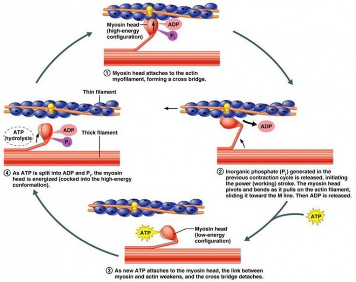
What happens during the relaxation phase in muscle contraction?
During the relaxation phase, calcium is sequestered in the sarcoplasmic reticulum. ATP binds to myosin, causing it to detach from actin, while the...
Why is excitation contraction coupling important?
Excitation contraction coupling is important for the movement of all muscles, including smooth, cardiac, and skeletal. ECC allows us to move our mu...
What is the coupling of skeletal muscle?
Excitation - contraction coupling in the skeletal muscle is the sequence of events through which the nerve fiber stimulates the skeletal muscle fiber causing its contraction. The cholinergic nerve fibers innervate the skeletal muscle fibers through the neuromuscular junctions, they release their neurotransmitters that cause activation ...
What does increased post synaptic surface area mean?
increase of the muscle fiber postsynaptic terminal surface area indicates increased number of ligand-gated ion-channel-coupled receptors on which the neurotransmitter released by the neuron can act on.
What happens when a neurotransmitter receptor complex is activated?
As soon as the neurotransmitter-receptors complex occurs it activates an integral protein coupled with the nicotinic receptor, undergoing conformational changes
Where do motor neuronal axons come in contact with the muscle membrane?
The neuromuscular junction is the site where the neuronal axon terminals (synaptic terminal) of a motor neuron come in contact with the plasma membrane of the skeletal muscle fiber (motor end plate). In reality there is no physical contact between the two membranes, but rather a small gap exists, being similarly functional to the synaptic cleft of a chemical synapse. The synaptic terminal of the neuron is of a sausage-like shape being embedded within the muscle fiber rather than lying over the the cell. The area of the muscle plasma membrane comes in relative contact with the synaptic neuronal terminal, presents a highly folded membrane forming a wavy pattern when it is cut perpendicularly to the longitudinal axis of the neuromuscular junction indicating many features of the excitation-contraction coupling of the skeletal muscle fibers.
What is the function of the folds in muscle fibers?
since the folds extend inside the muscle fiber it is presumed that it promotes contraction of the deeper sarcomeres when stimulation of the motor end plate occurs .
Which neurotransmitter is involved in the exocytotic process?
These cytoskeletal changes lead the exocytotic process concerning the synaptic vesicles containing acetylcholine (ACh) neurotransmitter
Where does action potential travel?
An action potential travels through the axon terminal and eventually reaches the synaptic terminal
Which muscle is involved in the excitation-contraction coupling mechanism?
The excitation–contraction coupling mechanism in skeletal muscle
What is the ECC in skeletal muscle?
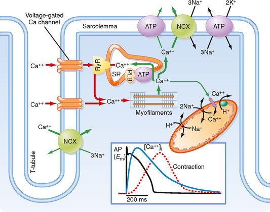
First coined by Alexander Sandow in 1952, the term excitation–contraction coupling (ECC) describes the rapid communication between electrical events occurring in the plasma membrane of skeletal muscle fibres and Ca2+release from the SR, which leads to contraction. The sequence of events in twitch skeletal muscle involves: (1) initiation and propagation of an action potential along the plasma membrane, (2) spread of the potential throughout the transverse tubule system (T-tubule system), (3) dihydropyridine receptors (DHPR)-mediated detection of changes in membrane potential, (4) allosteric interaction between DHPR and sarcoplasmic reticulum (SR) ryanodine receptors (RyR), (5) release of Ca2+from the SR and transient increase of Ca2+concentration in the myoplasm, (6) activation of the myoplasmic Ca2+buffering system and the contractile apparatus, followed by (7) Ca2+disappearance from the myoplasm mediated mainly by its reuptake by the SR through the SR Ca2+adenosine triphosphatase (SERCA), and under several conditions movement to the mitochondria and extrusion by the Na+/Ca2+exchanger (NCX). In this text, we review the basics of ECC in skeletal muscle and the techniques used to study it. Moreover, we highlight some recent advances and point out gaps in knowledge on particular issues related to ECC such as (1) DHPR-RyR molecular interaction, (2) differences regarding fibre types, (3) its alteration during muscle fatigue, (4) the role of mitochondria and store-operated Ca2+entry in the general ECC sequence, (5) contractile potentiators, and (6) Ca2+sparks.
What is the ECC phenomenon?
The ECC phenomenon represents a fast communication between electrical events occurring in the plasma membrane and Ca2+release from the SR, which leads to muscle contraction. The sequence of events in skeletal twitch muscle fibres involves: (1) initiation and propagation of an AP along the plasma membrane, (2) radial spread of the potential along the transverse tubule system (T-tubule system), (3) dihydropyridine receptors (DHPR, L-type Ca2+channel CaV1.1)-mediated detection of changes in membrane potential, (4) allosteric interaction of the DHPR with the sarcoplasmic reticulum (SR) ryanodine receptors (RyR), (5) release of Ca2+from the SR and transient increase of Ca2+concentration in the myoplasm, (6) transient activation of the myoplasmic Ca2+buffering system and the contractile apparatus, followed by (7) disappearance of Ca2+from the myoplasm mediated by its movement to the mitochondria, its transport by the Na+/Ca2+exchanger (NCX) and its final reuptake by the SR through the SR Ca2+adenosine triphosphatase (SERCA) (Sandow 1952; Caputo 1983; Fill and Copello 2002; Calderón-Vélez and Figueroa-Gordon 2009).
How are muscle fibres attached to their tendons?
2003; Edman 2005). Therefore, muscle fibres remain attached to their tendons. Isolated fibres or bundles are horizontally mounted on the experimental chamber and attached with small aluminum clips, on one end to a tension transducer and on the other to a hook attached to the chamber. Contractions are produced by suprathreshold stimulation through electrodes. Simultaneous measurements of Ca2+and tension can be obtained (Westerblad and Allen 1991; Bruton et al. 2003; Baylor and Hollingworth 2003; Calderón et al. 2011). This allows to calculate, for instance, myofibrils’ sensitivity to Ca2+in intact fibres, or to follow the changes of both variables during muscle fatigue. Recently, a biological adhesive was successfully used to attach dissociated fibres to a tension transducer (Ward et al. 2011) ,opening up a large number of possibilities with this cellular preparation.
What is the resting potential of a twitch muscle cell?
In twitch skeletal muscle cells, both the differential and selective conductance and the ion distribution across the membrane generate a resting potential of about –85 mV, with the interior of the cell negative compared to the exterior (Horowicz 1961; Luff and Atwood 1972). The acetylcholine neurotransmitter released into the motor plate by the inferior motor neuron acts as an initiator for the AP in muscle fibres causing the transmembrane potential change to reach values of up to 100 mV, through voltage-dependent ionic conductance changes (Horowicz 1961; Hodgkin and Huxley 1952; Luff and Atwood 1972). Since the AP is a regulator of ECC, its modifications (see below) may affect the kinetics of muscle contraction (Hodgkin and Horowicz 1960; Sandow et al. 1965). Experimentally, membrane depolarization can be achieved by replacing neurotransmitters with direct electrical stimulation or by increasing the extracellular K+concentration (Hodgkin and Horowicz 1959). Additionally, contractile activation can be induced bypassing the membrane depolarization step, for instance with the help of caffeine (Axelsson and Thesleff 1958; Endo 1975).
How to obtain muscle fibres?
Intact muscle fibres for physiological experiments can be obtained by means of enzymatic dissociation and manual isolation. In the first technique, described by Bekoff and Betz (Bekoff and Betz 1977) and modified by others (Caputo et al. 2004; Calderón et al. 2009; Calderón 2013), different rat or mouse muscles (mainly flexor digitorum brevis (FDB), extensor digitorum longus –EDL-, soleus and interossei) are subjected to an enzymatic dissociation with collagenase to digest the connective tissue surrounding the fibres, and subsequently subjected to mechanical dissociation through the use of glass-pipettes. The procedure yields complete, tendon-free muscle fibres. Once obtained, about 85 % of the fibres contract immediately and remain excitable for up to 24-36 hours when kept in Tyrode solution or culture medium (Calderón et al. 2009, 2010; Calderón 2013). One limitation with the use of dissociated fibres is their susceptibility to movement artifacts when recording Ca2+transients. This drawback, however, has been overcome with the use of N-benzyl-p-toluene sulphonamide (BTS), butanedione monoxime (BDM) (Sun et al. 2001) and laminin. BTS is a small molecule which inhibits myosin type II and avoids shortening of the fibres (Cheung et al. 2002; Shaw et al. 2003; Calderón et al. 2009, 2010). Laminin works as a substrate to which muscle fibres adhere, limiting their movement and allowing the recording of movement artifacts-free Ca2+transients with the advantage of working for all fibre types (Calderón et al. 2009, 2010).
What is ECC in biology?
The excitation–contraction coupling (ECC) phenomenon was defined by Alexander Sandow as the series of events occurring from the generation of the action potential (AP) in the skeletal muscle fibres to the beginning of muscle tension (Kahn and Sandow 1950; Sandow 1952). It has been more than 60 years since his early work on skeletal muscle, during which the temporal and spatial resolution of the techniques to study ECC have greatly improved, reaching a capacity for discrimination at a molecular level. Since then, a great amount of information on ECC morphological basis, physiological importance, and pharmacological modulation, initially in amphibians and more recently in mammalians, has been gathered.
Which molecule binds to actin and inhibits contraction?
Troponin I: bind s to actin and inhibits contraction
Is a coiled sock elastic?
Basically: both are elastic and coiled (they resist stretching but then help with rebounding if stretching does occur)
Neuromuscular Junction
Excitation Sequence of Skeletal Muscle
- An action potential travels through the axon terminal and eventually reaches the synaptic terminal
- The depolarization of the synaptic terminal from the action potential induces opening of the Ca2+voltage gated channels
- Opening of these channels allows flux of Ca2+ions inside the neuron
- An action potential travels through the axon terminal and eventually reaches the synaptic terminal
- The depolarization of the synaptic terminal from the action potential induces opening of the Ca2+voltage gated channels
- Opening of these channels allows flux of Ca2+ions inside the neuron
- Increase of the intracellular Ca2+concentration introduces conformational changes of the microtubular component of the neuronal synaptic terminal cytoskeleton
Contraction Sequence of Skeletal Muscle
- The Ca2+ that accumulates after a skeletal muscle cell depolarization is the reason for the initiation and the maintenance of the contraction of the sarcomere, thus increasing the Ca2+inside the ce...
- The free Ca2+binds with the troponin C protein component of the thin actin filaments introducing the active calcium-troponin complex
- This binding causes the conformational change of the troponin C
- The conformational change of the troponin C induces the alteration of the conformation of the tropomyosin protein component of the thin actin filaments
Removal of Ca2+ from The Smooth Muscle Cell
- Na+/Ca2+ antiporter: located in the plasma membrane, through which 3 Na+ ions are exchanged for a single Ca2+ ion. This type of Ca2+ transport occurs not directly through ATP cleavage but indirectl...
- Ca2+ ATPase pump: located in the membrane of the sarcoplasmic reticulum that transports Ca2+ from the cytosol into the reticulum using ATP. This type of Ca2+ transport is referred t…
- Na+/Ca2+ antiporter: located in the plasma membrane, through which 3 Na+ ions are exchanged for a single Ca2+ ion. This type of Ca2+ transport occurs not directly through ATP cleavage but indirectl...
- Ca2+ ATPase pump: located in the membrane of the sarcoplasmic reticulum that transports Ca2+ from the cytosol into the reticulum using ATP. This type of Ca2+ transport is referred to as the primary...
Links
- Bibliography
1. HALL, John E. – GUYTON, Arthur C. Guyton and Hall Textbook of Medical Physiology. 12. edition. Saunders/Elsevier, 2010. ISBN 1416045740. 1. Lecture Notes: Prof. MUDr. Jaroslav Pokorný DrSc.