Where is facial in anatomy?
The facial nerve is the seventh of 12 cranial nerves in your nervous system. You have two facial nerves, one on each side of your head. The facial nerve: Starts in your brainstem.
Why is it important to understand facial anatomy?
The knowledge of crucial facial anatomical structures allows injectors to provide services in a reproducible manner to optimize patient safety and results. As a practitioner of these treatments, you need to know the important anatomical relationships to offer your clients improved outcomes.
What are the 7 facial muscles?
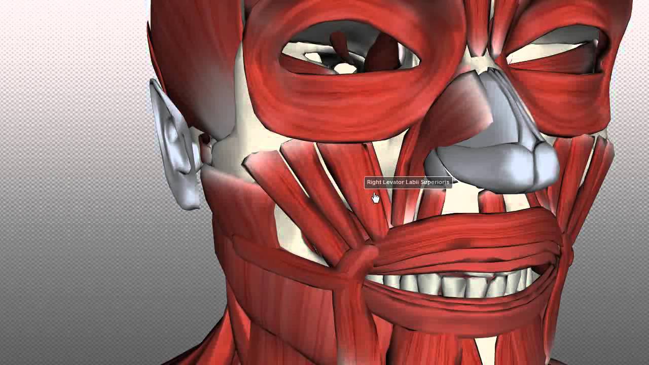
Facial Muscles:frontalis: the forehead.corrugator: the brow.nasalis: the nose.obicularis oculi: around the eye.levator labii: raises the upper lip.masseter: closes the jaw.Obicularis oris: purses the lips.risoris: draws the lips in a smile.More items...•
What are the 4 main muscles of the face?
Muscles of facial expression include frontalis, orbicularis oris, laris oculi, buccinator, and zygomaticus. These muscles of facial expressions are identified in the illustration below. There are four pairs of muscles that are responsible for chewing movements or mastication.
Where is the facial nerve?
The facial nerve exits the base of the skull at the stylomastoid foramen, which is an opening in the bone located near the base of the ear.
What are the branches of the facial artery?
On the face, four main vessels arise from the trunk of the facial artery: the inferior labial artery, superior labial artery, lateral nasal branch (to the nasalis), and the angular artery.
What are the 12 cranial nerves names?
In higher vertebrates (reptiles, birds, mammals) there are 12 pairs of cranial nerves: olfactory (CN I), optic (CN II), oculomotor (CN III), trochlear (CN IV), trigeminal (CN V), abducent (or abducens; CN VI), facial (CN VII), vestibulocochlear (CN VIII), glossopharyngeal (CN IX), vagus (CN X), accessory (CN XI), and ...
What is the facial nerve?
The facial nerve provides motor innervation of facial muscles that are responsible for facial expression, parasympathetic innervation of the glands of the oral cavity and the lacrimal gland, and sensory innervation of the anterior two-thirds of the tongue.
What muscles close the eye?
Overview. The orbicularis oculi muscles circle the eyes and are located just under the skin. Parts of this muscle act to open and close the eyelids and are important muscles in facial expression.
What is the largest bone in the face?
mandibleDescription. The mandible (lower jaw, the largest and strongest bone of the face, serves for the reception of the lower teeth.
What is the kissing muscle called?
Orbicularis oris muscleOrbicularis oris muscle, also known as musculus orbicularis oris is a complex, multi-layered muscle which attaches through a thin, superficial musculoaponeurotic system to the dermis of the upper lip and lower lip and serves as an attachment site for many other facial muscles around the oral region.
How many bones are in the face?
14 bonesThe facial skeleton consists of 14 bones with different anatomic structures and embryological formations (2 unpaired and 6 paired). These bones comprise the paired nasal bones, inferior nasal conchae, palatine bones, maxillae, zygomatic bones, lacrimal bones, and the unpaired mandible and vomer.
What are facial muscles?
The facial muscles are striated muscles that link the skin of the face to the bone of the skull to perform important functions for daily life, including mastication and expression of emotion.
How many facial muscles are there?
What are the facial muscles? Your face has almost 20 flat skeletal muscles that attach to different places on your skull. The craniofacial muscles are essential to chewing and making facial expressions. They originate from bone or fascia and insert into your skin.
What are the five muscles of facial expression?
The facial muscles include:Occipitofrontalis muscle.Temporoparietalis muscle.Procerus muscle.Nasalis muscle.Depressor septi nasi muscle.Orbicularis oculi muscle.Corrugator supercilii muscle.Depressor supercilii muscle.More items...
How many face muscles do you use to smile?
Most of the 43 muscles we use to smile are under the control of the seventh cranial nerve. This is also referred to as the facial nerve.
What are the features of the face?
The various features found on the human face have different embryological origins: 1 The upper lip develops from the maxillary prominence and the medial nasal prominence. 2 The lower lip derives from the mandibular prominence which is a direct result of the development of the first pharyngeal arch. 3 The lacrimal sac and the nasolacrimal duct are the mature structures of the nasolacrimal groove that separates the lateral nasal prominence and the maxillary prominence. 4 The nose consists of a triad of embryonic structures including the frontonasal prominence, the medial nasal prominence and the lateral nasal prominence. 5 Unlike the nose, the cheeks stem forms a single structure known as the maxillary prominence which arises from the first pharyngeal arch, just like the mandibular prominence. 6 The intermaxillary segment ends up as the philtrum, the primary palate and the upper jaw containing the central and lateral incisors.
What is the embryological background of the face?
Embryological background. The development of the face starts with the oral (or anterior) portion of the alimentary canal, the stomatodeum. It is surrounded by the three different swellings of the face divulging from the cells of the neural crest. the mandibular prominence.
What is a cleft lip?
Cleft lip - a partial or complete lack of fusion of the maxillary prominence with the medial nasal prominence on one or both sides. Depending on the severity of the lack of fusion, this can result in a partial or complete, unilateral or bilateral cleft lip.
What is the primary deformity of a cleft palate?
The primary (or anterior) cleft deformities include lateral cleft lip, upper cleft jaw and a cleft between the primary and secondary palates. Behind the incisive foramen the clefts can either be of the secondary palate or known as a cleft uvula. Cleft palates result from a lack of fusion between the palatine shelves. Rarely, a cleft will run from the lip to the secondary palate.
What is a medial cleft?
Median (or midline) cleft: This type of anomaly occurs with the incomplete fusion of the two medial nasal prominences in the midline. This particular defect can have much more serious consequences that the others it is associated with cognitive disabilities and brain abnormalities.
Where does the frontonasal prominence form?
Both the maxillary and mandibular prominence develop from the first pharyngeal arch whereas the frontonasal prominence is formed from the mesenchyme. Two ectodermal layers start to take shape just lateral of the frontonasal prominence and become the two nasal placodes.
Which nerve supplies the anterior scalp, forehead, and nasal dorsum?
The ophthalmic nerve (V1) which comes from the frontonasal prominence supplies the anterior scalp, forehead and nasal dorsum. Deriving from the maxillary prominence the maxillary nerve (V2) provides mainly the anterior cheek, the lateral face, the upper lip, the side of the nosa and the lower eyelid. The mandibular nerve (V3) ...
Why is anatomy important for face surgery?
The knowledge of the anatomy of the face can guide clinicians in the avoidance of damage to crucial structures in the face during surgeries and injections in the face.
What are the three main parts of the face?
The anatomy of the face can divide into three main regions: upper face, middle face, and lower face . The entire face is covered by skin superficially, while the deep anatomy contains muscles, fat pads, nerves, vessels, and bones.
What muscle is the forehead?
The muscular layer of the upper face is underneath the fat pads. The procerus muscle occipitofrontalis muscle, depressor supercilii muscle, and corrugator supercilii muscle form the majority of the forehead, while the temporal part contains the temporalis muscle. The procerus muscle is shaped like a pyramid and spans from the inferior part of the nasal bone to the middle part of the forehead. The procerus muscle is situated between the eyebrows and attaches to the frontalis muscle. The contraction of the procerus muscle allows for the elevation of the eyebrows. The occipitofrontalis muscle spans the majority of the forehead. The occipitofrontalis muscle originated from the galea aponeurosis superiorly and inserts and blends into the orbicularis oculi muscle. When the occipitofrontalis muscle contracts, it elevates the eyebrows and wrinkles the forehead. The depressor supercilii muscle originates from the medial orbital rim and inserts at the medial part of the bony orbit. The action of the depressor supercilii muscle is to depress the eyebrows. The corrugator supercilii muscle is a small muscle that originated from the supraorbital ridge and inserts on the skin of the forehead close to the eyebrows. The contraction of the corrugator supercilii muscle results in the wrinkling of the forehead. The temporalis muscle originates from the parietal and sphenoidal bone. The temporalis muscle inserts on the coronoid process and the retromolar fossa. The contraction of the temporalis muscle results in the elevation and retraction of the mandible.
How does cheek augmentation work?
The augmentation of the cheeks can be done by implantation of cheek implants or the injection of fillers to add volume to the cheeks. The addition of implants or injected fillers will increase the volume of the cheeks, usually resulting in less sagging and wrinkling of the cheeks.
How does the face develop?
The face derives from the first two pharyngeal arches, neural crest cells, frontonasal prominence, medial nasal prominence, oropharyngeal membrane, and lateral nasal prominence. During week four of development, the oropharyngeal membrane breaks down to create the oral cavity. The frontonasal prominences develop into the forehead, bridge of the nose, medial nasal prominences, and lateral nasal prominences. The medial nasal prominences will further develop into the primary palate, philtrum, upper four incisors, and parts of the jaw. The lateral nasal prominences develop into the sides of the nose. The first pharyngeal arch will form the cheeks, lateral upper lip, lateral upper jaw, and secondary palate. The second pharyngeal arch will form the lower lip and jaw. All these structures form bilaterally and migrate toward the midline before fusing. [2]
What is the most anterior region of the head?
The most anterior region of the head is the face . The human face is a unique aspect of each individual. The face contains many structures that contribute to the display of emotions, feeding, seeing, smelling, and communicating. One of the most distinguishing qualities of the face is that it is used for personal identity from person to person. Identity is essential since the face is usually the first aspect of a human that is noticeable during encounters with other individuals.
Why is the face variable?
The face is one of the most variable structures in humans. The face represents the identity of each person. Unfortunately, some individuals are born with congenital defects that alter the appearance of their face. And others suffer injuries or diseases that affect the way their face appears. And others alter the appearance of their faces with cosmetics products and procedures. The human face can also change over time as a person ages. The environment and lifestyle of an individual can manifest in changes in the face. For instance, a person exposed to UV light will have darkening of the skin, while an individual that consumes an excessive amount of calories may gain fat content in their face. For this reason, the human face is extremely variable, but the standard features such as the eyes, nose, and mouth are consistently present in most individuals.
What is the skin on the face?
The skin varies in thickness, pigmentation, dermal appendages, and adherence to the subcutaneous tissues between different areas of the face . The skin is thickest over the mentum and in the region of the forehead and eyebrows, and is thinnest over the eyelids. In the infraorbital region and medial to the midpupillary line, the skin is thin and usually contains no subcutaneous fat. The skin is firmly attached to the underlying muscles of the oral commissure and in the region of the nasal tip, while it is loosely adherent to the underlying soft tissue in the eyelids and at the root of the nose. As the dermis of the skin thins with age, the underlying muscles cause rhytids, which generally are perpendicular to the direction of facial muscle contraction ( Fig. 2.3 ). The amount of skin wrinkling is variable, and relates to the skin thickness and elasticity.
Which layer of the face is adherent to the skin?
Layer III: Superficial Fascia. The superficial fascia of the face is fairly adherent to the overlying skin and subcutaneous tissues of the face in most regions, and therefore must be dissected sharply from the skin in order to surgically separate these tissue planes.
What is the deep fascia of the lateral midface?
The deep fascia of the lateral midface is termed the parotidomasseteric fascia. This fascia covers the parotid gland and the parotid duct, and includes the buccal branches of the facial nerve. As this fascia travels towards the temple over the zygomatic bone, it becomes continuous with the superficial lamina of the DTF. The deep fascia of the neck is designated deep investing fascia. All deep fascial layers (V) are relatively fixed to the structures they overlie, and become good fixation points for repositioning of soft tissues and surgical procedures such as rhytidectomy or browlifting.
What is the superficial fascia?
In the forehead, the superficial fascia is called the galea aponeurosis. This layer envelopes the frontalis muscle in the forehead, and splits to encompass the occipitalis muscle at the posterior aspect of the scalp. In the temple, the superficial fascial layer is termed the temporoparietal fascia (TPF; see Fig. 2.1 ).
What is the layer IV of the head?
Layer IV: Deep Areolar Layer. In all regions of the head and neck, the superficial fascia (III) is connected to the deep fascia (V) by a loose areolar layer. This connection is variable and differs by age, genetics, and body weight.
What is the deep investing fascia?
As this fascia travels towards the temple over the zygomatic bone, it becomes continuous with the superficial lamina of the DTF. The deep fascia of the neck is designated deep investing fascia.
What is the deep fascia?
It is connected to the overlying mobile superficial fascia (III) by loose connective tissue (IV). The deep fascia of the forehead is the frontal bone periosteum. The periosteum is densely adherent to the frontal bone, and becomes continuous at the superior orbital rim with the septum orbitale. The thickening at the orbital rim is termed the arcus marginale. The deep fascia of the temple is designated deep temporal fascia (DTF) or temporalis muscle fascia. This fascia overlies, and is densely adherent to, the underlying temporalis muscle (see Fig. 2.1 ) .
What is the prenatal development of the face?
prenatal development: Fetal development. The face rapidly acquires a fairly human appearance; eyes, ears, and jaws are prominent. The eyes, previously located on the sides of the head, become directed forward. The nose lacks a bridge and so is of the “pug” type, with the nostrils directed forward instead of….
How does the face change in age?
In individual development the human face and braincase follow different patterns of growth. The brain and braincase attain 90 percent of adult size by the age of 6 years, while the face grows more slowly in concurrencewith the enlargement of the nasal passages and the eruption of both sets of teeth. Viewed in profile, the face at birth is less than one-fifth the size of the braincase; by adulthood it has increased to nearly half. Facial dimensions increase most in depth, next in height (length), and least in width. During adolescence, facial musculature increases and the facial sinuses enlarge, in general to a greater extent in males than in females.
Is the skull bigger than the face?
No consensus has developed on exactly where this…. skull. …large in comparison with the face. In most other animals the facial portion of the skull, including the upper teeth and the nose, is larger than the cranium. In humans the skull is supported by the highest vertebra, called the atlas, permitting nodding motion.
Do facial dimensions increase in height?
Facial dimensions increase most in depth, next in height (length), and least in width. During adolescence, facial musculature increases and the facial sinuses enlarge, in general to a greater extent in males than in females. Learn More in these related Britannica articles: prenatal development: Fetal development.
Where are facial muscles located?
Facial muscles are located throughout your face. They can be categorized by general location:
Why do facial muscles work?
Your facial muscles work together to control the parts of your face. They are essential to chewing, facial expressions and other functions. Weakness or paralysis of your face muscles can be a temporary condition or a serious medical problem. See a healthcare provider right away if you have facial palsy or any trouble smiling, talking or eating.
How many muscles are there on the face?
Your face has almost 20 flat skeletal muscles that attach to different places on your skull. The craniofacial muscles are essential to chewing and making facial expressions. They originate from bone or fascia and insert into your skin. Craniofacial muscles work together to control movements in your:
What muscle controls movement in the lower lip?
Depressor labii inferioris, a muscle in your chin that helps control movement in your lower lip.
What is the term for the inability to move parts of the face?
Facial paralysis (inability to move parts of the face).
What muscle pulls your eyebrows downward?
Procerus, a muscle between your eyebrows that can pull your brows downward and help flare your nostrils.
What is the muscle that holds your cheeks toward your teeth?
Buccinator, a thin muscle in your cheek that holds each cheek toward your teeth.
How many zones are there in the face?
The face can be split into 3 zones – the upper 1/3 rd, the middle 1/3 rd and the lower 1/3 rd. Knowing the important vessels and nerves in each zone as well as their corresponding depth in the skin is crucial in order to minimize injury and utilize a safe technique when injecting neurotoxins and fillers.
What are the landmarks of the face?
Important Landmarks for the Middle and Lower 1/3rd of the Face 1 The superficial temporal artery exits anteriorly to the tragus and runs superiorly towards the temple. It becomes more superficial as it traverses over the zygoma and then branches to anastomose with the supraorbital and supratrochlear vessels. 2 The area along the temple, where the superficial temporal artery is most vulnerable due to its superficial depth, is considered a danger zone. Keep injections deep in this area while avoiding excess pressure as the bone here is very thin. 3 The transverse artery runs across the medial cheek but is an end-vessel with no anastomoses. However, it does supply small branches to the infraorbital area. 4 The infraorbital foramen lies about 8-10 mm below the inferior orbital rim along the medial limbus. The infraorbital NVB emerges through this aperture. However, the depth of vessels in this area is variable and for safety, a cannula is preferred when injecting in this region. 5 The facial artery lies medial to the masseter and runs deep in a tortuous fashion about 1.5 cm lateral to the oral commissure. It supplies the superior and inferior labial arteries which lie deep in the skin. 6 The facial artery gives rise to the angular arteries which run along the nasolabial folds most commonly. However, about 30% of individuals may have angular arteries that lie as lateral as near the infraorbital foramen. For safety, a cannula is preferred when injecting this region.
How can dermatologists become more confident in injectables?
Adhering to these simple concepts and establishing a sound foundation in anatomy can help dermatologists become more confident in using injectables safely and effectively to achieve the best cosmetic results for their patients.
Which artery runs across the medial cheek?
The transverse artery runs across the medial cheek but is an end-vessel with no anastomoses. However, it does supply small branches to the infraorbital area.
Which artery runs deep in the skin?
The facial artery lies medial to the masseter and runs deep in a tortuous fashion about 1.5 cm lateral to the oral commissure. It supplies the superior and inferior labial arteries which lie deep in the skin.
What is the line drawn vertically along the medial limbus?
A line drawn vertically along the medial limbus denotes the anatomical locations of the supraorbital, infraorbital and mental foramen.
Overview
The facial nerve is a pathway from your brain to certain muscles in your face. It controls muscles that help you make expressions like raising an eyebrow, smiling or frowning. This nerve is also responsible for most of your tongue’s taste sensations.
Function
The facial nerve performs these motor (movement) and sensory functions:
Anatomy
The facial nerve is the seventh of 12 cranial nerves in your nervous system. You have two facial nerves, one on each side of your head.
Conditions and Disorders
Several conditions can cause weakness or paralysis of the facial nerve, including:
