What is the midbrain, and what does it do?
What is the midbrain and what does it do? The midbrain or mesencephalon is the forward-most portion of the brainstem and is associated with vision, hearing, motor control, sleep and wakefulness, arousal (alertness), and temperature regulation. The name comes from the Greek mesos, “middle”, and enkephalos, “brain”.
What are the two main parts of the midbrain?
The midbrain
- Colliculi. At the top of the midbrain are the colliculi, which derives its name from the Latin word for ‘hill. ...
- Tegmentum. The tegmentum (Latin for ‘hood’) actually stretches down the length of the brainstem, but a portion of it forms a part of the midbrain.
- Cerebral peduncles. ...
Is the limbic system found in the midbrain?
The posterior limbic midbrain complex comprising the stria medullaris, central gray and dorsal and ventral nuclei of Gudden are also key elements in the limbic midbrain. Some of these formations will be discussed in terms of the neurochemical connectivity between them.
Is the midbrain involved in hearing?
Your midbrain (derived from the mesencephalon of the neural tube) is a part of the central nervous system, located below your cerebral cortex and at the topmost part of your brainstem. This tiny, but mighty, structure plays a crucial role in processing information related to hearing, vision, movement, pain, sleep, and arousal.
See more

What important structure is located in the midbrain?
midbrain, also called mesencephalon, region of the developing vertebrate brain that is composed of the tectum and tegmentum. The midbrain serves important functions in motor movement, particularly movements of the eye, and in auditory and visual processing.
What neurons are in the midbrain?
Dopaminergic (DA) neurons of the ventral midbrain (VM) play vital roles in the regulation of voluntary movement, emotion and reward. They are divided into the A8, A9 and A10 subgroups.
What nuclei are in the midbrain?
Notable midbrain nuclei include the superior and inferior colliculus nuclei, red nucleus, substantia nigra, oculomotor nuclear complex, and trochlear nucleus.
Is the thalamus in the midbrain?
Structure and Function The thalamus is a paired gray matter structure of the diencephalon located near the center of the brain. It is above the midbrain or mesencephalon, allowing for nerve fiber connections to the cerebral cortex in all directions — each thalamus connects to the other via the interthalamic adhesion.
Which cranial nerves are found in the mesencephalon midbrain )?
Midbrain. The third and fourth cranial nerves originate from the midbrain: oculomotor nerve (CN III) trochlear nerve (CN IV)
What is the midbrain structure critical to movement?
The ventral tegmental area is termed as the midbrain structure which is critical to movement. VTA is termed as a group of neurons which are located close to the midline on the floor of the midbrain. This is the origin of dopaminergic cell bodies of the mesocorticolimbic dopamine system.
Is the hippocampus in the midbrain?
The midbrain is the smallest region of the brain, and is located most centrally within the cranial cavity. Limbic System – the limbic system is often referred to as our “emotional brain”, or 'childish brain'. It is found buried within the cerebrum and contains the thalamus, hypothalamus, amygdala and hippocampus.
Is hypothalamus part of midbrain?
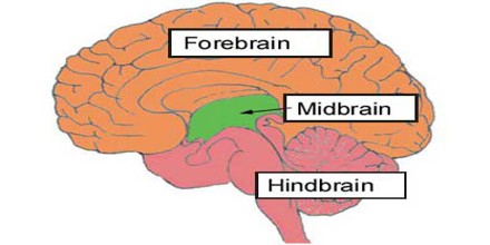
The forebrain, midbrain and hindbrain make up the three major parts of the brain. The structures in the forebrain include the cerebrum, thalamus, hypothalamus, pituitary gland, limbic system, and the olfactory bulb. The midbrain consists of various cranial nerve nuclei, tectum, tegmentum, colliculi, and crura cerebi.
What is the midbrain?
The midbrain, also known as the mesencephalon, is one of the primary divisions of the brainstem. Its functions extend to many different parts of the central and peripheral nervous systems, from motor to sensory and cognitive abilities.
Which two areas of the midbrain are named by color?
The portion of the tegmentum that constitutes this specific region of the midbrain is segmented into two areas, named by color: the red nucleus and the periaqueductal gray.
What is the tectum made of?
The tectum is made up entirely of the superior and inferior colliculi at the dorsal end of the midbrain. With these two structures, the tectum controls the “ master coordinate system ” for other sensory afferent nerves including auditory and somatosensory.
Where is the cerebral aqueduct located?
The cerebral aqueduct is located centrally to the cerebral peduncles. The midbrain can be further divided into two main regions which lie superior to the crus cerebri and substantia nigra: the tegmentum and tectum. Other regions of this section of the brainstem are the cerebral peduncles and the colliculi.
Where is the periaqueductal gray located?
The periaqueductal gray – boy, that’s a mouthful, we’re just going to call it the PG – is another structure located within the tegmentum of the midbrain. The PG is a mass of gray matter that surrounds the cerebral aqueduct.
Which part of the brain is connected to the cerebrum?
The midbrain is the most superior portion of the brainstem, connecting the brainstem to the cerebrum by the cerebral peduncles (not to be confused with the cerebellar peduncles which connect the brainstem to the cerebellum). Just the two cerebral hemispheres, there are also a right and a left cerebral peduncle.
What is the tegmentum?
The tegmentum is an area of gray matter surrounding the cerebral aqueduct and is one of the most superior regions of the midbrain, positioned anteriorly to the tectum. This structure actually extends down the entire length of the brainstem, but a portion of it forms the midbrain. The portion of the tegmentum that constitutes this specific region ...
Where is the midbrain located?
It is located within the brainstem and between the two other developmental regions of the brain, the forebrain and the hindbrain; compared with those regions, the midbrain is relatively small. structures of the human brain. Sagittal section of the human brain, showing structures of the cerebellum, brainstem, and cerebral ventricles.
What is the midbrain?
Midbrain, also called mesencephalon, region of the developing vertebrate brain that is composed of the tectum and tegmentum. The midbrain serves important functions in motor movement, particularly movements of the eye, and in auditory and visual processing.
What is the midbrain affected by?
The midbrain is affected by certain developmental disorders, including cobblestone lissencephaly (type II lissencephaly), in which neurons fail to migrate between the 12th and the 24th week of gestation, resulting in a lack of formation of grooves and folds in the brain surface.
What is the periaqueductal gray region?
Subscribe Now. The periaqueductal gray region of the tegmentum is made up of gray matter (neural tissue with relatively few axons covered in myelin) and surrounds the cerebral aqueduct, a short canal that runs between the third and fourth ventricles of the brain.
What are the two structures that make up the striatum?
These two structures, in addition to the globus pallidus, form the striatum. By inhibiting the action of neurons in the caudate nucleus and the putamen, the dopaminergic cells of the pars compacta influence the neuronal output of the neurotransmitter GABA ( gamma-aminobutyric acid ).
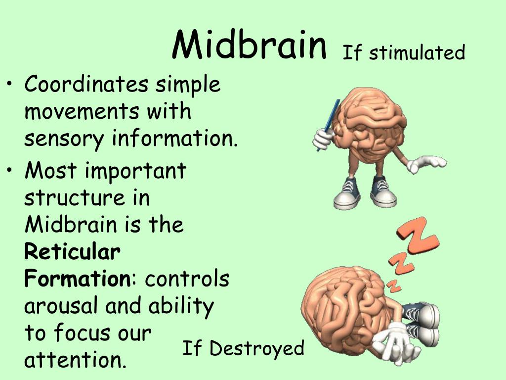
Which part of the brain is responsible for arousal?
The midbrain also contains a portion of the reticular formation, a neural network that is involved in arousal and alertness. Cranial nerves in the midbrain that stimulate the muscles controlling eye movement, lens shape, and pupil diameter form the nuclear complex of the oculomotor nerve and the trochlear nucleus.
Which cells are involved in the production of dopamine?
Cells of the pars compacta contain the dark pigment melanin; these cells synthesize dopamine and project to either the caudate nucleus or the putamen, both of which are structures of the basal ganglia and are involved in mediating movement and motor coordination.
What are the two parts of the midbrain?
The midbrain consists of two major parts: cerebral peduncles and tectum. The cerebral peduncles consist of the crura cerebri and tegmentum. They are separated from each other by a darkened stripe called the substantia nigra. The dorsal part of the tegmentum is traversed by the cerebral aqueduct, which connects the third and fourth ventricles of the brain. The tectum lies dorsal to the tegmentum and cerebral aqueduct, and it contains the nuclei of the superior and inferior colliculi.
What are the peduncles of the midbrain?
Cerebral peduncles. On the cross-section of the midbrain, we can see that the cerebral peduncles consist of the ventral and dorsal regions. The ventral region of each crus is called the crus cerebri, and contains the white matter from the cortex.
What is the posterior surface of the midbrain called?
The posterior surface of the midbrain is called the tectum , or roof, of the midbrain. The tectum features four tubercles on its surface which lie inferior to the pineal gland . The upper pair of tubercles are the left and right superior colliculi, while the lower pair are the left and right inferior colliculi.
What is the pretectal area?
Tegmentum (Pretectal area) The mesencephalic tegmentum, also known as the pretectum, is the central part of the midbrain. It contains the reticular and cranial nerve nuclei, as well as several neural pathways. These structures span on various levels of the midbrain, so let’s analyze their anatomy and location.
How many sensory pathways are there in the midbrain?
The midbrain contains five sensory pathways; one fasciculus and four lemnisci. Namely, they are the medial longitudinal fasciculus (MLF) and the medial, trigeminal, spinal, and lateral lemnisci. The medial longitudinal fasciculus is located just dorsal to the decussation of the superior cerebellar peduncle.
What is the most rostral part of the brainstem that connects the pons and cerebellum?
Midbrain (Mesencephalon) The midbrain, or mesencephalon , is the most rostral part of the brainstem that connects the pons and cerebellum with the forebrain. For most of its part, the midbrain sits in the posterior cranial fossa, traversing the hiatus of the tentorium cerebelli. The midbrain is the shortest part of the brainstem.
Where are the nuclei of the oculomotor and trigeminal nerves located?
The nuclei of the oculomotor (CN III), trochlear (CN IV), and trigeminal nerves (CN V) are located near the periaqueductal gray matter.
Midbrain Anatomy
The midbrain is the topmost part of the brainstem, which connects the brain with the cervical spinal cord. The Midbrain parts consist of three parts, namely, the colliculi, the tegmentum, and the cerebral peduncles.
Midbrain Blood Supply
The midbrain anatomy is stuck between the thalamus and pons, measuring around 1.5 centimetres; the lateral side of the midbrain is enclosed and hidden by the hippocampal gyri of the brain, as shown in the midbrain diagram inserted above. The cerebellar artery (Superior) supplies blood to the tectum of the midbrain.
Location and Function
Midbrain location is at the base of the skull; though it is the smallest area of the brain, it is an important processing centre for visual and auditory signals. Midbrain function involves free movement of body and head, as it provides passage for downward pathways for the cerebral cortex.
What are the four rounded prominences in the midbrain called?
The tectum of the midbrain houses four rounded prominences called colliculi. Where are they located in relation to the pineal gland?
What are the three areas of the brainstem?
2.1 Level of the Inferior Colliculus. 2.2 Level of the Superior Colliculus. 3 Vasculature. The midbrain (also known as the mesencephalon) is the most superior of the three regions of the brainstem.
What is the pathway between the superior colliculus and the retina?
Superior quadrigeminal brachium forms a pathway between the superior colliculus and the retina of the eye.
What is the smallest part of the brain?
The midbrain is the smallest of the three regions of the brainstem, measuring around 2cm in length. As it ascends, the midbrain travels through the opening in the tentorium cerebelli. It can be divided into two main parts: Tectum – located posterior to the cerebral aqueduct.
Where is the tectum located?
Tectum – located posterior to the cerebral aqueduct. Paired cerebral peduncles – located anteriorly and laterally. Internally, the cerebral peduncles are further separated by the substania nigra into the crus cerebri (anterior) and the tegmentum (posterior).
What is the aqueduct of the trigeminal nerve?
The cerebral aqueduct (see ventricles) is a midline structure surrounded by central gray matter – the periaqueductal gray matter. Within this gray matter lies the mesencephalic nucleus of the trigeminal nerve, as well as the trochlear nucleus with its fibres continuing around the gray matter to exit the midbrain.
Which structure houses four rounded prominences?
The tectum houses four rounded prominences named colliculi (collectively the corpora quadrigemina) which sit directly inferior to the pineal gland. The colliculi are separated by the cruciform sulcus; there are two superior and two inferior colliculi.
What is the Midbrain?
The midbrain is an area of the brain that, as you might have guessed, is in the middle of two other regions: the forebrain and the hindbrain. The forebrain is the 'front' (fore) brain and is composed of the cerebral cortex, the area that most people think of as the 'brain'; it's the 'supercomputer' of the human body. The hindbrain, or 'back' (hind) brain, is composed of the cerebellum and the pons and the medulla oblongata (or medulla, for short) of the brainstem; it is evolutionarily the oldest part of our brain, controlling primal instincts and automated actions of the body, such as our 'fight or flight' response and heart rate.
What are the three main structures of the midbrain?
Structures of the Midbrain. The midbrain is formed by three main structures: the cerebral peduncle (peduncle meaning 'foot' or 'base' of the cerebrum), the corpora quadrigemina (meaning 'quadruplet bodies' since it has four mound or hill-like structures), and the cerebral aqueduct, which is a canal dividing the two structures.
What is the canal that separates the cerebral peduncle from the corpora quadrigemina?
Cerebral Aqueduct. The cerebral aqueduct is a little canal that separates the cerebral peduncle from the corpora quadrigemina. It's part of a larger system of canals that circulates and distributes cerebrospinal fluid throughout your brain ('cerebro') and down your spinal cord ('spinal').
What is the function of the cerebral peduncle?
The main function of the cerebral peduncle is to transfer motor signals from the brain down to the brainstem. It's made up of a thick bundle of nerve fibers, called the corticospinal tracts, which carry motor signals from your brain to your muscles.
Where is the corpora quadrigemina located?
Structures of the corpora quadrigemina. The corpora quadrigemina is a structure located on the back side of the brainstem and hidden by the cerebellum. It's actually a funny-looking little structure because, if you were to flip the cerebellum down, it would almost look as if the brain were 'mooning' you!
What part of the brain does the cerebellum communicate with?
What's important to remember is that the cerebellum, while not a portion of the midbrain, does communicate with the cerebral peduncles through something called the red nucleus. This communication results in the fine-tuning of your motor movements by way of something called your sense of proprioception.
What is the back of the brain?
The hindbrain, or 'back' (hind) brain, is composed of the cerebellum and the pons and the medulla oblongata (or medulla, for short) of the brainstem; it is evolutionarily the oldest part of our brain, controlling primal instincts and automated actions of the body, such as our 'fight or flight' response and heart rate.
What is the midbrain?
Regina Bailey. Updated March 16, 2018. The mesencephalon or midbrain is the portion of the brainstem that connects the hindbrain and the forebrain. A number of nerve tracts run through the midbrain that connect the cerebrum with the cerebellum and other hindbrain structures.
What is the function of the midbrain?
A major function of the midbrain is to aid in movement as well as visual and auditory processing. Damage to certain areas of the mesencephalon have been linked to the development of Parkinson's disease.
What are the divisions of the brain?
Divisions of the Brain 1 Forebrain - encompasses the cerebral cortex and brain lobes. 2 Midbrain - connects the forebrain to the hindbrain. 3 Hindbrain - regulates autonomic functions and coordinates movement.
Which part of the brain is responsible for motor coordination?
The substantia nigra has nerve connections with the frontal lobes and other areas of the brain involved in motor function. Cells in the substantia nigra also produce dopamine, a chemical messenger that helps to coordinate muscle movement.
Where is the mesencephalon located?
The mesencephalon is the most rostral portion of the brainstem. It is located between the forebrain and the hindbrain.
What are the parts of the midbrain?
Parts Of The Midbrain. The midbrain can be divided into four different parts or regions. These regions are the tegmentum, the tectum, the cerebral aqueduct, and the cerebral peduncles. The tectum and its connections.
What is the function of the midbrain?
These functions are the regulation of temperature, control of vision and hearing, motor control, controlling the sleep-wake cycle, and arousal. The brain operates with assistance from ...
What are the two cranial nerves that control the eye?
The cerebral aqueduct contains the nuclei of two pairs of cranial nerves, the oculomotor nuclei and the trochlear nuclei. The oculomotor nuclei are responsible for controlling most eye movements and including the movement of the eyelids. The oculomotor nuclei are found alongside the superior colliculus. In contrast, the trochlear nuclei are found at the level of the inferior colliculus and they help refine vision, focusing the eyes on proximal objects. The oculomotor nerve runs the ventral width of the tegmentum, emerging out of the nucleus. Similarly, the tectum is also near the point of emergence for the trochlear nerve. The trochlear nerve exits the brainstem dorsally, being the only cranial nerve to do so. The size of the pupil, as well as the shape of the lens, is controlled by a structure known as the Edinger-Westphal nucleus, found between the cerebral aqueduct in the oculomotor nucleus.
What is the right cerebral aqueduct?
Right cerebral aqueduct is surrounded by the periaqueductal gray and is found in between the tegmentum and the tectum. The periaqueductal grey is responsible for bonding, analgesia, and quiescence among other things. The superior and inferior colliculi are in the yellow regions.
What are the four lobes of the tectum?
Four lobes or lumps found on the tectum are referred to as the corpora quadrigemina. The upper or superior to lobes are called the superior colliculi and they are responsible for the processing of visual information. They also help control certain eye movements and interact with fibers of the optic nerve.
Where is the substantia nigra located?
The substantia nigra is part of the midbrain that is linked to the motor system located in the basal ganglia. The midbrain is located above the hindbrain, the cerebral cortex, and situated near the center of the brain overall. The brain and spinal cord link together to enable the various functions of the midbrain.
What are the effects of midbrain damage?
Damage to structures in the midbrain can result in issues such as difficulty hearing, seeing, and recalling memories. Damage to certain areas of the midbrain has even been linked with Parkinsons’ disease.