What does the fovea let you see?
The fovea is responsible for sharp central vision (also called foveal vision), which is necessary in humans for reading, driving, and any activity where visual detail is of primary importance. The fovea is surrounded by the parafovea belt, and the perifovea outer region.
What are the five accessory structures of the eye?
- Crista ampullaris;
- macula;
- macula;
- crista ampullaris;
- crista ampullaris;
- macula.
What are the basic parts of the eye?
The outer layer is made up of the following:
- Conjunctiva
- Cornea
- Sclera
What are the structures of the eye?
There are two kinds of vision cells:
- The rods (light-dark vision, active in evening or darkness)
- The cones (accountable for color vision) Three different kinds of cone cells are essential for color vision:
- Pins for red-visibility (about 46% of all pins)
- Cones for green vision (about 46% of all cones)
Is the fovea the blind spot?
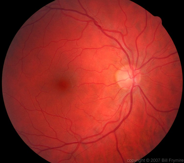
The back of the eyeball's coated by a specialized membrane known as the retina. This dimpled portion of the retina is known as the fovea, and the part of the retina directly in front of where the optic nerve exits the back of the eye is actually known as the blind spot.
What is a fovea simple definition?
: a small depression in the center of the macula that contains only cones and constitutes the area of maximum visual acuity.
What is the difference between retina and fovea?
The retina is the paper-thin tissue that lines the back of the eye and contains the photoreceptor (light sensing) cells (rods and cones) that send visual signals to the brain. The pit or depression within the macula, called the fovea, provides the greatest visual acuity.
What is fovea made of?
The mature human fovea consists of 3 spectral types of cone: red or long wavelength sensitive cones, L-cones; green or medium wavelength cones, M-cones; and blue or short wavelength cones, S-cones.
What happens if fovea is damaged?
When the fovea is compromised by disease or injury, the brain works, subconsciously, to find a position in the retina that it can use to develop a new fixation point — a pseudofovea — in a region of the retina with surviving photoreceptors.
Is fovea a retina?
The shallow depression in the center of the macular region is the fovea, or central fovea of the retina (fovea centralis retinae). This depression is formed because the retinal neurons are displaced, leaving only photoreceptors in the center. The fovea has a horizontal diameter of approximately 1.5 mm.
What is the main function of fovea?
Structure and Function As the fovea is responsible for high-acuity vision it is densely saturated with cone photoreceptors.
What is fovea function?
The fovea is responsible for sharp central vision (also called foveal vision), which is necessary in humans for reading, driving, and any activity where visual detail is of primary importance.
What is the blind spot of the eye called?
scotomaSimilarly, your eyes have a blind spot, called scotoma. The optic nerve carries info from the eyeball to the brain, then, spreads nerve fibers across the back of the eye, or retina. The small round spot where the nerve enters the back of your eye is called the optic disc.
Why is it called fovea?
The name “fovea” comes from the Greek word meaning “small pit.” This is an appropriate name, as the fovea is a tiny depression (or pit) in the macula, a small structure located in the center of the retina, the light-sensitive tissue that lines the back of the eye.
Why is vision best at the fovea?
Because of the layers that are swept away, there is less scattering of light in the fovea, allowing for the visual acuity to be higher in the fovea. It is the foveae of the retinae that give humans our excellent visual acuity. By visual acuity, we mean the clarity of vision.
How thick is the fovea?
The foveal thickness never exceeded 252 μm in any of the healthy eyes. As expected, macular thickness was thinnest at the center, thickest within 3-mm diameter of the center, and diminished toward the periphery of the macula. The temporal quadrant was thinner than the nasal quadrant.
How large is the fovea?
about 1.5 mmThe actual fovea is about 1.5 mm in diameter and the central fovea consists of a foveal pit (umbo) that is a mere 0.15 mm across (Figure 2B). This foveal pit is almost devoid of all layers of the retina beneath the cone photoreceptors.
How many fovea do humans have?
Total Number of Cones in Fovea approximately 200,000.
Can the fovea be repaired?
Conclusion: The authors describe an effective surgical approach for the correction of retinal folds involving the fovea. Prompt treatment as well as gentle surgical manipulation are key points to obtain an improvement in visual acuity.
What would damage to the fovea most strongly affect?
Answer and Explanation: Damage to the fovea of the eye would interfere with the ability to focus an image. The fovea is the only space on the retina that allows light to directly hit the cone cells.
How can I strengthen my retina?
How to Improve the Health of the RetinaHealthy and balanced diet. ... Avoiding unhealthy foods and drinks. ... Drinking plenty of water. ... Regular exercise. ... Wearing sunglass when out in the sun. ... Quitting smoking. ... Wearing eye protection. ... Regular eye check-up.
What is the fovea for kids?
The fovea is a small pit near the center of the macula. The fovea contains a high density of retinal cones. The fovea provides high-acuity vision due to the high acuity, or sharpness, of vision that cones provide.
What is the fovea of the eye quizlet?
The fovea centralis is a small, central pit composed of closely packed cones in the eye. It is located in the center of the macula lutea of the retina. The fovea is responsible for sharp central vision such as reading and driving.
What is fovea biology?
The fovea is a depression in the inner retinal surface, about 1.5 mm wide, the photoreceptor layer of which is entirely cones and which is specialized for maximum visual acuity. Within the fovea is a region of 0.5mm diameter called the foveal avascular zone (an area without any blood vessels).
Why is it called fovea?
The name “fovea” comes from the Greek word meaning “small pit.” This is an appropriate name, as the fovea is a tiny depression (or pit) in the macula, a small structure located in the center of the retina, the light-sensitive tissue that lines the back of the eye.
What is the term for thinning of the macula?
Macular degeneration (both wet and dry forms) — Age-related thinning and abnormal protein growth on the macula (dry) or macular scarring due to abnormal blood vessel growth and leakage (wet).
Why is the fovea anatomy so tricky?
Fovea anatomy can be tricky because the retina and macula are also light-sensitive parts of the eye that create sharp vision. So, where does the fovea come into play, and how is it different from the macula and retina?
What is the condition that affects the retina?
Cytomegalovirus retinitis — Viral infection that affects the retina. Retinitis pigmentosa — Genetic condition that affects how the retina responds to light; commonly seen in individuals with Usher syndrome. Macular telangiectasia — A condition that causes blood vessels around the fovea to dilate and leak.
Why is the fovea important?
Because the fovea is such an essential part of a person’s vision, it’s important to prevent and/or monitor the conditions that may jeopardize its function. Conditions that may affect the fovea include:
What is the fovea centralis?
Fovea centralis. The fovea is a tiny part of the eye’s anatomy that makes a huge difference in our eyesight. Resting inside the macula, the fovea (also called “fovea centralis”) provides our absolute sharpest vision.
What is the term for fluid buildup in the macula?
Macular edema — Fluid buildup in the macula. Retinal detachment — When the retina lifts or tears away from the back of the eye. Retinoblastoma — Cancer of the eye that begins in the retina. Retinal vein occlusion — When a vein or artery in the retina becomes blocked.
What are cone cells responsible for?
Cone cells are responsible for producing color and fine details , while rods provide peripheral vision, movement and shades of grey. Rods are mostly located outside the macula, and the cones are located inside. The fovea eye pit does not have any rods or other neurons, only millions of tightly packed cones.
What is the depression in the very center of the macula called?
The depression in the very center of the macula where eyesight is sharpest. It is also called the fovea centralis. The depression in the very center of the macula where eyesight is sharpest. It is also called the fovea centralis.
What are the problems with the fovea?
fovea-related problems include: Branch retinal vein occlusion. Central retinal vein occlusion. Central serous retinopathy.
How do pigments enhance the acuity of the fovea?
The pigments also enhance the acuity of the fovea by reducing the sensitivity of the fovea to short wavelengths and counteracting the effect of chromatic aberration. This is also accompanied by a lower density of blue cones at the center of the fovea. The maximum density of blue cones occurs in a ring about the fovea.
What is the size of the fovea?
Structure. The fovea is a depression in the inner retinal surface, about 1.5 mm wide, the photoreceptor layer of which is entirely cones and which is specialized for maximum visual acuity. Within the fovea is a region of 0.5mm diameter called the foveal avascular zone (an area without any blood vessels).
Why is the foveal focus dark?
The combined effects of the macular pigment and the distribution of short wavelength cones results in the fovea having a lower sensitivity to blue light (blue light scotoma). Though this is not visible under normal circumstances due to "filling in" of information by the brain, under certain patterns of blue light illumination, a dark spot is visible at the point of focus. Also, if mixture of red and blue light is viewed (by viewing white light through a dichroic filter), the point of foveal focus will have a central red spot surrounded by a few red fringes. This is called the Maxwell's spot after James Clerk Maxwell who discovered it.
What is the fovea?
The fovea is responsible for sharp central vision (also called foveal vision), which is necessary in humans for activities for which visual detail is of primary importance , such as reading and driving. The fovea is surrounded by the parafovea belt and the perifovea outer region.
How far is the Perifovea from the Fovea?
The parafovea extends to a radius of 1.25 mm from the central fovea, and the perifovea is found at a 2.75 mm radius from the fovea centralis. The term fovea comes from the from Latin foves 'pit'.
How many cone cells are there in the fovea?
On average, each square millimeter (mm) of the fovea contains approximately 147,000 cone cells, or 383 cones per millimeter. The average focal length of the eye, i.e. the distance between the lens and fovea, is 17.1 mm. From these values, one can calculate the average angle of view of a single sensor (cone cell), which is approximately 31.46 arc seconds .
Why is the Fovea not sensitive to dim light?
Since the fovea does not have rods, it is not sensitive to dim lighting. Hence, in order to observe dim stars, astronomers use averted vision, looking out of the side of their eyes where the density of rods is greater, and hence dim objects are more easily visible.
Where is the fovea located?
It is located in the center of the posterior portion of the retina on the temporal side of the optic nerve.
Why is the fovea important?
The fovea is repsonsible for sharp central vision that is necessary for humans to complete activities where detail is important , such as reading and driving.
Which layer of the retina spreads apart at the fovea?
The ganglion and bipolar layers of the retina spread apart at the fovea to give light a direct path to the cones for the sharpest vision.
Why is the resolution sharp?
The resolution or sharpness in vision is because of the high concentration of cone cells in the fovea.
What are the blue cones in the fovea?
Blue cones are sparse, even largely missing in the foveal center while occurring at somewhat higher density than elsewhere in the cone mosaic of the foveal slope. Signals from blue cones have different pathways to ganglion cells. The best understood is through an ON-type blue-cone-selecting bipolar cell to a non-midget, small bistratified ganglion cell. An OFF-center blue midget bipolar is known to be present in the fovea and connects to a blue OFF midget ganglion cell. Another OFF blue message is sent to a giant melanopsin ganglion cell that is present in the foveal rim area, but the circuitry driving that is less certain and possibly involves an intermediate amacrine cell. The H2 horizontal cells are thought to be feedback neurons primarily of the blue cone system.
How long does it take for the foveal retina to form?
The foveal retina sections of a human from (a) postnatal 8 days (P8d), through (b) 15 months, to fully formed (d) 13 years. (c) At 15 months the cones are thin, have outer segments and squash together and, except for the central bouquet, send axons radially outwards as the Henle fiber layer.
What is the function of the foveal pit?
The foveal pit is devoid of rod photoreceptors and of secondary and tertiary neurons, allowing light to directly stimulate cones and give us maximal visual acuity. The circuitry underlying the transmission to the brain occurs at the rim of the fovea.
How do S-cones and M-cones differ?
S-cones and M/L-cones differ in the time course of mitotic differentiation and expression of opsins. According to Xiao and Hendickson (25), S-opsin and various synaptic proteins are detectable at fetal week 11, while various synaptic and transduction proteins appear in M/L cone subclasses before their opsin visual pigments are detected at fetal week 13 (26). It is clear that S-cones develop in a different mosaic than M/L-cones. Ahnelt and coworkers (7) have noted that cones likely to be short wavelength sensitive tend to occur in irregular positions in both, foveal and peripheral areas. Figure 21A shows an opsin labeled S-cone (asterisk) positioned between seemingly linear series of unlabeled M/L-cone inner segments. Thus in the foveal all-cone mosaic, S-cones appear to interrupt the linear beads of L/M cone-cell inner segments and clearly do not belong to the mosaic of M- and L-cones (6).
How big is the fovea?
As we have illustrated in Figure 2B, the whole fovea is roughly 1.5 mm across and so any cell found within 750 µm of the foveal center is considered a foveal associated cell. It has been hard to get good staining of horizontal cells (HC) of the fovea but some Golgi impregnated human retinas in our possession did allow us to see a few within the 750 µm of eccentricity around the central foveal pit (Figure 22) (28).
What pathways are in the fovea?
So far, we have dealt with the architecture of the predominant cone spectral and high acuity pathways in the fovea namely the midget pathways for the green M-cones and red L-cones. What of the blue S-cone pathways? As we have seen above the S-cones have a small presence in the foveal cone mosaic (3% of cones generally) but reach their peak number for the whole retina at 12% of cones in foveal slope and rim at approximately 1 degree of eccentricity (Figure 13, Figure 14, Figure 15).
What is the central point of the visual field ahead of us?
The central point of the visual field ahead of us is the image falling on the fovea in the human retina. This is the area of our visually sensitive retina where the cone photoreceptors are tightly packed, where rod photoreceptors are excluded and where all intervening layers of the retina are pushed aside concentrically to allow light to reach the densely packed sensory cones with minimum scatter from overlying tissues. The fovea is where focusing on fine detail in the image is perfected, allowing us to read, discriminate colors well and sense three-dimensional depth.
How much retina is in the fovea?
Each nerve fiber coming from the fovea represents 2 square micrometers of retina; each nerve fiber coming from the peripheral field represents, on average, about 1 square millimeter of retina—half a million times as much retinal area. Thus you can see why your peripheral vision is low-res and your foveal vision is high-res. It’s comparable to comparing a low-res and a high-res image on a computer screen where the pixels of the low-res image are 500,000 times as big as the pixels of the high-res image.
How many receptor cells are in each nerve fiber?
Moving slightly away from the fovea, the receptor cells become larger and more widely spaced, and 600 or more receptor cells converge on each outgoing nerve fiber, so each nerve fiber carrying a sig
Which part of the retina has the largest concentration of cone shaped nerve endings that convert light to electrical impulses sent?
The fovea is the part of the retina where there is the largest concentration of cone shaped nerve endings that convert light to electrical impulses sent to the brain. A good analogy is that the fovea has more mega pixels (nerves) than in the rest of the retina. When a person looks directly at an object or a person’s face, that image falls on the fovea .
Which part of the eye is the sharpest?
The fovea is that part of the eye where we have our sharpest vision. It is aligned with the visual axis of the lens of the eye, so when we look directly at something, its image falls on the fovea. Here, the retinal receptor cells are very tiny, very closely spaced, and each one has its own nerve fiber (“private line”) to the brain. This gives us high-resolution, or fine-grained, vision in the fovea .
What is the membrane that covers the sclera?
The conjunctiva is the membrane covering the sclera (white portion of your eye). The conjunctiva also covers the interior of your eyelids.
What is the white part of the eye?
The sclera is sometimes known as the "whites" of the eye. It covers more than 80% of the eyeball's surface. 2
What is the episclera?
The episclera is a thin layer of tissue that lies on top of the sclera. The episclera has tiny blood vessels that supply the sclera with nutrients.
How many fibers are in the optic nerve?
The optic nerve is a bundle of about 1.2 million nerve fibers that transmit visual information to the central nervous system (brain). 7
How do eyes work?
The eyes work in the same way as cameras. When you focus on an object, light is reflected and enters the eye through the cornea. As the light passes through, the dome-shaped nature of the cornea bends light , enabling the eye to focus on fine details.
Where are light rays focused?
Light rays are focused on the macula lutea when an eye is looking directly at an object.
How many muscles are there in the eye?
The eye has six muscles. These muscles arise from the eye socket (orbit) and work to move the eye up and down, side to side, or in a circular motion.
Overview
Structure
The fovea is a depression in the inner retinal surface, about 1.5 mm wide, the photoreceptor layer of which is entirely cones and which is specialized for maximum visual acuity. Within the fovea is a region of 0.5mm diameter called the foveal avascular zone (an area without any blood vessels). This allows the light to be sensed without any dispersion or loss. This anatomy is responsible for the depression in the center of the fovea. The foveal pit is surrounded by the foveal rim that cont…
Function
In the primate fovea (including humans) the ratios of ganglion cells to photoreceptors is about 2.5; almost every ganglion cell receives data from a single cone, and each cone feeds onto between one and 3 ganglion cells. Therefore, the acuity of foveal vision is limited only by the density of the cone mosaic, and the fovea is the area of the eye with the highest sensitivity to fine details. Cones in the central fovea express opsins that are sensitive to green and red light. These cones are the …
Other animals
The fovea is also a pit in the surface of the retinas of many types of fish, reptiles, and birds. Among mammals, it is found only in simian primates. The retinal fovea takes slightly different forms in different types of animals. For example, in primates, cone photoreceptors line the base of the foveal pit, the cells that elsewhere in the retina form more superficial layers having been displaced away from the foveal region during late fetal and early postnatal life. Other foveae may show only …
Additional images
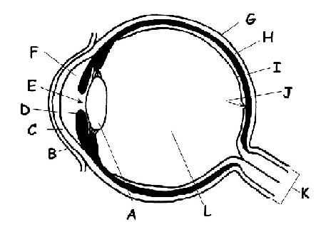
• Illustration showing main structures of the eye including the fovea
• Structures of the eye labeled
• This image shows another labeled view of the structures of the eye
• Schematic diagram of the macula lutea of the retina, showing perifovea, parafovea, fovea, and clinical macula
See also
• Eye movement
• Gaze-contingency paradigm
• Macular degeneration
• Foveated imaging