
See more

Is Haemophilus Parasuis a bacteria?
The Gram-negative bacterium Haemophilus parasuis is the causative agent of Glässer's disease in pigs and is one of the most important pathogen in the modern swine industry. The disease is characterized by pneumonia, meningitis, arthritis, polyserositis, and septicemia.
What is Haemophilus parasuis in pigs?
Haemophilus parasuis is an early colonizer of the porcine upper respiratory tract and is the etiological agent of Glasser's disease. The factors responsible for H. parasuis colonization and systemic infection are not yet well understood, while prevention and control of Glasser's disease continues to be challenging.
What causes Glasser's disease?
Glässer disease is caused by infection with Glaesserella (Haemophilus) parasuis. The most common form is characterized by fibrinous polyserositis and polyarthritis, but septicemia with sudden death and bronchopneumonia also can occur. Diagnosis is based on clinical signs and bacterial isolation or PCR.
What causes Glassers disease in pigs?
Glasser's disease is caused by a bacterium called Haemophilus parasuis. Acute outbreaks can cause a high mortality, while it is also an important cause of pleurisy found in slaughter pigs. It mainly affects 8-10 week old weaners, although infection of a naive herd can cause clinical signs in any age including adults.
What disease does Haemophilus cause?
Haemophilus influenzae type b (Hib) was once the most common cause of bacterial infection in children. Hib causes a variety of diseases including meningitis (inflammation of the coverings of the spinal column and brain), bacteremia (infection of the blood), and pneumonia (infection of the lungs).
How contagious is Haemophilus?
Carriers of Hib are infectious as long as organisms are present in the nasopharynx, which may be for a prolonged period even without nasal discharge. Transmission from person to person occurs through respiratory droplets, but infection may also be acquired through contact with infected respiratory discharges.
Why do pigs ears turn purple?
Classical swine fever (CSF) is a highly contagious and often fatal viral disease of swine. Infected pigs develop fever, hemorrhages, lethargy, yellowish diarrhea, vomiting, and a purple skin discoloration of the ears, lower abdomen, and legs.
How do you treat greasy pig disease?
Treatment. Determine the antibiotic sensitivity and inject affected piglets daily for five days, or on alternate days with a long-acting antibiotic to which the organism is sensitive to. Antibiotics include: amoxycillin, OTC, ceftiofur, cephalexin, gentamycin, lincomycin or penicillin.
Why has Glasser's disease increased in both incidence and severity in recent years?
Glässer disease is seen worldwide, and its incidence appears to have increased since the emergence of porcine reproductive and respiratory syndrome Porcine Reproductive and Respiratory Syndrome .
Can humans get PRRS from pigs?
No! With the exception of mallard ducks, PRRS virus only infects swine. It poses no threat to humans or other animals and in no way makes eating pork a threat to human health.
What causes blue eye in swine?
Porcine rubulavirus (PoRV) is an enveloped RNA virus in the family Paramyxoviridae, genus Rubulavirus. PoRV emerged in the 1980s, and its distribution appears to be limited to Mexico. PoRV causes a disease commonly referred to as “blue eye disease” because of the corneal opacity seen in some affected pigs.
How do you prevent meningitis in pigs?
Management control and prevention It is possible to vaccinate the sow using an autogenous vaccine to improve the colostral immunity. The sow can be injected with long-acting penicillin just before farrowing. The litter can be injected with long-acting penicillin in anticipation of disease.
What are the symptoms of Haemophilus?
Symptoms of bloodstream infection usually include:Fever and chills.Excessive tiredness.Pain in the belly.Nausea with or without vomiting.Diarrhea.Anxiety.Shortness of breath or difficulty breathing.Altered mental status (confusion)
What are the symptoms of circovirus in pigs?
Pigs affected may experience increased mortality, poor growth, and weight loss, progressing to the level of severe thinning, and weakness between 5 to 14 weeks of age. Also pigs can develop enlarged lymph nodes, skin rashes, difficulty breathing, jaundice, fever, stomach ulcers, diarrhea, or sudden death may occur.
What are the symptoms of PRRS in pigs?
Breeding age gilts, sows, and boars: Clinical signs may include a period of anorexia, fever, lethargy, depression, and perhaps respiratory distress or vomiting. Mild cyanosis of the ears, abdomen and vulva has been reported in some outbreaks.
Is Haemophilus serious?
Haemophilus influenzae disease is a name for any illness caused by bacteria called H. influenzae. Some of these illnesses, like ear infections, are mild while others, like bloodstream infections, are very serious.
What causes pigs to have atrophic rhinitis?
A common worldwide disease of pigs, atrophic rhinitis (progressive atrophic rhinitis) is characterized by inflammation and atrophy of nasal conchae (turbinates). In severe cases, atrophy of the conchae may cause a striking facial deformity in growing pigs because of deviation of the nasal septum and nasal bones. The etiopathogenesis of atrophic rhinitis is complex and has been a matter of controversy for many years. Pathogens historically associated with atrophic rhinitis include Bordetella bronchiseptica, Pasteurella multocida, Haemophilus parasuis, and viral infections such as porcine cytomegalovirus (inclusion body rhinitis). In addition, predisposing factors have included genetic makeup, environment, and nutritional deficiencies. The cause of atrophic rhinitis is currently believed to be a combined infection by specific strains of Bordetella bronchiseptica producing dermonecrotic toxin and toxigenic strains of Pasteurella multocida. The only lesion associated with infection with Bordetella bronchiseptica alone is a mild to moderate turbinate atrophy (nonprogressive atrophic rhinitis), but this bacterium actively promotes the colonization of the nasal cavity by Pasteurella multocida. The toxigenic strains of Pasteurella multocida produce potent cytotoxins that inhibit osteoblastic activity and promote osteoclastic reabsorption in nasal bones, particularly in the ventral nasal conchae, where abnormal bone remodeling results in progressive atrophy of conchae.
What is tildipirosin antimicrobial?
Tildipirosin is an antibacterial of the macrolide class. It is a 16-membered ring (tilmicosin also is a 16-member ring molecule) macrolide antimicrobial with three charged nitrogen atoms (tulathromycin also has 3 charged nitrogen atoms). Like other macrolides, it inhibits bacterial protein synthesis by binding to the ribosomal 50S subunit, specifically, the 23S rRNA within the 50S subunit. After binding, tildipirosin interacts with ribosomal RNA (rRNA) and ribosomal proteins adjacent to the peptidyl-transferase enzyme. Thus like other macrolides, it inhibits protein synthesis by blocking the prolongation and release of developing polypeptides. Tildipirosin has a spectrum of activity that is limited to gram-positive bacteria and some gram-negative bacteria that cause respiratory diseases in cattle and pigs (e.g., Mannheimia haemolytica, Mycoplasma, Pasteurella multocida, Actinobacillus pleuropneumoniae, Bordetella bronchiseptica, and Haemophilus parasuis). Escherichia coli and Pseudomonas aeruginosa are resistant. Some Staphylococcus spp. and Streptococcus spp. may be susceptible.
What are the signs of neurological disease in pigs?
Common CNS signs in pigs include behavioral abnormalities (most commonly stupor), ataxia, loss of righting, seizures or seizure-like activity (paddling), nystagmus, and blindness. Musculoskeletal disorders may clinically confuse or complicate perceived PNS signs and must be differentiated from each other.
What are the growth factors of Haemophilus?
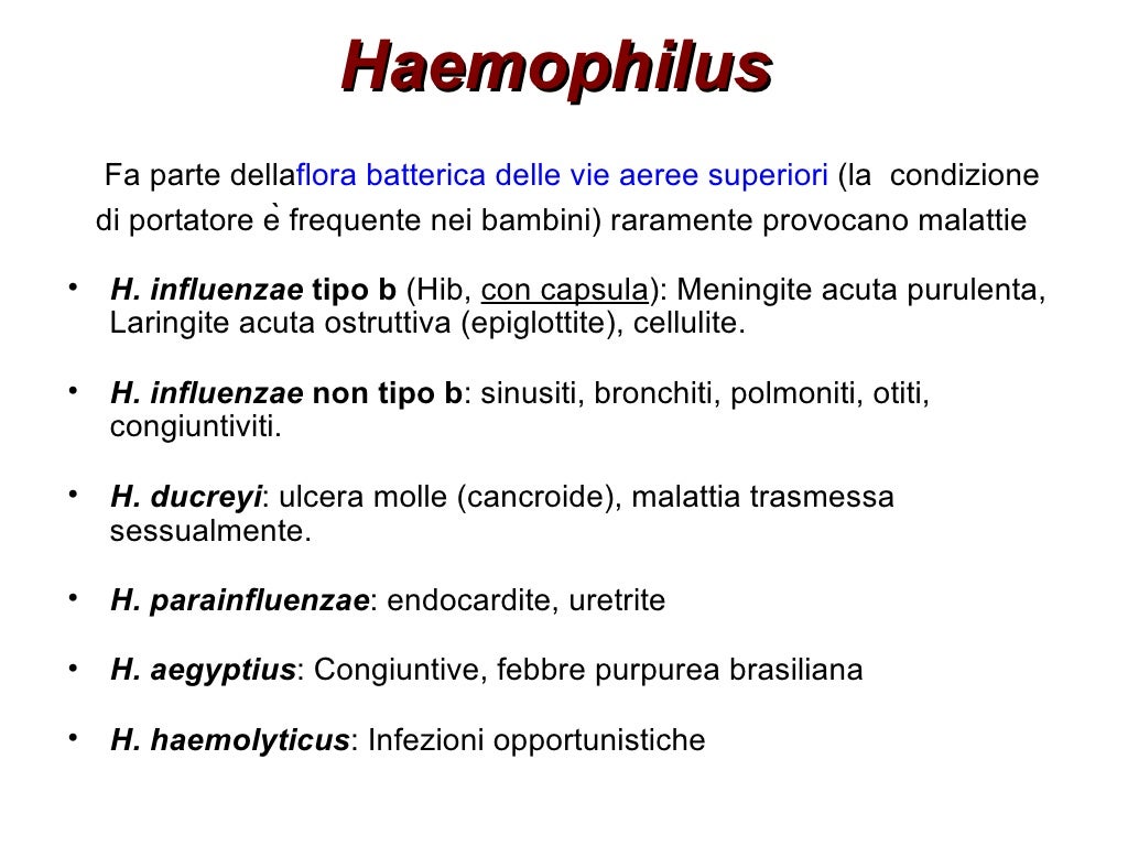
The genus Haemophilus consists of small, nonmotile, facultatively anaerobic, and gram-negative pleomorphic rods or coccobacilli , which require one or both of the two defined growth factors commonly supplied as hemin and nicotinamide adenine dinucleotide . Any organism corresponding to that description is assigned to this genus. Organisms corresponding to the traditional description of Haemophilus occur in many species, most commonly as apparent commensals on mucous membranes—especially of the upper respiratory and lower genital tracts. Some have a potential for pathogenicity but few are consistently pathogenic. Haemophilus parasuis, Haemophilus gallinarum, Haemophilus haemoglobinophilus, Haemophilus paracuniculus, and Haemophilus aphrophilus have been encountered in animals. Haemophilus parasuis is a common V factor-requiring commensal of the porcine upper respiratory tract. Haemophilus gallinarum is the agent of infectious coryza, which is an upper respiratory infection of chickens. Haemophilus haemoglobinophilus—also known as Haemophilus canis—is a fairly common commensal parasite of the lower canine genital tract, particularly of males. Taylorella equigenitalis causes an acute, self-limiting, suppurative metritis in mares that leaves no permanent effects on the breeding efficiency of recovered animals. The disease is sexually transmitted, but no clinical signs develop in stallions. Both sexes remain asymptomatic carriers for extended periods of time. The chapter also illustrates the pathogenicity, isolation, identification, and cultural characteristics of Haemophilus and Taylorella.
What is ceftiofur used for?
Ceftiofur crystalline-free acid is indicated for treatment of swine respiratory disease (SRD) caused by A. pleuropneumoniae, P. multocida, S. choleraesuis, Haemophilus parasuis, and Streptococcus suis. In cattle, it is used for treatment of BRD caused by Mannheimia haemolytica, P. multocida, and Histophilus somni (formerly Haemophilus somnus ). It also can be administered to control respiratory disease in cattle at high risk of developing BRD (metaphylaxis) associated with M. haemolytica, P. multocida, and H. somnus. This formulation also is approved for treating foot rot in cattle (interdigital necrobacillosis) caused by Fusobacterium necrophorum, Porphyromonas levii, and Bacteriodes melaninogenicus. It is approved for treatment of acute metritis in dairy cattle via a two-dose regimen. It is approved for treatment and control of swine respiratory disease (SRD) associated with Actinobacillus pleuropneumoniae, Pasteurella multocida, Haemophilus parasuis, and Streptococcus suis. This formulation also is approved for use in horses for treatment of respiratory tract infections caused by susceptible Streptococcus equi (S. zooepidemicus) after administration of two injections. Ceftiofur hydrochloride and ceftiofur crystalline-free acid have also been administered extralabel intramammary to dairy cattle. However, there are specific products designed for intramammary use (Spectramast).
How long after exposure to H. parasuis can you see symptoms?
In conventional herds where H. parasuis is enzootic, the clinical signs will be mild with low morbidity. In susceptible herds, the clinical signs occur within a week after exposure and consist of some or all of the following: pyrexia 40–41.7°C, anorexia, coughing, depression, swollen joints with lameness, neurological signs, dyspnea, and sudden death. A markedly increased WBC and decreased packed cell volume (PCV) have been reported in experimentally infected SPF piglets ( Wiegand et al., 1997 ). Long-term sequelae include abortion and chronic arthritis. An oligonucleotide-specific capture plate hybridization assay has been developed and is reported to be specific and more sensitive than culturing for H. parasuis from lesions and nasal swabs ( Calsamiglia et al., 1999 ). Differentials include Mycoplasma hyorhinis and other bacterial septicemic conditions that affect swine including Erysipelothrix rhusiopathiae, Salmonella choleraesuis, and Streptococcus suis.
What are the most common causes of abscesses in animals?
Streptococci and staphylococci are the most common causes of abscesses involving the skin and subcutis of most animal species. Actinomyces bovis and Actinobacillus lignièresii occur rarely as causes of abscesses in species other than the bovine. Likewise Nocardia asteroides is an infrequent cause of abscesses in domestic animals other than the dog and cat. Pseudomonas aeruginosa may be associated with abscesses in all of the domestic animals.
What is the epidemiology of H. parasuis?
Another interesting feature of the epidemiology of H. parasuis is the fact that no matter how many sources are commingled in the nursery, only a few prevalent strains (usually 2-3) will predominate in the herd [1]. Several within herd studies using genotyping by ERIC-PCR have confirmed this fact [1,4,13]. Another interesting feature of the epidemiology of H. parasuis between herds is the fact that although many herds are affected by the same serovar group, each herd has a different prevalent strain. This was observed in herds affected by strains from serovars 2, 4, and 12 [1].
How long after weaning do pigs get Streptococcus suis?
The epidemiology of Streptococcus suis infection is very similar to H. parasuis, which may explain why these two agents often affect nursery pigs at 4 to 6 weeks after weaning [4]. PRRSV infection, however, has a very distinct role in H. parasuis infection.
What causes pigs to die in the nursery?
Haemophilus parasuis is still one of the main causes of nursery mortality in most U.S. herds [1]. Mortality rates due to Haemophilus parasuis can be as high as 10% [2], which makes this agent one of the most costly pathogens in swine production. Although a few herds experience nursery mortality solely due to H. parasuis, disease caused by this agent may occur at the same time as other bacterial and viral infections. Streptococcus suis and Porcine Reproductive and Respiratory Syndrome Virus (PRRSV) are two of the agents that are frequently isolated from pigs showing polyserositis due to H. parasuis [3]. The epidemiology of Streptococcus suis infection is very similar to H. parasuis, which may explain why these two agents often affect nursery pigs at 4 to 6 weeks after weaning [4]. PRRSV infection, however, has a very distinct role in H. parasuis infection. A recent study suggests that pigs colonized by a virulent H. parasuis strain are susceptible to development of systemic infection by this agent following PRRSV challenge [5]. Although the interaction between PRRSV and H. parasuis is now evident both experimentally and in the field, the mechanisms involved in such interaction are still unclear.
How long does it take for pigs to colonize?
Colonization of piglets by H. parasuis occurs a few days after birth, possible through nose to nose contact with sows and gilts. Gilts appear to shed fewer organisms than sows [11]. If this is true, gilt piglets are at higher risk of developing systemic infection by virulent H. parasuis strains after weaning. Another factor that appears to influence colonization of piglets by H. parasuis is weaning age. Pigs weaned at 14 days tend to have lower levels of colonization compared with piglets weaned at 28 days [11]. Piglets that were colonized in the farrowing house in the presence of maternal immunity may develop active immunity against virulent strains and be protected against systemic infection after weaning and commingling. Piglets that were not colonized prior to weaning are naive animals that may develop systemic infection when commingled with colonized pigs. Systemic infection usually happens around 4 to 6 weeks after weaning, when maternal immunity is no longer protective. This hypothesis drives today’s control strategies to reduce nursery mortality due to H. parasuis infection, as it will be discussed in the control section [12].
What are the clinical signs of H. parasuis?
The clinical signs characteristic of H. parasuis infection are certainly not unique to this agent, and a differential diagnosis with other pathogens such as S. suis is necessary. The clinical presentation of H. parasuis systemic infection may vary with the virulence of the strain and the immune status of the pig. Three main presentations may be observed in the field: super-acute, acute, and chronic. Super-acute infections are characterized by sudden death with lack of clinical signs and gross lesions in most cases. Some animals may show a slight increase of fluids in the pericardial sac, pleural, and abdominal cavities. Fibrin may or may not be observed. In these cases, H. parasuis may be isolated from the blood, which confirms septicemia. Super-acute infections usually occur in naive herds (H. parasuis-free) or due to infection by a highly virulent strain. Acute infections are more commonly observed in the field and are characterized by fibrinous polyserositis (Figure 1). Clinical signs will usually appear 2-3 days following infection. Affected pigs may present fever (106 – 107 º F), labored breathing (abdominal breathing), coughing, swollen joints, and central nervous system (CNS) signs, which are characterized by lateral decubitus, paddling, and trembling. When samples are properly collected, H. parasuis can be easily isolated from these animals. Acute infection may also be characterized by development of either pneumonia or CNS signs. These clinical presentations may be associated with specific groups of strains. Some strains seem to have a tropism to the brain, causing only CNS signs. Other strains may be found in lungs causing pneumonia without systemic infection. Further studies are necessary to clarify the association between specific strains and clinical presentations in the field. Chronically affected animals (6-7 weeks after weaning) are survivals of the peak of nursery mortality. These animals probably received antibiotic treatments and were able to survive systemic infection. In most chronically affected animals, H. parasuis can no longer be isolated from lesions. These animals usually present poor growth performance throughout late nursery and early finisher and may die from complications of fibrosis in the thoracic cavity.3, 6
How to isolate H. parasuis?
Successful isolation of H. parasuis from clinical samples can be achieved by sampling acutely affected, non-treated animals. Isolation is easier when clinically affected animals are euthanized and fresh samples are submitted to a diagnostic laboratory as soon as possible. Sample collection may be performed using a sterile swab containing Stuart media. It is very important to collect the fibrin on the surface of affected organs, as H. parasuis will be mostly concentrated in this material. Tissues may also be collected for isolation, and should be submitted in separate sterile bags to the laboratory. It is known that different strains can affect one pig at the same time. Special care should be taken to separate brain tissue or swabs from other samples, as some H. parasuis strains seem to have a tropism to the brain [3]. When isolation is negative, polymerase chain reaction (PCR) may be used to detect the presence of H. parasuis in tissues and swabs [8]. PCR is useful to define the role of H. parasuis in nursery mortality, but isolation is still necessary for further characterization of strains by serotyping and genotyping. A new technique, more sensitive and specific, has been recently developed to serotype H. parasuis. Indirect hemaglutination (IHA) is now the technique of choice for H. parasuis serotyping. It apparently reduces the percentage of non-typable isolates compared with traditional serotyping by agar gel precipitation test (AGPT) [9]. Genotyping by Enterobacterial Repetitive Intergenic Consensus-based PCR or ERIC-PCR has been extensively used to characterize and compare H. parasuis field strains [1,10]. This technique is more discriminatory than serotyping and can be used to detect prevalent strains in affected herds (Figure 2). Both techniques (serotyping by IHA and genotyping by ERICPCR) are important for the development of control strategies.
How long does it take for pigs to die from H. parasuis?
The majority of swine herds experience the peak of mortality due to H. parasuis infection at 4 to 6 weeks after weaning. This period corresponds to the decrease of maternal immunity. In these cases, piglet vaccination at weaning and 2 weeks later may control mortality [1,3,6]..
What is a gram negative bacteria?
A genus of aerobic to facultatively anaerobic, nonmotile, parasitic bacteria containing minute, gram-negative, rod-shaped cells; occur in various lesions and secretions, as well as in normal respiratory tracts, of vertebrates.
What is the genus of small Gram-NEGATIVE rod-shaped micro-organisms?
A genus of small GRAM NEGATIVE rod-shaped micro-organisms that includes H. influenzae which can cause MENINGITIS, H. haemolyticus which is often found in the throat, and the causative organism of CHANCROID, H. ducreyi .
What is the genus of bacteria?
A genus of aerobic to facultatively anaerobic, nonmotile bacteria (family Brucellaceae) containing minute, gram-negative, rod-shaped cells that sometimes form threads and are pleomorphic. These organisms are strictly parasitic, growing best, or only, on media containing blood. They may or may not be pathogenic. They occur in various lesions and secretions, as well as in normal respiratory tracts, of vertebrates. The type species is H. influenzae.
What is the genus of hemophilic gram negative bacteria?
Haemophilus. a genus of hemophilic gram-negative bacteria. H. aphro´philus, H. parainfluen´zae, and H. paraphro´-philus are part of the normal oral flora and are occasionally associated with endocarditis.
Why do piglets have antibodies?
Since the colonization of Glaesserella parasuis occurs while the piglets are still receiving colostrum, they also receive maternal antibodies to protect against disease. Poor management practices that disrupt the balance between antibody protection and bacterial load cause the development of clinical signs.
What is Glaesserella parasuis?
Glaesserella parasuis is one of the main pathogens of importance in nursery pigs, causing polyserositis and lameness. The disease is ubiquitous and affects every country with pork production.
Why do pigs die?
Pigs dying due to the peracute form of this disease often do not display any lesions due to sudden onset and subsequent death. Pigs that have been acutely affected will display lesions on the brain, heart, and joints. Polyserositis with meningitis, pericarditis, pleurisy, peritonitis and arthritis.
How long do piglets stay infected?
Piglets are infected very early in life. They come in contact with the bacteria when nursing the sow for colostrum. Research has proven that removing piglets before they can nurse keeps them naive of Glaesserella parasuis. Healthy carriers can shed the bacteria in their nasal discharge for up to 6 months, mixing pigs of different provenance should be done carefully.
What is the condition of fibrin in the thoracic cavity?
The large quantity of fibrin present in the thoracic and abdominal cavities is typical of Glassër’s disease. Severe and large adherences between abdominal organs or between lungs and pleura make the necropsy more difficult. Arthritis accompanied by green to grey exudate is often found in the joints.
Is Glaesserella parasuis Gram negative?
Glaesserella parasuis is a Gram negative bacillus that is commensal in the upper respiratory system. 15 serotypes of Glaesserella parasuis have been described, with varying degrees of virulence. Multiple strains can infect one single herd. Piglets are infected very early in life. They come in contact with the bacteria when nursing ...
Why are animals condemned to slaughter?
They will usually be condemned at slaughter due to polyserositis.
Swine Software
MTech Systems USA, Inc. introduces SMTS, the Swine Management Tools Series, bringing the leading computer software advantages that have benefitted poultry producers of the world to the swine industry.
Waterproof Jackets
Udder Tech has created waterproof jackets with the thumb hole sleeve sewn right in. The sleeves extend past the wrist and have a loop to slide the thumb through. When worn with nitrile gloves, the jacket eliminates the problem of water and debris getting into the sleeve, protecting wrists that may be sensitive to moisture and manure.
Utility Tractor
Massey Ferguson announces the expansion of the company's 2600 Series utility tractor line to include a new turbocharged model — the Massey Ferguson 2635 with 74 gross engine horsepower.
What is the genus of Haemophilus influenzae?
Haemophilus influenzae on a Chocolate agar plate. Haemophilus is a genus of Gram-negative, pleomorphic, coccobacilli bacteria belonging to the family Pasteurellaceae.
What factors do hemophilus not need to grow?
Members of the genus Haemophilus will not grow on blood agar plates, as all species require at least one of these blood factors for growth: hemin (X-factor) and/or nicotinamide adenine dinucleotide (V-factor). They are unable to synthesize important parts of the cytochrome system needed for respiration, and they obtain these substances from the heme fraction, known as the X factor, of blood hemoglobin. The culture medium must also supply the cofactor nicotinamide adenine dinucleotide (from either NAD+ or NADP+), which is known as the V factor. Clinical laboratories use tests for the requirement of the X and V factors to identify the isolates as Haemophilus species.
Is H. paraphrohaemolyticus a Gram-negative bacteria?
H. paraphrohaemolyticus. H. pittmaniae. H. piscium. H. segnis. H. sputorum. Haemophilus is a genus of Gram-negative, pleomorphic, coccobacilli bacteria belonging to the family Pasteurellaceae. While Haemophilus bacteria are typically small coccobacilli, they are categorized as pleomorphic bacteria because of the wide range ...
Is chocolate agar good for hemophilus?
Clinical laboratories use tests for the requirement of the X and V factors to identify the isolates as Haemophilus species. Chocolate agar is an excellent Haemophilus growth medium , as it allows for increased accessibility to these factors.
Introduction

Objectives
- Discuss features regarding diagnosis and epidemiology of Haemophilus parasuis.
- Describe methods of control of Haemophilus parasuis in the nursery.
Etiological Agent
- Haemophilus parasuis is the etiological agent (cause) of the syndrome currently known as “Glässer’s disease”. This organism is an early colonizer of the upper respiratory tract and may be normally isolated from the nasal cavity, tonsil, and trachea of healthy pigs. Although non-pathogenic strains predominate in the upper respiratory tract , some animals may harbor virulen…
Clinical Signs and Lesions
- The clinical signs characteristic of H. parasuis infection are certainly not unique to this agent, and a differential diagnosis with other pathogens such as S. suis is necessary. The clinical presentation of H. parasuis systemic infection may vary with the virulence of the strain and the immune status of the pig. Three main presentations may be observed in the field: super-acute, a…
Diagnosis and Strain Characterization
- The diagnosis of H. parasuis systemic infection is based on the association between clinical history and isolation of the agent from characteristic lesions. Successful isolation of H. parasuis from clinical samples can be achieved by sampling acutely affected, non-treated animals. Isolation is easier when clinically affected animals are euthanized and fresh samples are submit…
Epidemiology
- Colonization of piglets by H. parasuis occurs a few days after birth, possible through nose to nose contact with sows and gilts. Gilts appear to shed fewer organisms than sows . If this is true, gilt piglets are at higher risk of developing systemic infection by virulent H. parasuis strains after weaning. Another factor that appears to influence colonization of piglets by H. parasuis is weani…
Co-Infection with Other Pathogens
- As mentioned before, S. suis has a very similar within-herd epidemiology compared with H. parasuis. Both organisms are early colonizers of the respiratory tract and the hypothesis for colonization dynamics for S. suis appear to be similar to that described for H. parasuis . Disease in the nursery caused by S. suis seems to be also associated with the commingling of colonized …
Control
- The correct diagnosis of H. parasuis systemic infection in nursery and the evaluation of the withinherd epidemiology is critical for control of nursery mortality caused by this agent. Sample collection from clinically affected, non-treated, euthanized pigs is very important. If isolation is not accomplished, PCR can help to define if H. parasuis is involved in nursery mortality. When isolati…
Summary
- Correct diagnosis of H. parasuis systemic infection in the nursery is key for control of this costly pathogen. It is very important to consider the differential diagnosis with S. suis infections and to evaluate the role of PRRSV concurrent infections in the nursery. Detection of H. parasuis in clinical samples may be performed by PCR, although isolation is still recommended for further characte…
References
- 1. Oliveira, S., Blackall, PJ, Pijoan, C. Characterization of the diversity of Haemophilus parasuis field isolates by serotyping and genotyping. Am J Vet Res 2003;64(4):435-442. 2. Oliveira, S, Pijoan, C., Morrison, R. Comparison of Haemophilus parasuis control in the nursery using vaccination and controlled exposure. Journal of Swine Health and Production. (Accepted). 2004…