What is Hyperchromic and Hypochromic?
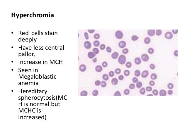
Erythrocytes containing the normal amount of hemoglobin (normal MCHC) are called normochromic. When the MCHC is abnormally low they are called hypochromic, and when the MCHC is abnormally high, hyperchromic. The terms above are used together to describe different forms of anemia.
What is Hyperchromic RBC?
Hyperchromic means that the red blood cells have more hemoglobin than normal. High levels of hemoglobin in your red blood cells makes them a deeper hue of red than normal. Congenital spherocytic anemia: Hyperchromic microcytic anemias are rare.
What is the meaning of Hypochromic?
Hypochromia means that the red blood cells have less color than normal when examined under a microscope. This usually occurs when there is not enough of the pigment that carries oxygen (hemoglobin) in the red blood cells.
What is Hyperchromic effect in chemistry?
The hyperchromic effect is the striking increase in absorbance of DNA upon denaturation. The two strands of DNA are bound together mainly by the stacking interactions, hydrogen bonds and hydrophobic effect between the complementary bases.
Why do you get Hyperchromic anemia?
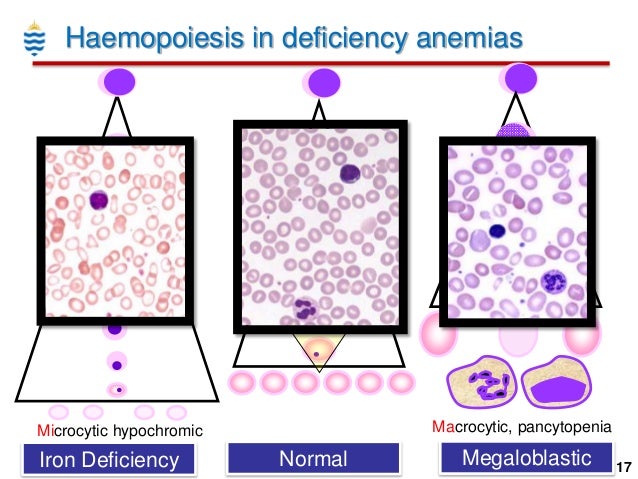
Macrocytic or magaloblastic anemia is caused by disturbances of DNA synthesis. It occurs, for example, in both folic acid and vitamin B12 deficiencies.
What happens if hypochromia is high?
Hypochromic microcytic anemia with iron overload can lead to pale skin (pallor), tiredness (fatigue), and slow growth. In hypochromic microcytic anemia with iron overload, the iron that is not used by red blood cells accumulates in the liver, which can impair its function over time.
How is hypochromia diagnosed?
Diagnosis of microcytic hypochromia is done by measuring certain characteristics changes in the count of blood cell and related indices. Complete blood count test (CBC) is the common process for measuring these characteristic changes.
What are the causes of Hypochromasia?
There are several causes of Hypochromasia but the most common are listed below:Iron Deficiency in the body.Poisoning especially lead poisoning.Excessive blood loss maybe in case of an injury or Leukemia.Deficiency of Vitamin B6.Ulcers.Piles.Gastric bleeding.Hermoloids and Gene Mutations.
How is Hypochromic anemia diagnosed?
Diagnosis and Tests Complete blood count (CBC): This test gives providers information about your hemoglobin levels and aspects of your blood. Peripheral blood smear: Providers use this test to examine your blood cells.
How is the hyperchromic effect measured?
2) can be calculated by using an absorbance coefficient of 15.5 for a 1% solution. At the end of the analysis, the increase in absorbance (as percentage of maximum increase) at 260 nm is plotted as a function of temperature.
What is hyperchromic shift in UV spectroscopy?
HYPSOCHROMIC SHIFT. The shift of absorption to a shorter wavelength due to substitution or solvent effect (a blue shift).
What is DNA hyperchromic shift?
The phenomenon of UV absorbance increasing as DNA is denatured is known as the hyperchromic shift. The purine and pyrimidine bases in DNA strongly absorb ultraviolet light. Double-stranded DNA absorbs less strongly than denatured DNA due to the stacking interactions between the bases.
Does Hyperchromic anemia exist?
hyperchromic anemia Anemia in which mean corpuscular hemoglobin concentration is higher than normal. The red blood cells are darker staining than normal.
What level of RBC is concerning?
A high red blood cell count is generally considered to be anything above 6.1 million red blood cells for males, 5.4 million for females, and 5.5 for children. Additional tests will help your healthcare provider determine the cause of your high red blood cell count and next steps in your care.
Is Hyperchromic a thing?
Hyperchromia means increase in color. The only cells that are truly hyperchromic are spherocytes. Spherocytes are the only cells that contain more hemoglobin than normal in relation to the cell volume.
What is hypochromic Microcytic Anaemia?
Microcytic, hypochromic anemia, as the name suggests, is the type of anemia in which the circulating RBCs are smaller than the usual size of RBCs (microcytic) and have decreased red color (hypochromic).
Why does hyperchromic anemia happen?
Although other rare causes exist (Table 26), almost all patients with hyperchromic anemia suffer from vitamin B12 and/or folic acid deficiency. Since a deficiency of these essential metabolic building blocks suppresses DNA synthesis not only in erythropoiesis, but in the other cell series as well, over time more or less severe pancytopenia will develop.
Which is the first item in the differential diagnosis of hyperchromic anemia?
In older patients, myelodysplastic syndrome should be the first item in the differential diagnosis of hyperchromic anemias a
What does it mean when you have large erythrocytes?
Conspicuous large erythrocytes suggest hyperchromic macrocytic anemia, usually megaloblastic in the bone marrow a c
Why is pernicious anemia less common than megaloblastic anemia?
Table 26 lists possible causes. It should be emphasized that "genuine" pernicious anemia is less common than megaloblastic anemia due to vitamin B12 deficiency. In pernicious anemia a stomach biopsy shows atrophic gastritis and usually also serum antibodies to parietal cells and intrinsic factor.
What are the anomalies of granylocytic series?
In the granylocytic series, anomalies become obvious at the myelocyte stage; characteristic giant cells with loosely structured nuclei develop which may tend to be classified as myelocytes /stab cells , but which in fact probably are myelocytes in which the maturation process has been disturbed. As in peripheral blood smears, segmented granulocytes are often hypersegmented. Megakaryocytes also show hypersegmentation of their nuclei or many individual nuclei. Iron staining reveals increased number of iron-containing reticular cells and sideroblasts, and a few ring sidero-blasts may develop. All these changes disappear after vitamin B12 supplementation, after just three days in the erythrocyte series and within one week in the granulocyte series. In the differential diagnosis, in relation to the causes listed in Table 26, the following should be highlighted: toxic alcohol damage (vacuolized proerythroblasts), hemolytic anemia (elevated reticulocyte count), myelodysplasia (for bone marrow morphology see Fig.37, p. 109).
What is hypersegmented in blood smears?
As in peripheral blood smears, segmented granulocytes are often hypersegmented. Megakaryocytes also show hypersegmentation of their nuclei or many individual nuclei. Iron staining reveals increased number of iron-containing reticular cells and sideroblasts, and a few ring sidero-blasts may develop.
What is hypersegmentation in granulocytes?
With hypersegmentation, i.e. > 4-5 segments/nucleus, the segmented granulocytes show all the indications of a maturation disorder. In addition, the reticulocyte count is increased (but it may also be normal), and the iron content is elevated or normal.