
Hindgut The hindgut (or epigaster) is the posterior (caudal) part of the alimentary canal
Gastrointestinal tract
The gastrointestinal tract is an organ system within humans and other animals which takes in food, digests it to extract and absorb energy and nutrients, and expels the remaining waste as feces. The mouth, esophagus, stomach and intestines are part of the gastrointestinal tract. Gastrointestinal is an adjective meaning of or pertaining to the stomach and intestines. A tract is a collection of related anatomic st…
What is the hindgut called?
(Hindgut labeled at upper right.) The hindgut (or epigaster) is the posterior ( caudal) part of the alimentary canal. In mammals, it includes the distal third of the transverse colon and the splenic flexure, the descending colon, sigmoid colon and upto ano-rectal junction. In zoology, the term hindgut refers also to the cecum and ascending colon .
What does the hindgut give rise to?
The hindgut gives rise to the distal third of the transverse colon, the descending colon, the sigmoid colon, the rectum, and the upper portion of the anal canal. The hindgut endoderm also lines the bladder and the urethra.
Is the hindgut part of the alimentary canal?
The hindgut (or epigaster) is the posterior ( caudal) part of the alimentary canal. In mammals, it includes the distal third of the transverse colon and the splenic flexure, the descending colon, sigmoid colon and upto ano-rectal junction.
What is hindgut digestion in horses?
Horses rely on fermentation for optimal digestion of feedstuffs and energy production. Hindgut digestion, which occurs in the cecum and large colon, progresses most efficiently when horses are allowed continual access to forage and limited access to feedstuffs that could upset the pH of the cecum, including large grain meals.
What is digested in the hindgut?
Hindgut fermentation is a digestive process seen in monogastric herbivores, animals with a simple, single-chambered stomach. Cellulose is digested with the aid of symbiotic bacteria. The microbial fermentation occurs in the digestive organs that follow the small intestine: the large intestine and cecum.
What is digested in the hindgut of a horse?
The major functions of the hindgut are the microbial digestion (fermentation) of dietary fiber (structural carbohydrates primarily from forages in the horse's diet).
What does the midgut consist of?
The midgut is defined as the remainder of the small intestine, along with the cecum, appendix, and ascending and transverse colon.
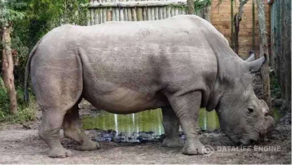
Which are hindgut fermenters?
What is Hindgut Fermentation? Rhinos, rabbits, some rodents, koalas and horses are all hindgut fermenters. To break down this term, you must first understand the anatomy of the equine digestive system.
What organs are in the hindgut?
The hindgut is composed of the cecum, large colon, small colon and the rectum.
Why can't horses vomit?
Horses can't vomit because they possess a valve at the entrance of the stomach called cardias or " Swiss tie ", the muscles of this valve are so strong that they prevent food from returning to the mouth.
What organs are part of the midgut?
The midgut consists of the distal half of the duodenum, jejunum, ileum, cecum, ascending colon, and the proximal half of the transverse colon (Figure 10-1A).
What is the mid gut and the hindgut?
The midgut is from the mid-duodenum to the initial two-thirds of the transverse colon. The hindgut is from the later one-third transverse colon to the upper portion of the anus.
Where does the hindgut start and end?
1 Gross Anatomy The hindgut of Homarus americanus constitutes a muscular rectum that begins in the sixth abdominal segment and continues to the anus (Fig. 1).
What is the difference between hindgut and foregut?
By definition, a foregut fermenter has a pre-gastric fermentation chamber whereas a hindgut fermenter has enlarged fermentation compartments in the cecum and/or colon (Stevens and Hume, 1998). The cow rumen is the most thoroughly studied foregut ecosystem.
What do hindgut fermenters eat?
Hindgut fermenters are evolved to eat a herbivorous diet. Such a diet includes large quantities of insoluble plant carbohydrates, such as cellulose. Mammals cannot digest these insoluble carbohydrates as they lack the essential enzymes, such as cellulase.
What is the difference between hindgut fermenters and ruminants?
Hindgut fermenters have a shorter passage time than ruminants, and hence are less efficient in cellulose digestion, for which they compensate with a higher intake of food (Clauss et al.
What do hindgut fermenters eat?
Hindgut fermenters are evolved to eat a herbivorous diet. Such a diet includes large quantities of insoluble plant carbohydrates, such as cellulose. Mammals cannot digest these insoluble carbohydrates as they lack the essential enzymes, such as cellulase.
Where is hay digested in horses?
hind gutFibrous sources such as oat hulls, soy hulls, beet pulp, hay and pasture are digested in the hind gut.
Where does digestion take place in the horse?
The saliva of a horse contains only small amounts of amylase and there is little actual digestion that occurs in the stomach of most horses. Most digestion therefore occurs in the small and large intestines. Although the intestine itself secretes some enzymes, the pancreas releases by far the greatest amount.
Why are horses hind gut fermenters?
Being a hindgut fermenter is a huge advantage to horses because it essentially gives them a second chance to process energy from feed that has already passed through the small intestine.
Embryology of the Anorectum
The hindgut gives rise to the distal third of the transverse colon, the descending colon, the sigmoid colon, the rectum, and the upper portion of the anal canal. The hindgut endoderm also lines the bladder and the urethra.
Metabolic Systems
The insect hindgut, along with the Malpighian tubules, is concerned primarily with osmoregulation and will be discussed in more detail in Chapter 8. The Malpighian tubules produce a primary iso-osmotic urine that is rich in potassium and low in sodium, and contains various ions, amino acids, and waste materials.
Invertebrate Kinins
Kinins stimulate hindgut contractions, but their effects on the foregut and oviduct are significantly lower than on the hindgut, and the heart does not respond to any of the kinins in insects [2]. Kinins are also associated with diuretic activity in insects.
Hedgehog Signaling in Gastrointestinal Morphogenesis and Morphostasis
Willemijn A. van Dop, Gijs R. van den Brink, in Physiology of the Gastrointestinal Tract (Fifth Edition), 2012
Water and Ion Balance, Hormonal Control of
Hormones acting on the hindgut appear to stimulate recovery of fluid and ions from the primary urine, leading to cycling of materials through the excretory system and hemolymph, and clearance of wastes. The majority of our information about regulation of hindgut transport comes from studies on the locust.
Human Pluripotent Stem Cell Derived Organoid Models
Mid/hindgut spheroids can develop into intestine-like tissue if cultured in conditions developed for growth of mouse intestinal organoids (McCracken, Howell, Wells, & Spence, 2011; Sato et al., 2009; Spence et al., 2011 ).
Gastrointestinal physiology
In the equine hindgut, the mRNA expression of different proteins involved in transcellular calcium transport have been reported: the transient receptor potential vanilloid member 6 (TRPV6) and member 5 (TRPV5), the calbindin D9k (CB9) and D28k (CB28), the sodium calcium exchanger 1 (NCX1) and the plasma membrane calcium ATPase 1 (PMCA1).
Metabolic Systems
The insect hindgut, along with the Malpighian tubules, is concerned primarily with osmoregulation and will be discussed in more detail in Chapter 8. The Malpighian tubules produce a primary iso-osmotic urine that is rich in potassium and low in sodium, and contains various ions, amino acids, and waste materials.
Invertebrate Kinins
Kinins stimulate hindgut contractions, but their effects on the foregut and oviduct are significantly lower than on the hindgut, and the heart does not respond to any of the kinins in insects [2]. Kinins are also associated with diuretic activity in insects.
Hedgehog Signaling in Gastrointestinal Morphogenesis and Morphostasis
Willemijn A. van Dop, Gijs R. van den Brink, in Physiology of the Gastrointestinal Tract (Fifth Edition), 2012
Gastrointestinal Physiology and Nutrition of Rabbits
Susan M. Smith PhD, in Ferrets, Rabbits, and Rodents (Fourth Edition), 2020
Water and Ion Balance, Hormonal Control of
Hormones acting on the hindgut appear to stimulate recovery of fluid and ions from the primary urine, leading to cycling of materials through the excretory system and hemolymph, and clearance of wastes. The majority of our information about regulation of hindgut transport comes from studies on the locust.
Datura species and related plants
As hindgut fermenters, it is not surprising that pigs are susceptible to tropane alkaloid toxicity because the toxin is preferentially absorbed from the upper gastrointestinal tract. It is also not surprising that pigs are commonly involved in toxicity because their diets are intensive and consist typically of maize in the ration.
Human Pluripotent Stem Cell Derived Organoid Models
Mid/hindgut spheroids can develop into intestine-like tissue if cultured in conditions developed for growth of mouse intestinal organoids (McCracken, Howell, Wells, & Spence, 2011; Sato et al., 2009; Spence et al., 2011 ).
Big Picture
The midgut consists of the distal half of the duodenum, jejunum, ileum, cecum, ascending colon, and the proximal half of the transverse colon ( Figure 10-1A ). Branches of the superior mesenteric arteries and veins provide the primary (but not exclusive) vascular supply for the midgut ( Figure 10-1B ).
Distal Half of the Duodenum
The duodenum is the first part of the small intestine. The chemical digestion of food (i.e., carbohydrates to simple sugars; fats to fatty acids and glycerol; proteins to amino acids) primarily occurs in the duodenum because of the secretion of pancreatic enzymes.
Jejunum and Ileum
The jejunum is the second part of the small intestine and has the most highly developed circular folds lining the lumen, thereby increasing the surface area of the mucosal lining for absorption. In contrast to the ileum, the jejunum also has a greater number of vasa recti.
