What is'in-situ'keratinocyte atypia?
Jan 18, 2020 · What is keratinocyte atypia? Actinic keratosis with cytologic atypia of keratinocytes, primarily involving the basal and suprabasal layers of the epidermis. The atrophic form of actinic keratosis is characterized by a thinned epidermis in which the basal cells are replaced by a layer of atypical keratinocytes.
What is atrophic keratinocyte carcinoma?
Background: Actinic Keratosis (AK) is the clinical manifestation of cutaneous dysplasia of epidermal keratinocytes, with progressive trend towards squamous cell carcinoma. Objective: To evaluate the strength of the correlation between keratinocyte atypia, as detected by Reflectance Confocal Microscopy (RCM) and histopathology, and to develop a more objective atypia …
How to grade keratinocyte atypia in actinic keratosis?
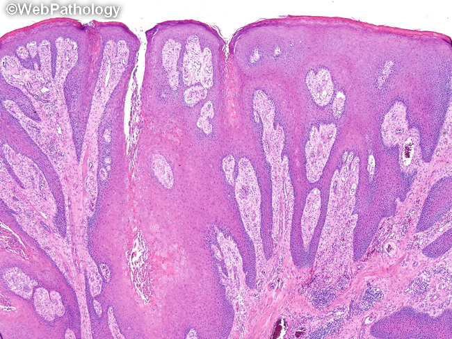
Feb 01, 2012 · The histological features of keratinocytic intraepidermal neoplasia I (KIN I) are cellular atypia of basal keratinocytes confined to the lower third of the epidermis. KIN II shows atypical keratinocytes occupying the lower two-thirds of the epidermis, and KIN III shows atypical keratinocytes throughout the epidermis; this latter stage is equivalent to carcinoma in situ ( 11 ).
What is a keratinocyte?
Sep 17, 2008 · Keratinocytes are the most common type of skin cells. They make keratin, a protein that provides strength to skin, hair, and nails. These cells form in the deep basal-cell layer of the skin, and take about a month to reach the surface. It is …
Are keratinocytes cancerous?
Abstract. Keratinocyte carcinoma (KC) (also referred to as nonmelanoma skin cancer) is by far the most common form of human cancer.
What type of skin cancer arises from keratinocytes?
Keratinocyte cancers and precancers arise in keratinocytes, the most common type of skin cells in the epidermis. They include the two most common skin cancers, basal cell carcinoma (BCC) and cutaneous squamous cell carcinoma (cSCC), and actinic keratosis (AK), the most common skin precancer.
Are atypical keratinocytes cancerous?
Squamous cell carcinoma in situ is an ''intraepidermal carcinoma'' made up of atypical keratinocytes throughout the full thickness of the epidermis. Bowen's disease is a term initially historically used for squamous cell carcinoma in situ of ''non–sun- exposed'' skin.
What is actinic atypia?
Actinic keratosis is an erythematous scaly papule or plaque that develops on sun-damaged skin as a result of chronic exposure to ultraviolet radiation, typically in elderly patients with lighter skin types.Jul 7, 2020
What does a keratinocyte do?
Function. Keratinocytes are highly specialized. They play an essential role in protection, as they form a tight barrier that prevents foreign substances from entering the body, while minimizing the loss of moisture, heat, and other constituents.
What do keratinocytes produce?
Keratinocytes are the predominant cell type of epidermis and originate in the basal layer, produce keratin, and are responsible for the formation of the epidermal water barrier by making and secreting lipids.Nov 19, 2021
Are keratinocytes squamous cells?
The Squamous Cell Layer Keratinocytes produce keratin, a tough, protective protein that makes up the majority of the structure of the skin, hair, and nails. The squamous cell layer is the thickest layer of the epidermis, and is involved in the transfer of certain substances in and out of the body.
How can malignant neoplasms be prevented?
Consider these cancer-prevention tips.Don't use tobacco. Using any type of tobacco puts you on a collision course with cancer. ... Eat a healthy diet. ... Maintain a healthy weight and be physically active. ... Protect yourself from the sun. ... Get vaccinated. ... Avoid risky behaviors. ... Get regular medical care.
What is squamous atypia?
Atypical squamous cells of undetermined significance is the most common abnormal finding in a Pap test. It may be a sign of infection with certain types of human papillomavirus (HPV) or other types of infection, such as a yeast infection.
What do precancerous lesions look like?
Visible signs of precancerous skin Crustiness or bleeding. Diameter of less than one inch. Discoloration, often appearing brown, pink, gray, red, yellow, or white. Flat or slightly raised.
Are all actinic keratosis precancerous?
It's true an AK is a precancer and may never become malignant. But there's no way of knowing for sure that a lesion on your skin is an AK, and not a skin cancer. Even if it is just an actinic keratosis, assuming it won't become cancerous is an unnecessary gamble.Apr 23, 2021
Can you scratch off actinic keratosis?
While an actinic keratosis can sometimes resolve on its own, it usually recurs after further sun exposure; if scratched or picked off, it will return as well.Apr 23, 2012
Basal Cell Carcinoma
Basal cell carcinoma is the far more common of this skin cancer type. 1 BCC accounts for some 80% of these cancers, though they rarely cause death. The other 20% are cSCC. A small percentage of people with cSCC will go on to see the cancer metastasize, or spread, and roughly 70% of that small subset will die, according to one 2019 study. 2
Symptoms of Basal Cell Carcinoma
BCCs usually develop on sun-exposed parts of your body, especially your head and neck. A much smaller number occur on the torso and legs. Yet BCCs also may occur on parts of your body that are rarely exposed to sunlight. 3
Causes of Basal Cell Carcinoma
BCC occurs when one of the basal cells of the epidermis develops a mutation, or change, in its DNA. The process of creating new skin cells is controlled by a basal cell's DNA. A mutation in the DNA may cause a basal cell to multiply fast and continue growing when it would normally die.
Cutaneous Squamous Cell Carcinoma
After BCC, it's cutaneous squamous cell carcinoma (cSCC) that is the second most common kind of skin cancer. 8 Both are of the keratinocyte type. Though cSCC occurs less often, it may have more serious outcomes, including death.
Summary
There are two primary types of skin cancer that arise from keratinocytes, the most common type of skin cells. Basal cell carcinoma accounts for the vast majority of them. Cutaneous squamous cell carcinoma accounts for just one of every five cases but it often proves the more fatal of the two.
A Word From Verywell
If you've noticed skin changes, or symptoms like sores that won't heal, it may be time to find out why. Make an appointment with your healthcare provider if you have any signs or symptoms that worry you.
Background
Actinic Keratosis (AK) is the clinical manifestation of cutaneous dysplasia of epidermal keratinocytes, with progressive trend towards squamous cell carcinoma.
Objective
To evaluate the strength of the correlation between keratinocyte atypia, as detected by Reflectance Confocal Microscopy (RCM) and histopathology, and to develop a more objective atypia grading scale for RCM quantification, through a discrete ranking.
Methods
A total of 48 AKs and two control areas (photodamaged and non-photodamaged skin) were selected for this study. All these areas were documented by RCM and biopsied for histopathology. One representative image of the epidermis was selected for RCM and for histopathology and used for side-by-side comparison with purpose written software.
Results
Good interobserver correlation was obtained for RCM and histopathology grading, with high concordance between RCM and histopathology grading.
Conclusions
Expert rater scan consistently distinguish different grades of cytological atypia. Non-invasive RCM data from in vivo imaging can be graded for keratinocyte atypia, comparable to histopathological grading.
What is actinic keratosis?
Actinic keratoses are lesions that have epidermal basal layer nuclear atypia with variable hyperkeratosis and parakeratosis, and background dermal solar elastosis. Actinic keratoses may have several intraepidermal layers of atypical keratinocytes, even approaching full-thickness atypia.
What is a cutaneous squamous cell carcinoma in situ?
Cutaneous squamous cell carcinoma in situ (Bowen’s disease) Cutaneous squamous cell carcinoma in situ ( Bowen’s disease) refers to a erythematous patch or plaque (in sun-exposed or non-sun-exposed skin) with full thickness epidermal nuclear atypia that often extends down and replaces the follicular infundibular epithelium.
Is keratinocyte atypia invasive?
The variations of patterns of in situ keratinocyte atypia may uncommonly evolve into invasive cSCC and can be viewed as cSCC in situ. In practice, the term 'in-situ keratinocyte atypia' is most commonly used with Bowen’s disease .
What does cytologic atypia mean?
Hi James, Breaking it down etymologically, "cytologic" basically means "of or relating to cells" and "atypia" means, well, "atypical." In the setting of a nevus, this means that there wasn't a significant proportion of the cells that showed an abnormal appearance consistent with atypical growth, division of cells, or transition to a type of cell inconsistent with what was taken out (like finding gut cells in a skin mass, which is relatively rare). If you cut out skin from a variety of people, there would likely be some cells that look atypical , but as long as the relative proportion of atypical cells is small, there's nothing to do but watch closely, as the body generally does a really good job of killing of cells that are trying to act up and divide without being told to do so. Unfortunately, there's no such thing as a "100% guarantee" when it comes to the human body, but there being "no significant cytologic atypia" means that, per the guidelines of the pathologist, the nevus removed was benign.
Is there a 100% guarantee on cytologic atypia?
Unfortunately, there's no such thing as a "100% guarantee" when it comes to the human body, but there being "no significant cytologic atypia" means that, per the guidelines of the pathologist, the nevus removed was benign. Helpful. Richard W. Siy, MD.
Is nevus with no significant cytologic atypia benign?
On online forum is not really the place for this question since I have no clue about your particular case - no photos, not what was done, nothing. That is why I anticipate few if any people will respond to your question. The proper place to discuss this is in detail with your doctor. That being said, and making the disclosure that this is NOT IN ANY WAY a comment about your particular case, in general, a "nevus with no significant cytologic atypia" means it is likely a benign condition. Good luck and review with your doctor. Damon B. Chandler, MD Harvard-Penn Trained Oculofacial Plastic Surgeon