What is the function of the lumbosacral plexus?
Lumbar plexus. The lower limbs have the tremendous responsibility of mobilizing and stabilizing the human body. Muscles of the pelvic region, posterior abdominal wall, and the fifty-nine muscles of the lower limb, as well as their corresponding joints, are innervated by branches of the lumbosacral plexus. These nerve fibers originate ...
What is lumbosacral plexopathy?
The lumbosacral (LS) plexus is a network of nerves formed by the anterior rami of the lumbar and sacral spinal cord. LS plexopathy is an injury to the nerves in the lumbar and/or sacral plexus.
What are upper and lower lumbosacral plexus lesions?
Lumbosacral plexus lesions usually are divided clinically into those affecting the upper lumbar plexus and those affecting the lower lumbosacral plexus, analogous to the underlying anatomic division.
What causes pain in the lumbosacral plexus?
Problems with lumbosacral plexus can occur due to trauma at the pelvic level that damages the roots or nerves, and can be due to birth defects/trauma or lumbosacral (carcinomatous) neuropathy. Carcinoma of the intestines, bladder, or prostate can invade the lumbosacral plexus.
Where is lumbosacral plexus?
Where is the lumbosacral plexus located? The lumbosacral plexus is formed by the anterior rami (i.e., branches) of spinal nerves L4 to L5 and S1 to S4. It is located on the posterolateral wall of the lesser pelvis, adjacent to the lumbar spine.
What causes lumbar plexus pain?
The most frequent causes are high-energy trauma, sports injuries, penetrating trauma, and surgery that results in damage to the plexus. In particular, injuries that cause damage between the spinal cord and spinal ganglion (proximal) lead to severe pain.
What nerves make up the lumbosacral plexus?
The lumbosacral plexus is formed by ventral rami of the lumbar and sacral nerves, T12 through S4. The lumbar part is formed by roots from T12 to L4 and the sacral component by L4–S4 roots. These divide into anterior and posterior divisions, which give rise to anterior and posterior branches, respectively.
What is a lumbosacral plexus MRI?
Magnetic resonance imaging is an invaluable tool for evaluation of the lumbosacral plexus due to its anatomic detail and sensitivity to pathologic changes. It can identify the cause for disability, indicate prognosis for improvement, and be a tool for delivery of interventions.
How do you treat lumbar plexus?
The treatment of lumbar radiculopathy also depends on the cause. In an acute setting, analgesics such as nonsteroidal anti-inflammatory drugs (NSAIDs) or acetaminophen and activity modification are the main treatments.
What happens if the lumbar plexus is damaged?
Malfunction of the lumbosacral plexus causes pain in the lower back and leg as well as weakness and loss of sensation in all or part of a leg (such as the foot or calf). Recovery depends on the cause.
What happens if the sacral plexus is damaged?
Sacral plexus lesions are usually unilateral and do not result in significant SD, unless the sensory symptoms are disruptive. Trauma may cause pudendal nerve injury, leading to loss of penile sensation, dysesthesias, pain syndromes, and dribbling ejaculation due to perineal muscle denervation.
What causes sacral nerve damage?
The most common causes of spinal cord injuries to the sacrum are: Motor vehicle accidents. Trauma. Falls.
Is the sciatic nerve part of the lumbar plexus?
The sciatic nerve is formed in the lower spine by the combination of motor and sensory fibers from spinal nerves L4 to S3. These spinal nerves belong to a larger group of nerves in the lower spine called the lumbosacral plexus.
What can a lumbar MRI detect?
A lumbar spine MRI can detect a variety of conditions in the lower back, including problems with the bones (vertebrae), soft tissues (such as the spinal cord), nerves, and disks.
What is the lumbosacral plexus?
The lumbosacral plexus is a network of nerve fibers, derived from the roots of lumbar and sacral spinal nerves that branch out to form the nerves s...
Where is the lumbosacral plexus located?
The lumbosacral plexus is formed by the anterior rami (i.e., branches) of spinal nerves L4 to L5 and S1 to S4. It is located on the posterolateral...
What nerves are found in the lumbosacral plexus?
The lumbosacral plexus includes nerves that arise from both the lumbar plexus and sacral plexus. From superior to inferior, the major nerves that b...
What is the clinical significance of the lumbosacral plexus?
The lumbosacral plexus can be damaged as a result of various conditions and mechanical trauma, but the most common cause of injury is a spinal disc...
What are the most important facts to know about the lumbosacral plexus?
The lumbosacral plexus is formed by the anterior rami of spinal nerves L4, L5, and S1 to S4, which are branches of the lumbar plexus and sacral ple...
What is the lumbar plexus?
Definition: The lumbosacral plexus is a network of nerves derived from lumbar and sacral roots with each one of them dividing into anterior and posterior branches. Their communications are called lumbar plexus (compare: brachial plexus). The anterior branches supply the flexor muscles of thigh and leg and posterior branches supply ...
Why does lumbosacral plexus damage?
Problems with lumbosacral plexus can occur due to trauma at the pelvic level that damages the roots or nerves, and can be due to birth defects/trauma or lumbosacral (carcinomatous) neuropathy. Carcinoma of the intestines, bladder, or prostate can invade the lumbosacral plexus.
What is the treatment for plexus malfunction?
Treatment varies depending on the cause of plexus malfunction. Corticosteroids are prescribed in the acute stage of an autoimmune reaction or inflammation/compression. Very limited benefit has been proven. Stretch injury needs time; transection needs operative repair with mixed results.
Where is the lumbar plexus located?
The lumbar plexus is situated within the upper two-thirds of the psoas major. The iliohypogastric, ilioinguinal, and genitofemoral nerves descend posterior to the iliac fascia and paraaortic and iliac lymph nodes. The sacral plexus lies within the pelvis adjacent to the rectum, colon, and ureter.
Which plexus provides innervations to the pelvis, buttocks, genitals,?
The lumbar plexus provides innervations to back-buttock, abdomen, groin, thighs, knees, and calves. The sacral plexus provides innervations to the pelvis, buttocks, genitals, thighs, calves, and feet. When there is dysfunction to the plexus, areas affected can be traced by dermatomes (numb/pain) or individual muscle dysfunction. Nerve root compression is due to intraspinal pathology
Where is pain located in the pelvis?
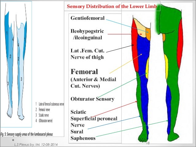
Pain, if present, usually is located in the pelvis with radiation into the anterior thigh. Sensory loss and paresthesias occur over the lateral, anterior, and medial thigh and may extend down the medial calf (Figure 32–5 ). Lesions of the lower lumbosacral plexus predominantly affect the L4–S3 nerve fibers.
What are the major nerves in the lower LP?
The major nerves from the lower LP include the superior gluteal nerve (L4-S1), the inferior gluteal nerve (L5-S2), the sciatic nerve (L4-S3), the posterior femoral cutaneous nerve (S1-S3), and the pudendal nerve (S1-S4). View chapter Purchase book. Read full chapter.
Where does the lumbar plexus originate?
The lumbar plexus originates from the anterior rami of spinal nerves L1-L4 and is formed largely within the posterior aspect of the psoas major muscle. The anterior ramus of spinal nerve T12 contributes to the formation of the lumbar plexus via the dorsolumbar nerve, which joins the anterior ramus of spinal nerve L1. Together, these roots (T12, L1) form a common trunk which gives rise to the iliohypogastric and ilioinguinal nerves. The anterior rami of L1 and L2 each give rise to a branch, which go on to merge with one another to form the genitofemoral nerve.
Which plexus gives rise to several branches which supply various muscles and regions of the posterior abdominal wall and lower limb?
The lumbar plexus gives rise to several branches which supply various muscles and regions of the posterior abdominal wall and lower limb . These branches include the iliohypogastric , ilioinguinal , genitofemoral , lateral femoral cutaneous , femoral and obturator nerves . In addition, the lumbar plexus gives off muscular branches from its roots, a branch to the lumbosacral trunk and occasionally an accessory obturator nerve.
What is the lateral cutaneous nerve?
The lateral cutaneous nerve of the thigh, also called the lateral femoral cutaneous nerve, is formed by fibers of the posterior divisions of the anterior rami of spinal nerves L2 and L3. It emerges from the lateral border of the psoas major muscle and courses inferolaterally to enter the iliac fossa, ultimately reaching the thigh. This nerve provides sensory innervation to the peritoneum of the iliac fossa and iliac fascia, and the skin of the anterior and lateral thigh (along the iliotibial tract ) to the knee .
What nerves are located in the anterior and posterior divisions of the spinal rami?
As the spinal rami of spinal nerves L2 - L4 course away from the lumbar vertebrae, they divide into anterior (ventral) and posterior (dorsal) divisions. The anterior divisions merge with one another to form the obturator nerve. Sometimes, the anterior divisions of L3 and L4 anterior rami may give branches that unite to form an accessory obturator nerve. Fibers from the posterior divisions, on the other hand, mainly unite to form the femoral nerve. The posterior divisions of L2 and L3 also give rise to branches that merge to form the lateral femoral cutaneous nerve.
What nerves are in the femoral canal?
The nerve continues its inferior course deep to the lateral part of the inguinal ligament and enters the femoral canal lateral to the femoral vessels. As it enters the canal, it supplies pectineus, and then divides into multiple parts. The branches of the femoral nerve are mostly named for the muscles they innervate. These include: 1 the nerve to pectineus 2 two nerves to rectus femoris (one of which also innervates the hip) 3 a nerve to vastus lateralis, vastus intermedius, and vastus medius 4 two nerves to sartorius, one of which becomes the intermediate femoral cutaneous nerve and the other, the medial femoral cutaneous nerve. The former gives cutaneous innervation to the fascia lata covering the anterior thigh to the knee as well as the skin in this region. The latter innervates the medial aspect of the thigh. 5 the saphenous nerve, which is responsible for giving cutaneous supply to the skin of the anteromedial part of the knee, over the medial malleolus and all the way down to the distal end of the first metatarsal bone.
What nerve is the lateral cutaneous femoral nerve?
Lateral cutaneous femoral nerve. The peritoneum of the iliac fossa, iliac fascia and the lateral side of the thigh (along the iliotibial tract) to the knee is supplied by an entirely sensory nerve called the lateral cutaneous femoral nerve. Proximally, the nerve is formed by fibers of the posterior division of L2 – L3.
Where is the i liohypogastric nerve located?
The i liohypogastric nerve is formed from the anteriorl ramus of spinal nerve L1 but may receive a contributory branch from the anterior rami of T12. It runs anterolaterally across the lower posterior abdominal wall after emerging from the superolateral border of the psoas major muscle. This nerve is a mixed nerve that provides both motor and sensory innervation to the internal oblique and transversus abdominis muscles and the skin of the posterolateral gluteal region and suprapubic region respectively.
What is the LS plexus?
The LS plexus is a combination of lumbar and sacral plexuses and encompasses the anterior rami of the L1 through S4 nerve roots of the peripheral nervous system, with a small contribution from T12 as well. Lumbar plexus lies above the pelvic brim and forms from L1 through L4 nerve roots, while the S1 through S4 nerve roots make up the sacral plexus, which lies below the pelvic brim.
What is LS plexopathy?
LS plexopathy is an injury to the nerves in the lumbar and/or sacral plexus. LS plexopathy is not an uncommon condition but can be difficult to diagnose and manage.[1] However, it is far less common than brachial plexopathy. Patients with LS plexopathy usually present with low back and/or leg pain. They can also experience motor weakness, other sensory symptoms of numbness, paresthesia, and/or sphincter dysfunction. [2][3] LS plexopathy can be caused by multiple etiologies, with diabetes mellitus, traumatic injury, neoplasms, and pregnancy being a few of the important causes. Treatment is often limited and varies significantly depending on the underlying pathology.[4] LS plexopathy can be debilitating, severely affecting a patient's quality of life. Early identification and management are critical in reducing morbidity and mortality. [5]
What are the symptoms of sacral plexopathy?
A straight leg raise test is positive in more than half of the patients. Asymmetric lower limb muscle weakness may be seen with asymmetrically absent or reduced deep tendon reflexes. Knee jerk reflex is affected in lumbar plexopathy and ankle jerk is affected in sacral plexopathy. Muscle weakness in hip flexion, knee extension, or adduction suggests a possible injury to the lumbar plexus. Sensory loss may be present in a dermatomal pattern in cases of proximal LS plexopathy involving the roots, or in the nerve distribution. Sensory changes to the medial thigh, anterior thigh, and medial leg can suggest lumbar plexus involvement; posterior thigh, dorsum of the foot, and perineum are likely related to sacral plexus involvement. Spinal point tenderness may be present, especially in cases of sacral fracture or infection. A rectal exam should be performed to assess rectal tone. Saddle anesthesia and bowel or bladder incontinence are rare and may be present, making it difficult to differentiate from cauda equina and conus medullaris syndromes. The inguinal region should also be palpated for suspected hematomas.
How can nerve repair help with pelvic fracture?
Surgical nerve repair techniques and nerve grafting have helped improve muscle function in pelvic fractures. One small study of 10 patients experiencing traumatic lumbosacral plexopathy, who underwent nerve grafting showed significant improvement of muscle function at 38 months follow-up. [34][35]
How long does it take to recover from a LS plexopathy?
Traumatic LS plexopathies are generally considered to have an unfavorable prognosis but a case-series of 72 patients with traumatic LS plexopathies demonstrated that more than two-thirds (about 70%) of patients recovered spontaneously within 18 months. [38]
What imaging is used to diagnose LS plexopathy?
Thus advanced imaging is often needed.[25] MRI is often ordered for the initial evaluation of neoplasm-associated LS plexopathy. Positron emission tomography (PET) is to determine the full extent of malignancy.[18] It also helps in the staging of the disease and subsequent treatment and prognosis.
What is the best imaging for LS plexopathy?
Neuroimaging , preferably, magnetic resonance imaging (MRI) of the LS spine and electrodiagnostic studies (nerve conduction study and electromyography) are important in confirmation of the diagnosis of LS plexopathy.
What is the plan of the lumbar plexus?
Plan of lumbar plexus. The lumbar plexus and its branches. The lumbar plexus is a web of nerves (a nervous plexus) in the lumbar region of the body which forms part of the larger lumbosacral plexus. It is formed by the divisions of the first four lumbar nerves (L1-L4) and from contributions of the subcostal nerve (T12), ...
Which nerve is the largest and longest of the plexus?
The femoral nerve is the largest and longest of the plexus' nerves. It gives motor innervation to iliopsoas, pectineus, sartorius, and quadriceps femoris; and sensory innervation to the anterior thigh, posterior lower leg, and hindfoot.
Where does the iliohypogastric nerve run?
The iliohypogastric nerve runs posterior to the psoas major on its proximal lateral border to run laterally and obliquely on the anterior side of quadratus lumborum. Lateral to this muscle, it pierces the transversus abdominis to run above the iliac crest between that muscle and abdominal internal oblique.
Which nerve pierces the psoas major?
In males it supplies motor innervation to the cremaster . The lateral cutaneous femoral nerve pierces psoas major on its lateral side and runs obliquely downward below the iliac fascia.
Where does the obturator nerve go?
The obturator nerve leaves the lumbar plexus and descends behind psoas major on it medial side, then follows the linea terminalis into the lesser pelvis, and finally leaves the pelvic area through the obturator canal. In the thigh, it sends motor branches to obturator externus before dividing into an anterior and a posterior branch, both of which continues distally. These branches are separated by adductor brevis and supply all thigh adductors with motor innervation: pectineus, adductor longus, adductor brevis, adductor magnus, adductor minimus, and gracilis. The anterior branch contributes a terminal, sensory branch which passes along the anterior border of gracilis and supplies the skin on the medial, distal part of the thigh.
Where do the motor branches of the psoas go?
Its smaller motor branches are distributed directly to psoas major, while the larger branches leave the muscle at various sites to run obliquely down through the pelvis to leave under the inguinal ligament with the exception of the obturator nerve which exits the pelvis through the obturator foramen.
Which lumbar nerve is the last thoracic nerve?
It is formed by the divisions of the first four lumbar nerves (L1-L4) and from contributions of the subcostal nerve (T12), which is the last thoracic nerve. Additionally, the ventral rami of the fourth lumbar nerve pass communicating branches, the lumbosacral trunk, to the sacral plexus.
What is lumbosacral plexopathy?
Lumbosacral Plexopathy. The lumbosacral (LS) plexus is a network of nerves formed by the anterior rami of the lumbar and sacral spinal cord. LS plexopathy is an injury to the nerves in the lumbar and/or sacral plexus. LS plexopathy is not an uncommon condition but can be difficult to diagnose and manage. However, it is far ….
What is LS plexopathy?
LS plexopathy is an injury to the nerves in the lumbar and/or sacral plexus.
Is LS plexopathy more common than brachial plexopathy?
However, it is far less common than brachial plexopathy. Patients with LS plexopathy usually present with low back and/or leg pain. They can also experience motor weakness, other sensory symptoms of numbness, paresthesia, and/or sphincter dysfunction.
Is LS plexopathy debilitating?
Treatment is often limited and varies significantly depending on the underlying pathology. LS plexopathy can be debilitating, severely affecting a patient's quality of life. Early identification and management are critical in reducing morbidity and mortality. Copyright © 2021, StatPearls Publishing LLC.
Is LS plexopathy uncommon?
LS plexopathy is not an uncommon condition but can be difficult to diagnose and manage. However, it is far …. The lumbosacral (LS) plexus is a network of nerves formed by the anterior rami of the lumbar and sacral spinal cord. LS plexopathy is an injury to the nerves in the lumbar and/or sacral plexus. LS plexopathy is not an uncommon condition but ...
