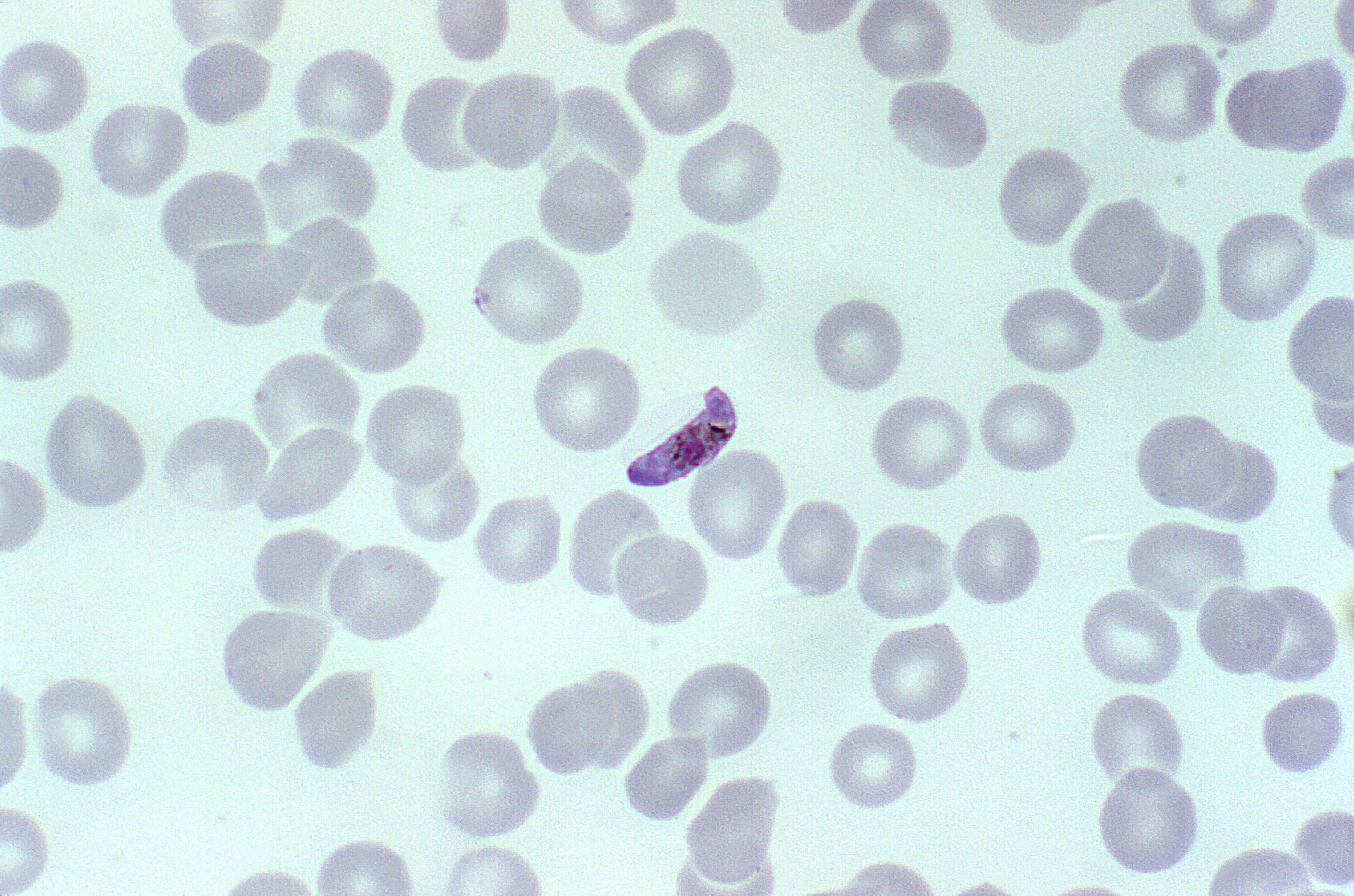
Microscopy is the method of choice for the investigation of malaria treatment failures. Giemsa is the classical stain used for malaria microscopy, and diagnosis requires examination of both thin and thick films from the same patient. Light microscopy is the diagnostic standard against which other diagnostic methods have traditionally been compared.
How is malaria diagnosed using light microscopy?
Light microscopy of thick and thin stained blood smears remains the standard method for diagnosing malaria. It involves collection of a blood smear, its staining with Romanowsky stains and examination of the Red Blood Cells for intracellular malarial parasites.
What is the best method for malaria diagnosis in endemic countries?
Microscopy is still considered the “gold standard” for malaria diagnosis in endemic countries. This method has a sensitivity of 50–500 parasites/μl [ 6 ], is inexpensive, and allows the identification of species and parasite density [ 7, 8 ].
What is the peripheral smear test for malaria?
Peripheral smear study for malarial parasites – The MP test. Thin smears allow one to identify malaria species (including the diagnosis of mixed infections), quantify parasitemia, and assess for the presence of schizonts, gametocytes, and malarial pigment in neutrophils and monocytes.
What is the purpose of the malaria microscopy training program?
It aims to improve competence in confirming malaria infection with optical microscopy and is intended for microscopists, laboratory technicians and trainers involved in teaching malaria microscopy in endemic countries as well as in malaria-free countries.

What is the importance of malarial microscopy?
Microscopy is important in both malaria diagnosis and research. It is used to differentiate between Plasmodium species and stages and to estimate parasite density in the blood – an important determinant of the severity of disease.
What microscope is used for malaria?
Blood slide microscopy makes it possible to count the number of parasites and is more useful than rapid diagnostic tests for monitoring the effectiveness of malaria treatment.
What is MP microscopy?
Peripheral smear study for malarial parasites – The MP test It involves collection of a blood smear, its staining with Romanowsky stains and examination of the Red Blood Cells for intracellular malarial parasites.
What is malaria microbiology?
Malaria is caused by protozoa of the genus Plasmodium. Four species cause disease in humans: P falciparum, P vivax, P ovale and P malariae. Other species of plasmodia infect reptiles, birds and other mammals. Malaria is spread to humans by the bite of female mosquitoes of the genus Anopheles.
What is the principle of malaria test?
The principles of tests stem from detection of malaria parasites' protein i.e. histidine. Where antibody method is used, it means detection of the presence of antibodies against histidine in the human serum and where whole blood is used, it implies detection of malaria parasites' histidine on the red blood cells[6].
Which test is best for malaria?
Your blood sample may be tested in one or both of the following ways.Blood smear test. In a blood smear, a drop of blood is put on a specially treated slide. ... Rapid diagnostic test. This test looks for proteins known as antigens, which are released by malaria parasites.
How is malaria microscopy done?
Malaria parasites can be identified by examining under the microscope a drop of the patient's blood, spread out as a “blood smear” on a microscope slide. Prior to examination, the specimen is stained (most often with the Giemsa stain) to give the parasites a distinctive appearance.
What dOES +++ mean in malaria test result?
These scores were used to estimate parasite densities: + = 10 to 90 parasites/μl; ++ = 100 to 1,000 parasites/μl, +++ = 1,000 to 10,000 parasites/μl; ++++ = >10,000 parasites/μl, assuming a white blood cell count of 8,000/μl. The species was identified using the thin smear.
What is the meaning of MP test?
Peripheral smear for Malarial parasite helps to detect the presence of the malarial parasite in the blood. The malarial parasite is detected when an individual is suffering from malaria.
What are the 5 types of malaria?
Five species of Plasmodium (single-celled parasites) can infect humans and cause illness:Plasmodium falciparum (or P. falciparum)Plasmodium malariae (or P. malariae)Plasmodium vivax (or P. vivax)Plasmodium ovale (or P. ovale)Plasmodium knowlesi (or P. knowlesi)
What is the scientific name for malaria?
Plasmodium falciparum is the type of malaria that most often causes severe and life-threatening malaria; this parasite is very common in many countries in Africa south of the Sahara desert.
What are the four causes of malaria?
There are four kinds of malaria parasites that can infect humans: Plasmodium vivax, P. ovale, P. malariae, and P. falciparum....Malaria is transmitted by blood, so it can also be transmitted through:an organ transplant.a transfusion.use of shared needles or syringes.
What equipment is needed for a malaria test?
Microscope slides and slide boxes for storage. Staining trays. Plastic measuring cylinders (100 and 500 mL) Plastic serologic pipettes (1, 5, and 10 mL)
Which microscope can be used to visualize the stages of development of malarial parasites?
Cellphone based microscope with a ball lens objective has been optimized for high resolution bright field imaging of malaria parasite in thin blood smears. Parasites in various stages of infection have been detected in sample infected smears.
How do you count malaria parasites under a microscope?
To quantify malaria parasites against RBCs, count the parasitized RBCs among 500-2,000 RBCs on the thin smear and express the results as % parasitemia.
How do you prepare a slide for malaria parasite?
Prepare at least 2 smears per patient! A thin smear being prepared. Place a small drop of blood on the pre-cleaned, labeled slide, near its frosted end. Bring another slide at a 30-45° angle up to the drop, allowing the drop to spread along the contact line of the 2 slides.
How to determine malaria parasites?
Within a few hours of collecting the blood, the microscopy test can provide valuable information. First and foremost it can determine that malaria parasites are present in the patient’s blood. Once the diagnosis is established – usually by detecting parasites in the thick smear – the laboratorian can examine the thin smear to determine the malaria species and the parasitemia, or the percentage of the patient’s red blood cells that are infected with malaria parasites. The thin and thick smears are able to provide all 3 of these vital pieces of information to the doctor to guide the initial treatment decisions that need to be made acutely.
What is the gold standard for malaria diagnosis?
Malaria Diagnosis (U.S.) - Microscopy. Microscopic examination remains the “gold standard” for laboratory confirmation of malaria. These tests should be performed immediately when ordered by a health-care provider. They should not be saved for the most qualified staff to perform or batched for convenience.
What is the RDT for malaria?
The laboratories associated with these health-care settings may now use an RDT to more rapidly determine if their patients are infected with malaria. BinaxNOW® Malaria Test, the only available RDT for malaria in the United States. Disadvantages. The use of the RDT does not eliminate the need for malaria microscopy.
How many species of malaria are there?
In addition to the four classic human species of malaria, there are more than 20 species of malaria parasites that naturally infect non-human primates. It was thought that natural infections of simian malaria in humans were rare and not of public health importance until recent reports from Asia have suggested that P. knowlesi, a simian malaria species, is emerging as a public health problem.
What is a rapid diagnostic test?
A Rapid Diagnostic Test (RDT) is an alternate way of quickly establishing the diagnosis of malaria infection by detecting specific malaria antigens in a person’s blood. RDTs have recently become available in the United States. Technique.
What is the blood stage of Plasmodium species?
Blood stage Plasmodium species schizonts (meronts) are used as antigen. The patient’s serum is exposed to the organisms; homologous antibody, if present, attaches to the antigen, forming an antigen-antibody (Ag-Ab) complex.
Can a negative RDT be used for malaria?
The use of the RDT does not eliminate the need for malaria microscopy. The RDT may not be able to detect some infections with lower numbers of malaria parasites circulating in the patient’s bloodstream. Also, there is insufficient data available to determine the ability of this test to detect the 2 less common species of malaria, P. ovale and P. malariae. Therefore all negative RDTs must be followed by microscopy to confirm the result.
Overview
This CD-ROM was developed by the United States Centers for Disease Control and Prevention (CDC) with technical contribution from an independent group of experts convened by WHO.
Downloading the CD-ROM
Because of its size, the CD-ROM can be downloaded as a single complete file or in 2 parts:
What is the diagnosis of malaria?
Diagnosis of malaria involves identification of malaria parasite or its antigens/products in the blood of the patient. Although this seems simple, the efficacy of the diagnosis is subject to many factors. The different forms of the four malaria species; the different stages of erythrocytic schizogony; the endemicity of different species;…
What questions do you need to ask a clinician who faces a case of fever?
A clinician who faces a case of fever would need answers to the following questions: Is it malaria? If yes; What is the species? Is it severe? Is it new/ recurrence? Is it active? At present, ONLY the peripheral smear can provide answers to ALL these questions on a single…
What is the diagnosis of malaria?
D iagnosis of malaria involves identification of malaria parasite or its antigens/products in the blood of the patient. Although this seems simple, the efficacy of the diagnosis is subject to many factors. The different forms of the four malaria species; the different stages of erythrocytic schizogony; the endemicity of different species; the population movements; the inter-relation between the levels of transmission, immunity, parasitemia, and the symptoms; the problems of recurrent malaria, drug resistance, persisting viable or non-viable parasitemia, and sequestration of the parasites in the deeper tissues; and the use of chemoprophylaxis or even presumptive treatment on the basis of clinical diagnosis can all have a bearing on the identification and interpretation of malaria parasitemia on a diagnostic test.
How is malaria diagnosed?
The diagnosis of malaria is confirmed by blood tests and can be divided into microscopic and non-microscopic tests.
How much parasitemia is in falciparum?
In nonfalciparum malaria, parasitemia rarely exceeds 2%, whereas it can be considerably higher (>50%) in falciparum malaria. In nonimmune individuals, hyperparasitemia (>5% parasitemia or >250 000 parasites/μl) is generally associated with severe disease. The smear can be prepared from blood collected by venipuncture, finger prick and ear lobe stab.
How often should you get a blood smear for malaria?
One negative blood smear makes the diagnosis of malaria very unlikely (especially the severe form); however, smears should be repeated every 6–12 hours for 48 hours if malaria is still suspected. [1-5] Sometimes no parasites can be found in peripheral blood smears from patients with malaria, even in severe infections.
How to prepare for a malaria smear?
Preparation of the smear: Use universal precautions while preparing the smears for malarial parasites – use gloves; use only disposable needles/lancets; wash hands; handle and dispose the sharp instruments and other materials contaminated with blood carefully to avoid injury. Take a clean glass slide.
How to detect malaria?
Light microscopy of thick and thin stained blood smears remains the standard method for diagnosing malaria. It involves collection of a blood smear, its staining with Romanowsky stains and examination of the Red Blood Cells for intracellular malarial parasites. Thick smears are 20–40 times more sensitive than thin smears for screening of Plasmodium parasites, with a detection limit of 10–50 trophozoites/μl. Thin smears allow one to identify malaria species (including the diagnosis of mixed infections), quantify parasitemia, and assess for the presence of schizonts, gametocytes, and malarial pigment in neutrophils and monocytes.
What is the best test for malaria?
The most commonly used microscopic tests include the peripheral smear study and the Quantitative Buffy Coat (QBC) test.
Why is microscopy important for malaria?
According to the Malaria Microscopy Quality Assurance (QA) Manual from the WHO [ 32 ], it is necessary to ensure that healthcare professionals and patients have full confidence in the laboratory result , and the diagnostic results benefit the patient and community. Hospitals and health centres require expert microscopy for the management of malaria cases; it is the gold standard in endemic countries for identifying mixed infections, treatment failures, and quantifying parasite density. In the present study, microscopy showed less sensitivity (55.3%) and specificity (81.28%) than the RDT (83.74% and 89.11%, respectively) using SnM-PCR as the reference technique. Taking into account the age groups, it was observed a decrease with age of the sensitivity in microscopy as well as in the RDTs. Parasite density might have determined the positive infections detected by microscopy and RDTs [ 33 ], as the parasite density (from moderate to low) decrease with age [ 34 ]. This decrease might be also related to the immunity status. In malaria endemic countries, as EG, acquired immunity in adult is associated with the presence of submicroscopic infections that are more likely to be undetected by microscopy and RDTs [ 33, 35 ]. On the other hand, it was observed that RDT specificity values were higher when sensitivity declined. A study carried out by Laurent et al. found that the specificity of RDT varied with age and was inversely related to the prevalence of positive blood films in different age groups, that is, the specificity decreased as the prevalence of malaria increased. Moreover, malaria prevalence was higher in children under 5 years of age, when less specificity is detected [ 36 ]. Abeku et al. [ 37] also detected a decrease in specificity with decreasing age and increased prevalence of malaria in symptomatic patients. Another study reported a similarly low RDT specificity (52%) in symptomatic children younger than 5 years in an area of intense transmission [ 38 ]. The RDT decrease of sensitivity with age while specificity increases have been also described by Siahaan et al. According to this paper, both parameters were influenced by parasite density related to the improvement of their immune system (antiparasite disease) with age [ 39 ].
What is the most sensitive method for detecting malaria?
Implementation of the molecular techniques as diagnostic methods in sub-Saharan Africa is complicated due to the equipment required, reagent maintenance, and the qualified personnel required. Polymerase chain reaction (PCR) is the most sensitive method available, detecting parasitemia as low as 2–5 parasites/μl. However, it is not appropriate for use in the field, as it is an expensive and complex method.
How many false positives were found in SNM-PCR?
According to SnM-PCR results, they were false positives in RDT and microscopy (102 positive samples by RDTs and 203 by microscopy).
Why is it important to reinforce microscopy training?
Owing to the high number of false negatives in microscopy, it is necessary to reinforce training in microscopy, the “Gold Standard” in endemic areas. A network of reference centres could potentially support ongoing diagnostic and control efforts made by malaria control programmes in the long term, as the National Centre of Tropical Medicine currently supports the National Programme against Malaria of Equatorial Guinea to perform all of the molecular studies necessary for disease control. Taking into account the results obtained with the RDTs, an exhaustive study of the deletion of the hrp2 gene must be done in EG to help choose the correct RDT for this area.
Why is it important to diagnose Plasmodium species?
Accurate diagnosis of Plasmodium species is important not only for establishing the correct treatment regimen , but also for applying effective malaria control strategies in endemic regions where the four species of malaria parasites exist, as in EG. Misidentification of the Plasmodium species could result in severe public health concerns due to inappropriate treatments, leading to recrudescence and even drug resistance [ 28 ]. Malaria control requires a high quality diagnostic method to detect the parasite before prescribing anti-malarial treatment following the WHO’s indications. Malaria parasitological diagnosis targets treatment, supports characterization of the treatment response, and enables early identification of the parasite [ 29 ].
What was the prevalence of malaria in 2016?
In 2016, the prevalence of malaria was 12.09% and malaria caused 15% of deaths among children under 5 years. In the Continental Region, 95.2% of malaria infections were Plasmodium falciparum, 9.5% Plasmodium vivax, and eight cases mixed infection in 2011.
How many false negatives were detected in PCR?
Among the negative samples detected by microscopy, 335 (19.4%) were false negatives. On the other hand, the negative samples detected by RDT, 128 (13.3%) were false negatives based on PCR. This finding is important, especially since it is a group of patients who did not receive antimalarial treatment.
