
Microlaryngeal surgery is a minimally invasive procedure used to biopsy or remove abnormal growths, such as granulomas or benign cysts, in the larynx. It is usually performed to correct voice disorders or to diagnose or treat laryngeal cancer.
What is microlaryngoscopy (Microlaryngeal surgery)?
Microscopic laryngeal surgery, otherwise known as microlaryngoscopy, is the most precise means of visualizing and operating on the vocal folds. It allows the use of the two most essential tool sets in laryngeal surgery: the operative microscope, and microlaryngeal dissection instruments.
What are the pros and cons of Microlaryngeal surgery?
The majority of microscopic voice surgery procedures, though performed on an outpatient basis, are performed under general anesthesia to minimize the danger of gagging or breathing problems. Patients who undergo microlaryngeal surgery typically recover more quickly than patients who undergo more traditional laryngeal surgery.
What is minimally invasive larynx surgery?
This is a Minimally Invasive Technique that helps in the correction of the voice disorder or problem in the larynx. It involves the removal of the unwanted and unnatural growth in the larynx such as cysts or polyps that may not respond to more conservative treatment and needs surgery.
What technology is used to perform microlaryngoscopy?
At present, microlaryngoscopy (and, in particular, surgery on the vocal cords) is generally performed with either the use of: (1) a binocular operating microscope, or (2) a magnified, rigid telescope. There are advantages and disadvantages to each technology, but the point is that laryngeal surgery is more precise with better magnification.
What is Microlaryngeal excision?
Microlaryngeal surgery is a minimally invasive procedure used to biopsy or remove abnormal growths, such as granulomas or benign cysts, in the larynx. It is usually performed to correct voice disorders or to diagnose or treat laryngeal cancer.
Is Microlaryngeal surgery safe?
Microlaryngeal surgery is a common and relatively safe otorhinolaryngological surgery. Its common complications include pain and numbness of the tongue, bruising of the lip, and chipped teeth. However, reports of subcutaneous emphysema of the neck with pneumomediastinum following microlaryngeal surgery are rare.
Why would you need Microlaryngoscopy?
Microlaryngoscopy is the examination of your larynx (voice box), using a microscope, while you are under general anaesthetic. You will therefore be asleep for the procedure. This operation is used to find out, and treat, any problems of the larynx and to help improve your voice.
Is Microlaryngoscopy surgery safe?
The surgery is usually safe and uncomplicated however it is important that you are aware of the risks of the procedure. General complications such as nausea, vomiting, sore throat and drowsiness may occur as a result of the anaesthetic. Serious drug reactions related to the anaesthetic are very rare.
What is a Microlaryngoscopy biopsy?
A microlaryngoscopy is performed under brief anesthesia and involves the insertion of an endoscope through the nose and into the throat. If swelling is present, the surgeon will perform a biopsy (take a tissue sample) inside the larynx. Since only a tiny piece of tissue is removed, stitches are not necessary.
What is the most common reason for a laryngectomy?
Most often, laryngectomy is done to treat cancer of the larynx. It is also done to treat: Severe trauma, such as a gunshot wound or other injury. Severe damage to the larynx from radiation treatment.
How long does it take to recover from a Microlaryngoscopy?
After microlaryngoscopy or endoscopy. You need 24 hours off work, sport and driving after a general anaesthetic. You will need four days of complete voice rest (no talking at all) and one to two weeks off work depending on your treatment plan. The better you eat, the better you heal, especially fruit and vegetables.
Can I talk after Microlaryngoscopy?
Recovery from Microlaryngoscopy with Excision Complete voice rest is a vital part of a full recovery. After the procedure, you should not speak for three to five days to allow your vocal cords to heal.
How is a Microlaryngoscopy performed?
During a microlaryngoscopy, your surgeon accesses your vocal cords through the mouth using a laryngoscope. This tool provides high-quality images that are magnified to show every detail of the vocal cords and surrounding areas. The surgeon then removes the lesion using tiny surgical instruments.
What happens if you talk after vocal cord surgery?
Your doctor may ask you to speak as little as you can for 1 to 2 weeks after the procedure. If you speak, use your normal tone of voice and do not talk for very long. Whispering or shouting can strain your vocal cords as they are trying to heal. Try to avoid coughing or clearing your throat while your throat heals.
How serious is vocal cord surgery?
Surgery for laryngeal disorders is considered very safe, and the risk depends on the procedure. With framework surgery and implants, such as a thyroplasty, there can be complications related to surgery and general anesthesia, such as bleeding or infection.
What does a Microlaryngoscopy mean?
When there are lesions on the vocal cord that need to be removed, such as polyps or cysts, this is performed during a surgery called “microlaryngoscopy”, which literally means to view the vocal cords using a microscope.
What can you eat after vocal cord polyp removal?
Start out with cool, clear liquids; flavoured ice pops; and ice cream. Next, try soft foods like pudding, yogurt, canned or cooked fruit, scrambled eggs, and mashed potatoes. Do not eat hard or scratchy foods such as chips or raw vegetables until your throat has healed.
What is the purpose of Phonosurgery?
Phonosurgery is composed of procedures that are intended to maintain or improve the quality of the voice by correcting defects in laryngeal sound production. These procedures are rooted in a rich medical history that dates back to the early 19th century.
How do they remove polyps from vocal cords?
Surgical excision of a vocal cord polyp is most precisely performed under general anesthesia using an operating microscope and a microflap technique, during which a small flap of tissue containing the polyp is carefully raised.
What causes polyps on the vocal cords?
Causes of Vocal Fold Nodules and Polyps Most of the time, vocal abuse or misuse causes nodules. Long-term vocal abuse can cause polyps, too. But polyps may happen after just one instance of vocal abuse, like yelling at a concert. Smoking cigarettes for a long time, thyroid problems, and reflux may also cause polyps.
What is microscopic laryngeal surgery?
Microscopic laryngeal surgery, otherwise known as microlaryngoscopy, is the most precise means of visualizing and operating on the vocal folds. It allows the use of the two most essential tool sets in laryngeal surgery: the operative microscope, and microlaryngeal dissection instruments. All surgery is done through a laryngoscope, an instrument inserted via the mouth, without the need to make skin incisions.
What is a microlaryngoscopy?
Microlaryngoscopy is a surgical technique used in the evaluation and removal of various lesions of the vocal folds, including (but not limited to): c ancer , cysts , papilloma , polyps, and Reinke’s edema.
How long does it take to go home after microlaryngoscopy?
Despite the use of general anesthesia, it remains an ambulatory procedure - allowing a patient to go home the same day as the procedure, which takes approximately one hour. Pain after surgery is not severe, and rarely requires more than over-the-counter pain relievers.
Can a carbon dioxide laser be used for vocal surgery?
The Carbon Dioxide Laser. The use of the carbon dioxide laser for surgery of the vocal fold is controversial. Most laryngologists prefer to avoid it, for although the cutting beam is precise, it is hypothesized that the underlying tissue reaction to the emitted heat is somewhat unpredictable, and may at times result in scarring.
Can microscopic laryngeal surgery cause tongue tingling?
Complications arising from microscopic laryngeal surgery are rare. They can include temporary numbness or tingling of the tongue, and damage to teeth - especially in the presence of crowns, caps and veneers, or if the teeth are in poor condition to begin with.
How is microlaryngeal surgery done?
Almost all microlaryngeal surgery is done with the patient totally asleep (usually called general anesthesia). The patient must be anesthetized strongly enough so that he does not gag with the laryngoscope in place, so that he doesn't feel anything, and that he have no memory of the surgery. During the surgery the patient must be given oxygen and the carbon dioxide produced in the lungs must be removed. This is usually accomplished by a combination of intravenous medications and the use of anesthetic gases. The gases and oxygen are given by first passing a plastic tube (called an endotrachial tube) through the vocal folds into the upper windpipe. This is called intubation.
What is the name of the procedure where the surgeon is holding the scope in his left hand and looking down the lary?
Note that the surgeon isholding the scope in his left hand and looking down the laryngoscope at the patient's vocal folds. This is called "direct laryngoscopy". The surgeon can then use instruments in his right hand, working through the laryngoscope.
What is the instrument used to bring the vocal folds into view?
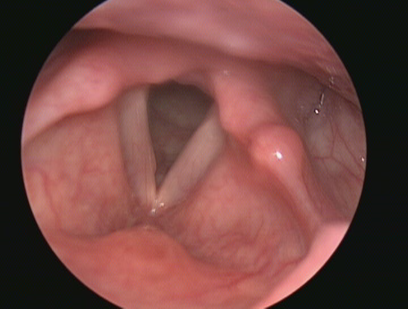
1. LARYNGOSCOPES . There are several types of instruments that are extremely important for microlaryngeal surgery. First, an instrument called a laryngoscope is used to bring the vocal folds into view. The patient undergoing surgery will be, in almost all cases, totally asleep and lying on one's back.
What is a laryngoscope attached to?
Note also that the laryngoscope is attached to a special stand that holds it in the proper position. This is called "suspension laryngoscopy" and it frees up the surgeon's other hand so both can be used during the operation. 2. MICROLARYNGEAL INSTRAMENTS.
Where can a laryngeal telescope be placed?
Special laryngeal telescopes can, in contrast, be placed just above the vocal folds and give unobstructed views of the folds. The photo to the right shows an example of microlaryngeal surgery using a microscope.
What is the best way to magnify vocal folds?
Delicate surgery on the vocal folds is best done under high magnification. Special operating microscopes and endoscopic telescopes are used to magnify the folds during surgery. The microscope has a large focal length and binocular eyepieces that allows a good three-dimensional view of the folds.
How small are laryngoscopes?
They have to be long and narrow since all surgery is done through the laryngoscope. The ends must be very small, usually only 1 or 2 mm, since the structures on the vocal folds are also tiny.
What is micro laryngeal surgery?
Micro-Laryngeal Surgery is done to address problem in the vocal chords or a voice disorder. This is a Minimally Invasive Technique that helps in the correction of the voice disorder or problem in the larynx. It involves the removal of the unwanted and unnatural growth in the larynx such as cysts or polyps that may not respond to more conservative treatment and needs surgery.
What is the best way to magnify vocal folds?
Delicate surgery on the vocal folds is best done under high magnification. Special operating Microscopes and Endoscopic telescopes are used to magnify the folds during surgery. The microscope has a large focal length and binocular eyepieces that allows a good three-dimensional view of the folds. One drawback with the microscope is that, since surgery is done through the narrow laryngoscope, the upper parts of the various surgical instruments can block one's view. Special laryngeal telescopes can, in contrast, be placed just above the vocal folds and give unobstructed views of the folds.
Is microlaryngeal surgery safe?
Microlaryngeal surgery is extremely safe. Like any surgery, there are some risks. There are slight risks of general anesthesia, especially in individuals with severe heart or lung problems. Specific risks from the laryngoscope include pain and numbness to the tongue due to pressure (fairly common), some bruising to the lips (also relatively common), and in a worse case scenario, a chipped tooth (quite rare, but still possible if it is hard to see the vocal folds).
Is SurgeryPlanet a medical service provider?
SurgeryPlanet is an Healthcare Facilitator and not a Medical service provider. The information provided in this website is not to be used for diagnosis or treatment of any medical condition or use for any medical purposes. We provide information solely for medical travel facilitation and do not endorse any particular health care provider, hospital, facility, destination or any healthcare service or treatment listed. We are not an agent for, or affiliated to any health care provider, or service listed in our website and is not responsible for health care services provided by them. Choice of hospital or doctor for your healthcare services is your independent decision. Consult your domestic licensed health care provider before seeking the services of any health care provider you learn about from our website.
Is SurgeryPlanet a hassle free service?
SurgeryPlanet facilitates a plethora of services to the medical treatment traveler also which includes, a hassle free and discounted travel option, a welcome hand at the airport on arrival, travel in an air-conditioned car, round the clock service & support. Your medical evaluation is pre arranged with the least of waiting time. Once your assessment is complete and found medically fit, the procedure is immediately scheduled without a waiting period. Please read through our Services and Testimonials to understand and select your best options.
Can you gag with a laryngoscope?
Almost all microlaryngeal surgery is done under general anesthesia. The patient must be anesthetized strongly enough so that he does not gag with the laryngoscope in place, so that he doesn't feel anything, and that he have no memory of the surgery.
How many instruments are used in microlaryngeal surgery?
In general, microlaryngeal surgery is performed with two instruments at a time, one in each of the surgeon’s hands.
What is the vertical segment of a microlaryngeal probe?
The vertical segment on this probe is 4 millimeters in length and is shown here next to a penny for a sense of scale.
What are the distal ends of laryngoscopes used for?
The distal ends of several different laryngoscopes used in microlaryngeal surgery in adult patients. Note the various sizes and shapes, which are necessary to accommodate the variety of voice boxes (larynges) that exist across the anatomical spectrum.
Why is the laryngoscope fixed?
It is typically then temporarily fixed into position in order to free the surgeon’s hands. The laryngoscope contains a light source, which illuminates the inner part of the laryngoscope, since room light is insufficient to view beyond the very outer portions of even the mouth.
What is a binocular microscope?
A binocular operating microscope provides high-powered magnification when working through the laryngoscope. Notice that it is crucial for the surgeon’s arms and wrists to be supported, in order to maximally stabilize the long instruments when performing laryngeal surgery.
What is the procedure to remove the voice box?
Microlaryngscopy is surgery done on the voice box (larynx) with the aid of magnification via a relatively small diameter, rigid metal tube (laryngoscope) through which microlaryngeal instruments and/or lasers are inserted.
What microscope do you use for vocal cord surgery?
At present, microlaryngoscopy (and, in particular, surgery on the vocal cords) is generally performed with either the use of: (1) a binocular operating microscope, or (2) a magnified, rigid telescope.
What is microlaryngeal surgery?
Microlaryngeal surgery encompasses a wide range of laryngeal procedures that can be organized in two broad categorie s: phonomicrosurgery (i.e., benign and malignant vocal cord lesions, laser laryngeal surgery, and vocal cord augmentation) and laryngeal framework surgery (i.e., vocal cord paralysis and motion disorders, scarring, stenosis of the glottic, subglottic, and tracheal areas, and laryngeal trauma).1For practical purposes, these may be further categorized as involving endoscopy alone, surgical excision, injection, dilation, or a combination of these approaches.
What is the name of the surgery that is performed as part of the diagnostic work-up for patients with head and neck?
A combination of these three interventions, called panendoscopy , is typically performed as part of the diagnostic work-up for patients with head and neck cancer, and is accompanied by surgical biopsies of the base of the tongue, piriform sinuses, nasopharynx, and other diseased or suspicious areas.11–13Additional surgical indications for bronchoscopy and esophagoscopy are discussed in the corresponding sections of this chapter, and management of pediatric patients is discussed separately from approaches to adults.
What are the risks of post-radiation changes in the neck and decreased mandibular protrusion?
Postradiation changes in the neck and decreased mandibular protrusion are important factors predicting the risk of impossible mask ventilation, difficult mask ventilation, and difficult intubation in patients at risk for these conditions (see Chapter 8),30,31and these risk factors may occur with increased frequency among patients presenting for microlaryngeal surgery or panendoscopy.12The pharyngeal space may also be reduced by limited submandibular compliance of the soft tissues (e.g., cancerous involvement, masses, inflammation, previous radiation therapy) (Fig. 38-2), which may result in difficult intubation or failed intubation due to the restriction of the space that accommodates the tongue during direct laryngoscopy.32
What chapter is the upper airway endoscopy?
Chapter 38Anesthetic and Airway Management of Microlaryngeal Surgery and Upper Airway Endoscopy
Is it necessary to use an ETT for a microlaryngeal surgery?
Gentle airway manipulation during direct laryngoscopy is essential. The use of a smaller-diameter endotracheal tube (ETT) is frequently warranted , and the absence of dental trauma should be documented before surgical instrumentation of the patient’s airway commences.
Can you have anemia with laryngeal surgery?
Many patients presenting for laryngeal surgery and panendoscopy have a long history of heavy smoking and drinking,17which are directly linked to the development of squamous cell carcinoma of the larynx, the second most common malignancy of the head and neck.18It is not uncommon for these patients to present with anemia.18Appropriate laboratory studies should be obtained, and the electrolyte and fluid status of these patients should be optimized preoperatively.
Can laryngoscopic surgery be performed in an office setting?
For selected patients, many laryngoscopic procedures can be safely and effectively performed in an office-based setting, including diagnostic endoscopy, laser surgery, panendoscopy for cancer screening and biopsies, and therapeutic vocal cord injections. 2–516 The key to success for office-based surgery remains adequate topical and regional anesthesia of the patient’s airway, which is usually performed by the surgeon and typically follows preparation of the patient for awake oral and nasal flexible fiberoptic intubation (see Chapter 19 ). Although highly motivated patients can undergo office-based laryngoscopic surgery strictly under local anesthesia, most desire sedation and amnesia. 3
Which laryngoscope is best for laser surgery?
Dedo laryngoscope: The "workhorse" provides adequate exposure of the glottis in most patients; limited for laser surgery by absence of smoke evacuation port. Ossoff-Karlan laryngoscopes: Good exposure but cannot be used in all patients because of larger size; best for laser surgery because of smoke evacuation port.
Which laryngoscope expands both proximally and distally to provide excellent exposure for supraglo?
Weerda laryngoscope: Expands both proximally and distally to provide excellent exposure for supraglottic surgery
When to remove nodules after resection?
Remove nodules only after behavior has been modified to diminish the risk of nodule recurrence after resection
Can you purchase a dental protector for a suspension laryngoscopy?
Less expensive dental protectors may be purchased and prepared by the patient - see Custom Dental Guards for Micro Direct Laryngoscopy (Suspension Laryngoscopy)
Does videotroboscopic imaging of the larynx decrease the use of microlaryngoscopy?
Use of videostroboscopic equipment with improved imaging of the larynx in the clinical setting has decreased the use of microlaryngoscopy for diagnostic purposes. The capacity to evaluate the undersurface of the vocal folds and to palpate, manipulate, and possibly offer a temporary (trial) injection maintains the value of microdirect laryngoscopy as a diagnostic tool.
