The neuromuscular system includes all the muscles in the body and the nerves serving them. Every movement the body makes requires communication between the brain and the muscles. The nervous system provides the link between thoughts and actions by relaying messages from the brain to other parts of the body.
What are the three components of the neuromuscular junction?
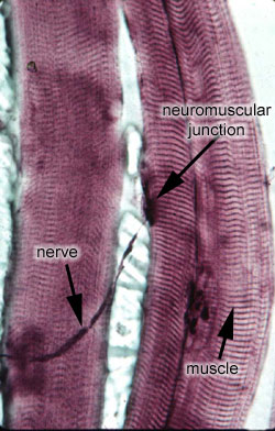
The anatomy of a neuromuscular junction can be divided into three parts:
- the presynaptic terminal (i.e. the motor neuron)
- the synaptic cleft
- the postsynaptic membrane (i.e. the membrane of the muscle cell ).
What are treatments for neuromuscular diseases?
While there’s no cure for MG, there are several treatments that can help you manage the condition:
- Medication, especially immunosuppressants
- Surgery to remove the thymus gland
- Infusions of immunoglobulin
- Plasma exchange
- Lifestyle changes, including dietary changes and avoiding getting overheated
Do I have a neuromuscular problem?
The most common sign of these diseases is muscle weakness. Mayo Clinic neurologists provide comprehensive evaluation of these diseases, including electrodiagnostic studies and other tests. Doctors are active in clinical and laboratory research to improve the diagnosis and treatment of people with neuromuscular diseases.
What is the breakdown of the nervous system?
The nervous system is divided into two major parts: (a) the Central Nervous System and (b) the Peripheral Nervous System. Peripheral Nervous System The peripheral nervous system is made up of thick bundles of axons, called nerves, carrying messages back and forth between the CNS and the muscles, organs, and senses in the periphery of the body (i.e., everything outside the CNS).
What are the main functions of the neuromuscular system?
The neuromuscular system involves our nervous system and muscles working together to control, direct and allow movement of the body.
What is the importance of the neuromuscular?
The neuromuscular junction plays a vital role in the function of skeletal muscle. It is responsible for transducting the excitatory electrical impulse from the nervous system to the muscle fiber, resulting in a muscle fiber action.
What are the parts of the neuromuscular system?
Physiological Anatomy of Neuromuscular Junction For convenience and understanding, the structure of NMJ can be divided into three main parts: a presynaptic part (nerve terminal), the postsynaptic part (motor endplate), and an area between the nerve terminal and motor endplate (synaptic cleft).
What happens to the neuromuscular system during exercise?
Nerve–muscle connections Increased recruitment of additional motor units, which respond in a simultaneous fashion to improve force production. There is an increased activation of synergistic muscles to assist force production for strength, power, speed and hypertrophy.
What is the most common neuromuscular disease?
The most common of these diseases is myasthenia gravis, an autoimmune disease where the immune system produces antibodies that attach themselves to the neuromuscular junction and prevent transmission of the nerve impulse to the muscle.
What are some common neuromuscular disorders?
Neuromuscular Disease OverviewAmyotrophic lateral sclerosis (ALS)Botulism.Congenital myasthenic syndromes.Congenital myopathies.Cramp-fasciculation syndrome.Elevated creatine kinase.Inclusion-body myositis.Lambert-Eaton syndrome.More items...
What are neuromuscular disorders?
Neuromuscular disorders include a wide-range of diseases affecting the peripheral nervous system, which consists of all the motor and sensory nerves that connect the brain and spinal cord to the rest of the body. Progressive muscle weakness is the predominant condition of these disorders.
What are the 7 steps of neuromuscular junction?
Match1) An AP travels down the axon. to the axon terminal.2) Electrical gated calcium channels open. ... 3) Calcium causes the vesicles to. ... 4) ACH diffuses across the synaptic cleft. ... 5) ACH binding opens ion channels. ... 6) If the muscle reaches the threshold (-55mv) at the motor end plate. ... 7) ACH is broken down by.
What are neuromuscular skills?
Neuromuscular performance can be regarded as the ability of the neuromuscular system to functionally control and drive movements by an appropriate integration, coordination and use of sensory feedback, reflex activity, central motor drive, muscle recruitment pattern, muscular excitation-contraction coupling ...
How can I improve my neurological strength?
A person can use exercise to improve the functioning of the nerves that serve the muscles and other peripheral parts of the body. Increasing the activity in the peripheral nervous system strengthens the nerves, in the same way that exercise strengthens the muscles.
How do you increase neuromuscular control?
Cross-exercise is the ability for exercise of 1 limb to cause an increase in strength of the contralateral, nonexercised limb. This mode of exercise is capable of enhancing neuromuscular control by selectively targeting neural pathways that are associated with altered movement patterns.
How do you build neurological strength?
Nervous System Training #1: Train Specific Overload in your Movement NeedsSprinting: Overload with overspeed or “plyosoidal” sprinting.Vertical Jump: Overload with depth jumps.Barbell Squat: Overload with supramaximal repetitions.Throwing: Overload with a lighter throwing implement (5-20% lighter)
What are the benefits of neuromuscular training?
The Benefits of Neuromuscular Training:Enhanced body movement mechanics.Increased muscle strength.Improved functional abilities.Increased speed and agility.Decreased risk of injury in sport.Increased VO2 max and endurance.Corrected muscular imbalances.
What are neuromuscular skills?
Neuromuscular performance can be regarded as the ability of the neuromuscular system to functionally control and drive movements by an appropriate integration, coordination and use of sensory feedback, reflex activity, central motor drive, muscle recruitment pattern, muscular excitation-contraction coupling ...
What is neuromuscular development?
The assembly of a complex neuromuscular circuit involves parallel development of muscles and of the motor and sensory neuronal connections necessary for locomotor functions. In the past, we focused on motor neurons, whereas in recent years, we progressively shifted towards muscles.
What is the neuromuscular system quizlet?
functional unit of the neuromuscular system. consists of single motor neuron together with all of the muscle fibers that its axon supplies. motor units vary in the number of muscle fibers supplied by one motor neuron. Precision movements (eyes and hands) have more motor neurons per muscle fiber.
What is the neuromuscular system?
Neuromuscular System. The neuromuscular system includes assessment of coordination, balance, and gait. Coordination is the ability to perform movements with appropriate speed, distance, direction, rhythm, and muscle tension. When assessing children, normal development of skill acquisition must be taken into consideration ...
What is the neuromuscular system responsible for?
The neuromuscular system is responsible for the generation of force required for motor performance. The aging process induces numerous structural and functional alterations to the neuromuscular system limiting force generation and power production.24,25 In the presence of disease and chronic illness, the loss of force-producing characteristics ...
Which system represents the biomechanical apparatus through which the CNS executes postural actions?
The neuromuscular system represents the biomechanical apparatus through which the CNS executes postural actions. Muscle strength, endurance, latency, torque and power, flexibility, range of motion (ROM), and postural alignment all affect the ability of a person to respond to balance perturbations effectively.
What causes muscle loss in older adults?
Numerous factors contribute to the complex process of age-associated muscle loss, including reductions in anabolic hormones, chronic inflammation, degradation of muscle contractile proteins, loss of regenerative capacity, altered neural activation, and mitochondrial dysfunction. 25 Median values of loss in skeletal muscle mass have been reported to occur at a rate of 0.47% per year in men and 0.37% per year in women when comparing younger (18–45 years) and older (> 65 years) adults. 26 After 75 years of age, the loss of skeletal muscle mass is accelerated in both men (0.80%–0.98% per year) and women (0.64–%0.70%). 26 In addition, older adults experience a preferential loss and marked atrophy in type II muscle fibers with relatively preserved type I fiber number and size. 24–26 Women are more vulnerable to loss of function secondary to type IIa muscle atrophy, 27 and women appear to experience greater declines in muscular strength and power (particularly in the upper extremities) than men. 3 Strength is lost at a disproportionate rate compared with skeletal muscle mass with losses in strength experienced at rates two to five times faster than loss of mass, especially in the legs. 26,28 Therefore, mechanisms other than muscle atrophy contribute to the declines in neuromuscular force. Alterations in neural control, increased fat and connective tissue accumulation, and changes in contractile units in addition to muscle atrophy have all been identified as possible mechanisms leading to reductions in neuromuscular force. 24–26
What are the components of the neuromuscular system?
The neuromuscular system is composed of a neural circuit including motor neurons, sensory neurons, and skeletal muscle fibers. The system is essential to movements of the body, the control of posture, and breathing. The motor nerve fiber makes synaptic contacts with the muscle fiber at the neuromuscular junction. The neuromuscular junction is composed of three cellular elements: the nerve terminal, glial cells (perisynaptic Schwann cells), and the muscle fiber . Inside the nerve terminal, there are numerous synaptic vesicles containing the neurotransmitter, acetylcholine. The electrical impulse at the nerve terminal increases an influx of calcium ions that trigger the exocytosis of synaptic vesicles and release of acetylcholine at the active zone. Acetylcholine binds acetylcholine receptors, which are concentrated at the neuromuscular junction, on the muscle surface. The unbound acetylcholine is rapidly removed by acetylcholinesterase. The binding of acetylcholine to the acetylcholine receptors results in membrane depolarization, which, in turn, triggers a release of calcium ions from the sarcoplasmic reticulum into muscle cytosol and initiates muscle contraction. Agrin and neuregulin play important roles in the aggregation and synthesis of acetylcholine receptors, respectively. The perisynaptic Schwann cells sprout profusely after nerve injury and the glial sprouts may lead and guide nerve terminal regeneration and sprouting. The muscle spindle and the Golgi tendon organ provide information on the muscle length and tension, respectively, to the spinal cord. The motor output and the sensory input constitute a circuit of the stretch reflex that maintains proper muscle fiber length. Neurotoxins, such as α-bungarotoxin, botulinum toxin, and nerve gases are deadly when they stop an animal's breathing by interfering with synaptic transmission in the diaphragm muscle. Diseases of the neuromuscular system include disorders affecting transmission, such as, myasthenia gravis; motor neuron degeneration, such as amyotrophic lateral sclerosis; and muscular dystrophies.
When do neuromuscular movements occur?
These movements should be occurring and actively observable by 6 months of age (Fig. 4.1 ).
What is the purpose of the CNS?
Disorders of the neuromuscular system only rarely escape notice during the history and review of systems; the purpose of the central nervous system (CNS) examination is primarily to assess the severity of the abnormality and the implications for anesthetic care.
What is the NM junction?
The NM junction or motor end plate consists of a presynaptic membrane of the axonal terminal of the motor neuron, the synaptic space, and the postsynaptic membrane and junctional sarcoplasm of the myofiber. The neurotransmitter released at the NM junction is acetylcholine (ACh), which binds to nicotinic receptors in the postsynaptic membrane and generates an end-plate potential. The end-plate potential can result in an action potential and contraction of the skeletal muscle. For more detailed information on anatomy and function of the NM system, the reader is referred to the Suggested Readings.
Where are motor neurons located?
Motor units consist of a single lower motor neuron located in the central nervous system (CNS), either in the cranial nerve nuclei of the brainstem (III to VII, IX to XII) or in the ventral horns of gray matter in the spinal cord. The motor neuron reaches the periphery through its axons, located in the ventral nerve roots and spinal nerves or in the cranial nerves. The axon, which is surrounded by supportive Schwann cells that produce myelin, terminates at the NM junction. Each muscle fiber (myofiber or myocyte) is innervated by a single α-motor neuron, but a single motor neuron may innervate from a few to thousands of myofibers, depending on the function of a given muscle. In muscles such as those of the eye, which are responsible for fine movements, one motor neuron supplies only a few myofibers. In contrast, in muscles involved in posture and locomotion, hundreds to thousands of myofibers are innervated by a single motor neuron. All motor neurons innervating skeletal muscle are excitatory, and a single discharge of a motor neuron results in contraction of all the myofibers that it innervates. However, interneurons within the spinal cord can have either excitatory or inhibitory effects on the motor neurons.
Which structure is responsible for force transmission in muscle?
Whereas adaptations in the force-generating capacity of muscle after training are relatively well characterized, adaptations in the passive (tendon) and active (cross bridges) structures of the series elastic component and the cytoskeletal structures responsible for force transmission within the contracting muscle are less understood.
What does it mean when your eye moves?
Spontaneous ocular movements are also common in patients with disorders of consciousness, specifically roving eye movements from side to side or up and down and blinking at rest or in response to light, sound, or threat. These eye movements do not indicate that the patient is moving toward a higher state of arousal or awareness. Rehabilitation clinicians should be aware that nystagmus is uncommon in patients who are in a coma or vegetative state. 6 Nystagmus is produced by the attempt to bring the eyes back to midline and is seen clinically as quick horizontal eye movements. This requires interaction of the brainstem with the cerebral cortex and this interaction is lacking in patients who are in a coma or vegetative state. Consistent visual tracking is one of the first indicators that a patient is no longer in a vegetative state and has achieved some level of awareness. 12
What are the neuromuscular systems of amphibians?
Amphibian embryos possess only a single major neuromuscular system: the segmental myotomes on the two sides of the trunk and tail and the motoneurones that drive them. Their repertoire of behavioural responses is therefore constrained by what they can do using this system and, in fact, probably all the most obvious possibilities for movement have been investigated over the small range of amphibian species that have been studied. In the past, some of these movements have been used to designate particular ‘physiological’ stages of development (Coghill, 1928 ). Broadly, though, they can be split into two groups: non-rhythmic behaviours and rhythmic behaviours.
What are non-rhythmic movements?
The simplest non-rhythmic movements seen in embryos of the urodele Triturus ( Soffe et al., 1983) and the anurans Rana and Xenopus ( Soffe, 1991a) are twitches, usually termed flexions. The smallest of these are probably driven by weak contractions of the myotomes on one side ( Fig. 5.1A,Bi ). Stronger flexion responses can involve a tight bending to one side (often termed a C-flexion), as in Triturus, or even coiling as in Rana. Again the responses are driven by contractions on one side only and may or may not produce a change of body direction. Triturus embryos can also show S-flexions. Here contraction of myotomes rostrally on one side is associated with contraction caudally on the opposite side.
How do androgens influence the skeletal system?
Androgens exert considerable control over neuromuscular systems that lead to the motivation of male vertebrates to identify, court and then copulate with female conspecifics. Androgens, largely acting through androgen receptors, promote courtship performance by actions on both the CNS and on the skeletal muscular systems ...
Why is electromyography important?
Electromyography also provides a useful common ground between behavioral and neurophysiological studies. Efforts at integration are important. Both behavioral and physiological investigators often forget that behavior is physiology. No one discipline has access to a higher truth.
Is thyroid dysfunction a neuromuscular disorder?
The close relationship between the thyroid gland and the neuromuscular system is indicated by the fact that overactivity of the gland may be associated with four different neurological syndromes—myasthenia gravis, periodic paralysis, and thyrotoxic ‘myopathy’ and ophthalmopathy. The first two syndromes have been discussed in chapters 18 and 15 respectively; it is the thyrotoxic ‘myopathy’ which is of present concern. The precise incidence of this last condition is difficult to determine. Clinically, most patients with hyperthyroidism tire easily and part of their loss of weight may be due to muscle atrophy. Frank weakness of muscles, particularly proximal ones of the limbs, occurs in rather more than half the patients ( Ramsay, 1969; Whitfield and Hudson, 1961 ). Much less common are spontaneous twitchings and cramps of affected muscles.
What is neuroelectrophysiological test?
Neuroelectrophysiological tests are tools that are used to further explore the neuromuscular system. It is not necessary to argue how many exact muscles fibers are involved in composing waveform spikes; instead, we are interested in whether the method can aid in clinical work or in research.
How does the SNB system organize sexual dimorphism?
(8) In the SNB neuromuscular system, sexual dimorphism in the spinal cord is organized through an androgen receptor-mediating mechanism.
Neuromuscular Disease Overview
Neuromuscular diseases affect the function of muscles due to problems with the nerves and muscles in your body. The most common sign of these diseases is muscle weakness.
Research advances that improve clinical care
Doctors are active in clinical and laboratory research to improve the diagnosis and treatment of people with neuromuscular diseases.
Nationally recognized expertise
Mayo Clinic in Rochester, Minn., and Mayo Clinic in Jacksonville, Fla., are ranked among the Best Hospitals for neurology and neurosurgery by U.S. News & World Report. Mayo Clinic in Phoenix/Scottsdale, Ariz., is ranked highly performing for neurology and neurosurgery by U.S. News & World Report.
What is the muscular system?
Definition. The muscular system is a set of tissues in the body with the ability to change shape. Muscle cells connect together and eventually to elements of the skeletal system. When the muscle cells contract, force is created as the muscles pull against the skeleton.
What is the second function of the muscular system?
Circulation. The second and less obvious function of the muscular system is to assist with circulation. Visceral and cardiac muscle tissues surround the blood vessels and lymph vessels that carry crucial nutrients and oxygen to the cells of the body. Cardiac muscle makes up the heart and supplies the main force for blood traveling through the body.
Why are visceral tissues different from skeletal muscles?
The difference in muscular system tissues is due to their very different uses. Skeletal muscles must be able to do a large amount of work quickly, therefore they consist of striated muscle cells, which can contract voluntarily. The smooth muscle tissue found in visceral tissues has fewer energy-producing mitochondria. These tissues are simply used to contract hollow organs and move the fluid inside. The stomach, intestines, and blood vessels are lined with visceral muscles. Cardiac muscle is striated because it needs to produce lots of force, although it is not controlled voluntarily.
What is the function of the head in the muscle cell?
The head releases the actin, reaches forward, and grips the actin again. This moves the protein filaments and contracts the fiber. Depending on the muscle cell, different forms of actin and myosin can be used. In some organisms, completely different proteins are used. Skeletal Muscle Contraction.
How does the muscular system move food?
Much like its ability to move fluids through vessels in the circulatory system, the muscular system also aids in moving food through the digestive system. Most digestive organs are surrounded by smooth muscle tissue. Although the tissue cannot be voluntarily contracted like skeletal muscles, it is controlled subconsciously. When food needs to be moved through the gut, the muscles contract in a synchronized fashion in a wave through the digestive system. These wave-like muscular contractions are called peristalsis.
How does ATP work in the muscular system?
These two components use ATP to pull against each other. They attach to each side of the cell, which shortens the cell as they move past each other. As seen in the graphic below, the muscular system contracts when energy from ATP is applied to the myosin heads of the myosin protein filament. The head releases the actin, reaches forward, ...
Which system relies on the coordination of millions of actin and myosin filaments pulling in the same direction?
In some organisms, completely different proteins are used. Skeletal Muscle Contraction. The muscular system relies on the coordinated action of millions of actin and myosin filaments pulling in the same direction at the same time. To achieve this coordination, muscles are innervated by the nervous system.
What is neuromuscular disorder?
Neuromuscular disorders are conditions that affect the nerves that send electrical signals to muscles to control movement. When the nerves are damaged, communication between the nerves and muscles becomes disrupted. This results in significant muscle weakness, wasting, and loss of function.
How do nerves communicate with muscles?
Nerves communicate with muscles through the release of neurotransmitters at the neuromuscular junction, the space between a nerve cell and a muscle fiber. Neuromuscular disorders can damage the nerve itself or the neuromuscular junction, where the signal is transmitted from a nerve to a muscle.
How many types of muscular dystrophy are there?
There are nine different types of muscular dystrophy, all caused by genetic mutations, but the most common forms are Duchenne muscular dystrophy and Becker muscular dystrophy.
What is a genetic disorder that causes a gradual loss of motor function?
Muscular dystrophies are a group of genetic diseases characterized by a gradual loss of motor function, muscle weakness and wasting away, gait problems, progressive respiratory failure, and cardiomyopathy.
What is MRI in medical terms?
Magnetic resonance imaging (MRI) of your brain and spinal cord to assess for damage