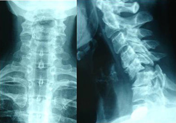
What is a normal cervical spine?
The neck is part of a long flexible column, known as the spinal column or backbone, which extends through most of the body. The cervical spine (neck region) consists of seven bones ( C1-C7 vertebrae ), which are separated from one another by intervertebral discs. These discs allow the spine to move freely and act as shock absorbers during activity.
What is normal cervical rotation?
- Flexion — 50 degrees
- Extension — 80 degrees
- Right lateral flexion — 45 degrees
- Left lateral flexion — 45 degrees
- Right rotation — 80 degrees
- Left rotation — 80 degrees
What is normal cervical lateral flexion?
What is normal cervical lateral flexion? The cervical spine’s range of motion is approximately 80° to 90° of flexion, 70° of extension, 20° to 45° of lateral flexion, and up to 90° of rotation to both sides. What does cervical flexion mean?
What is normal cervical rotation ROM?
The normal ranges of rotation of C1 on C2 are reported to be 50° to each side. Rotation of the atlas on the axis does not occur without a small degree of extension and lateral flexion and sometimes flexion. Cervical spine flexion and extension often create motion in the direction opposite that being experienced in the atlas.
What is the normal range of rotation for C1 on C2?
The normal ranges of rotation of C1 on C2 are reported to be 50° to each side. Rotation of the atlas on the axis does not occur without a small degree of extension and lateral flexion and sometimes flexion.
How do you find the range of motion of the cervical spine?
0:394:21Cervical Spine - Active Range of Motion testing / C1/C2 (AA Joint)YouTubeStart of suggested clipEnd of suggested clipChange position by the person slumping around in the shoulders you almost have a forward headMoreChange position by the person slumping around in the shoulders you almost have a forward head carriage. And then the middle to lower part of the cervical spine almost seems to get a little bit
What is considered normal mobility in the cervical side bending test?
During clinical evaluation, normal mobility on this test is approximately 30° combined rotation to each side, although there are no studies to have reported normal values or that have evaluated the reliability of this test.
How can I improve my neck range of motion?
Sit upright with your shoulder blades squeezed together. Pull your chin in gently (not fast or aggressively), keeping your neck and head straight with your eyes looking forwards (not tipping your head upward or downwards). Hold at the end position for 2 seconds and feel the stretch behind your neck.
How do you test for C1 and C2?
To perform this test, maximally flex the cervical spine followed by maximal rotation either left or right. Flexion is thought to lock out all vertebrae below allowing for rotation at C1-C2 only. The difficulty with the flexion-rotation test is maintaining flexion while maximally rotating the upper cervical spine.
Does C7 move with flexion?
The vertebra that showed no motion in the flexion-extension test was considered to be C7, according to the description of Shin et al. At this point, the evaluator determined whether the most prominent vertebra was coincident with the stationary vertebra determined by the test.
What are the four range of motions of the neck?
Range of Motion (ROM) The normal cervical ROM is as follows: extension, 55 degrees; flexion, 45 degrees; lateral bending, 40 degrees; rotation, 70 degrees.
What is cervical spine flexion?
Cervical flexion: bending the head forward towards the chest. Cervical extension: bending the head backward with the face towards the sky. Cervical rotation: turning the head to the left or the right. Cervical side-bending: tipping the head to the side or touching an ear to the shoulder of the same side.
How does goniometer measure cervical ROM?
0:241:38Cervical flexion and extension ROM using a goniometer - YouTubeYouTubeStart of suggested clipEnd of suggested clipThe instructions that i want to give my patient are to bring your chin to your chest. So i'm goingMoreThe instructions that i want to give my patient are to bring your chin to your chest. So i'm going to have her bring her chest her chin all the way down to her chest the fulcrum of the goniometer.
What are the four range of motions of the neck?
Range of Motion (ROM) The normal cervical ROM is as follows: extension, 55 degrees; flexion, 45 degrees; lateral bending, 40 degrees; rotation, 70 degrees.
How do you measure cervical ROM with inclinometer?
0:061:2444 Cervical Rotation Inclinometers and CROM Device - YouTubeYouTubeStart of suggested clipEnd of suggested clipAdjust the inclinometer dial to zero with the cervical spine at the end of passive. Range record theMoreAdjust the inclinometer dial to zero with the cervical spine at the end of passive. Range record the measurement on the inclinometer. In this example the measurement is 55 degrees of right cervical.
How is cervical lateral flexion measured?
0:051:3541 Cervical Lateral Flexion Goniometer and Tape MeasureYouTubeStart of suggested clipEnd of suggested clipRecord the end feel it is typically firmed for this motion. With a cervical spine positioned at theMoreRecord the end feel it is typically firmed for this motion. With a cervical spine positioned at the end of passive.
Why is there a difference in the spine?
If areas of the spine are misaligned or not moving freely in relationship to each other, tissues on one side of the spine (or even above or below the locked segment [s]) might be making up the difference. Another reason for this might be that there is ADHESED FASCIA present.
What is the position of your nose over your shoulder?
If so, you are engaging in Cervical Rotation. You should be able to get your nose over mid-shoulder (or at the very least, the front of your shoulder ). RIGHT AND LEFT LATERAL FLEXION: These are the movements you would use when you put your ear towards your shoulder (do not cheat and shrug your shoulder up towards your ear).
What causes loss of range of motion?
Besides frequently being related to pain, one of the biggest problems associated with loss of range of motion is DEGENERATIVE JOINTS caused by a loss of PROPRIOCEPTION. In other words, loss of normal range of motion causes deterioration of the joint, and deterioration of the joint causes loss of range of motion.
Can you have a normal ROM of your neck?
Just be sure to understand that it is quite possible to have a “normal” ROM of your neck and still be restricted (HERE). There are several reasons this can happen. For one, everyone is different. Some people are quite flexible, while others were born stiff and inflexible. This is why comparing right to left is so important. For another, you might have VERTEBRAL SUBLUXATION . If areas of the spine are misaligned or not moving freely in relationship to each other, tissues on one side of the spine (or even above or below the locked segment [s]) might be making up the difference. Another reason for this might be that there is ADHESED FASCIA present. In other words, ROM is a wonderful (simple) test, but does not necessarily tell the whole story.
How to assess cervical extension?
This position can be considered as 0°. To assess cervical flexion, ask the patient to nod forward and bring their chin towards their chest. Normal cervical flexion is usually approximately 80º. To assess cervical extension, ask the patient to look upwards as far as possible, until full extension of the neck is achieved . Normal cervical extension is usually 50°. Total range of cervical motion from full flexion to full extension should be 130° (4). Nevertheless, it is possible to gauge if the patient has normal cervical flexion if he/she is able to touch his/her chest with their chin.
What equipment is needed for cervical spine examination?
No specific equipment is required during the general inspection and physical examination of the cervical spine. However, for the range of motion assessment, utilising certain tools allows for an objective and standardised assessment during follow-up. Bedside instruments that can be used may include a measuring tape, compass, draughtsman’s flexible ruler (2), finger-floor method, goniometer, inclinometer and CROM instrument (3). Given that the aim of this paper is specific to bedside instruments, adjuncts such as X-rays, imaging and motorised or electrical equipment like digital goniometers and inclinometers, shall not be mentioned further. A link demonstrating the examination technique using bedside instruments can be viewed at the following link: https://youtu.be/3_ztdQBQMTI.
How to read a goniometer on a patient's head?
Note the reading of the goniometer on the side of the head, which should read at 0° at neutral position. Then, ask the patient to flex and extend his/her neck, at which point readings are recorded at each extreme of the motion (8).
What is the lowest end of the sternal notch?
The lowest end of the sternal notch should be identified as the fixed point or reference point. Then, ask the patient to flex and extend their neck. Measure the distance between the reference point to chin during maximal flexion and extension (5).
How to measure acromion?
Then, ask the patient to flex his/her neck sideways. Measure the distance between the fixed point to the lowest point of the earlobe during maximal lateral flexion. Repeat the same for the contralateral side (5) .
What is the function of the spinal vertebrae?
The function and significance of the spinal vertebrae cannot be overstated. Its roles include serving as anchor for limbs and the head, corralling the spinal cord , besides being a significant attachment to the rib cage and torso muscles. Hence deriving its common name in the literal and figurative sense, the backbone. The vast functions of the spine stems from changes of its structural anatomy across the vertebrae, giving rise to different functional anatomy and mobility properties (1). Thus, examining different parts along the vertebrae may provide different measurements and range of movement during clinical examination.
Where to place inclinometer?
Place the inclinometer device on the top of the patient’s head, along the sagittal plane and ensure the reading on the inclinometer is at 0°. Then, ask the patient to flex and extend the neck. Record readings of the inclinometer at each extreme of the motion (6).
What is the cervical spine?
Cervical Spine. Your cervical spine supports and enables you to move your head. It's made up of seven vertebrae and is shaped like an inward "C" called a lordotic curve. Flexion is dropping your chin to your chest, and the normal ROM is 45 degrees. Extension is dropping your head back and looking up.
What is the degree of spinal motion?
All movement starts from a neutral position, standing up straight, arms to your sides and eyes straight ahead. This is 0 degrees. The four movements measured are flexion, extension, lateral flexion and rotation.
How many vertebrae are in the thoracic spine?
The thoracic section of your spine is made up of 12 vertebrae and shaped like a backward C, called a kyphotic curve. The lumbar spine is made up of five vertebrae, and it curves in like the cervical spine in a lordotic curve. Together, these vertebrae allow you to perform the major movements of the spine.
What is the normal ROM for flexion?
Together, these vertebrae allow you to perform the major movements of the spine. The normal ROM for flexion or forward bending is 90 degrees. For extension, it's approximately 30 degrees. The normal ROM for side bending and rotation is also 30 degrees.
How many bones are there in the spine?
Structure of the Spine. Your spine is made up of 24 bones called vertebrae. These are divided into three groups: cervical, thoracic and lumbar or your neck, mid-back and low-back. In between each bone is a disc that acts as a cushion called the intervertebral disc. The bones are further connected by small muscles called multifidi ...
What is the spinal column made of?
Your spinal column is made up of vertebrae that allow movement.
What are the two regions of the cervical spine?
7, 8, 11, 13 The cervical region of the spine is made up of two anatomically and functionally distinct regions: the suboccipital region and the lower cervical region . The bones of the suboccipital region include the occipital bone and the first and second cervical vertebrae (C1-C2); the third through the seventh cervical vertebrae (C3-C7) make up the lower cervical region ( Fig. 9-1 ).
What connective tissue is found in the cervical spine?
A general overview of the connective tissue of the cervical spine includes the intervertebral discs, which connect the vertebral bodies starting with C2-C3 and below that form intervertebral cartilaginous joints. The supporting ligaments include: anterior longitudinal, posterior longitudinal, ligamentum flavum, interspinous, ligamentum nuchae, and the joint capsules of the facet joints (Fig. 9-4).
How much movement is needed for TMJ?
Stentpetery 14 defined functional mandibular depression (opening) as the minimal amount of movement needed in the TMJ to eat and speak without problems. This author defined functional mandibular depression as 25 to 30 mm. Kraus 10 described functional mandibular depression as the “patient’s ability to actively open his or her mouth to 40 mm.” Magee 12 suggested that “only 25 to 35 mm of opening is needed for everyday activity.” Friedman and Weisberg 6 suggested that the amount of functional opening varies according to the individual’s size, and that on average, an individual should be able to place two to two-and-a-half knuckles between the upper and lower incisors.
What is segmented motion?
Segmental motion occurs as the top vertebra slides onto the bottom vertebra (arthrokinematic movement), whereby the facet joints of the vertebrae contribute to and guide the motion. The direction of movement between two vertebrae is greatly influenced, in large part, by the orientation of the joint surfaces that make up the facet joints. Although movement at each segment of the cervical spine is somewhat small, the combined movement of all cervical segments produces a large, triaxial/triplanar range of motion (ROM) of the whole cervical spine, including the head. During measurement of cervical movement, the combined motions of all facet joints between the occiput and C7 are measured because segmented motion is very difficult to assess accurately. With the neutral, resting position of the head and neck as a point of reference, these multi-segment, osteokinematic movements are called flexion and extension (sagittal plane), right and left lateral flexion (frontal plane), and right and left rotation (transverse plane).
What are the bones of the suboccipital region?
The bones of the suboccipital region include the occipital bone and the first and second cervical vertebrae (C1-C2); the third through the seventh cervical vertebrae (C3-C7) make up the lower cervical region ( Fig. 9-1 ). Fig. 9-1 The cervical spine.
What is the movement of the Atlas and the Occipital Joint?
Movement between the atlas and the occiput (atlanto-occipital joint) is primarily a nodding motion in the sagittal plane about a medial-lateral axis. The axis (C2) has a vertical projection called the dens (also known as the odontoid process) that arises from the superior surface of the body. The dens of the axis fits into a ring formed by ...
Which ligament limits movement?
LIMITATIONS OF MOTION: TEMPOROMANDIBULAR JOINT. The temporomandibular, or lateral, ligament is a strong ligament that limits mandibular depression, protrusion, and lateral deviation. The limitation of protrusion is assisted by the stylomandibular ligament.
What is the cervical spine?
The cervical spine is made up of the seven bones in the neck. At the lower portion of the neck, the spine curves backwards (kyphosis) and becomes the thoracic spine. The thoracic spine consists of the 12 thoracic vertebra and the ribs located on each side. When checking cervical range of motion, the examiner tests the movement of the head, or skull, and neck in flexion, extension, lateral bending and rotation. Normal ranges of motion for the cervical spine include 50 degrees of flexion, 60 degrees of extension, 45 degrees of lateral, or side bending, and 80 degrees of rotation. The ranges of motion for the thoracic spine include 30 degrees of rotation and 50 degrees of kyphosis.
What are the ranges of motion of the shoulder?
Common upper extremity ranges of motions for the shoulder include 170 to 180 degrees of flexion, 50 to 60 degrees of extension, 170 to 180 degrees of abduction for moving the arm away from the body, 80 to 90 degrees of internal rotation, and 90 to 100 degrees of external rotation. Ranges of motion in the elbow and forearm include 90 degrees ...
How many vertebrae are in the lumbar spine?
The lumbar spine has five vertebrae and connects the spine to the pelvis. Normal lumbar ranges of motion include 60 degrees of flexion, 25 degrees of extension, and 25 degrees of lateral, or side, bending.
Where is range of motion measured?
Areas Commonly Tested for Range of Motion. Range of motion is commonly tested in the cervical spine, thoracic spine and lumbar spine. In many sports medicine clinics, range of motion in the upper and lower extremities are also tested. The measured degrees are compared with the expected norm and also from a healthy joint with an injured joint.
What are the ranges of motion of the lower extremities?
Lower extremity ranges of motion for the hip include 120 to 130 degrees of flexion, 10 to 20 degrees of extension, 45 degrees of abduction away from the body, 30 degrees of adduction toward body, 45 degrees of internal rotation, and 50 degrees of external rotation. Knee range of motion consists of the flexion and extension arc of motion which totals 135 to 145 degrees. Ankle range of motion includes 50 degrees of plantar-flexion, or toes pointing toward the ground, and 20 degrees of dorsi-flexion with the toes pointing toward head. It also includes 20 degrees of inversion and 5 degrees of eversion.
What is range of motion?
This movement occurs in the various areas of the body including the spine and extremities. Range of motion refers to the amount of movement that a particular joint or body part can move measured in degrees.
Which vertebrae are located on each side of the neck?
At the lower portion of the neck, the spine curves backwards (kyphosis) and becomes the thoracic spine. The thoracic spine consists of the 12 thoracic vertebra and the ribs located on each side. When checking cervical range of motion, the examiner tests the movement of the head, or skull, and neck in flexion, extension, lateral bending and rotation.
