What is the function of the oculomotor nerve?
The oculomotor nerve is the third cranial nerve (CN III). It allows movement of the eye muscles, constriction of the pupil, focusing the eyes and the position of the upper eyelid. Cranial nerve III works with other cranial nerves to control eye movements and support sensory functioning. Olfactory nerve (CN I) enables sense of smell.
What is oculomotor dysfunction and how is it treated?
However, Oculomotor Dysfunction is a result of an anomaly within the vision system. There are six muscles around each eye and these muscles work together with your brain to accurately control your eye movements. If a patient’s oculomotor muscles are impaired, several essential eye functions may be affected including:
Where is the oculomotor muscle located?
These muscles are located within the eye itself and are both supplied by parasympathetic fibers of the oculomotor nerve. They are actually the anterior extensions of the vascular layer of the eyeball. As such they don’t conform to the typical organization of other muscles with well defined origins and insertions.
Can oculomotor dysfunction be outgrown?
Oculomotor Dysfunction affects patients of all ages, both children and adults. It is not a condition that can be outgrown. If left untreated, Oculomotor Dysfunction patients will rely on compensatory techniques such as using a finger as a marker for reading or tilting the head to direct the eyes.
See more
What is ocular motor control?
Normal visual perception requires the proper functioning of ocular motor systems that control the position and movement of the eyes to focus the image of the object-of-interest (i.e., the visual target) on corresponding areas of the retinas of the two eyes.
What are oculomotor skills?
Ocular Motor Skills: There are three basic types of eye movements: Fixations: ability to hold eyes steady without moving off target. Saccades: the ability of our eyes to make accurate jumps as we change targets. Pursuits: the ability of our eyes to follow a moving targets.
What are oculomotor issues?
Oculomotor dysfunction is a medical condition in which the eyes are unable to work together while tracking. Symptoms can occur while following a moving object, or while moving the eyes between two objects or words. There is no known cause for oculomotor dysfunction.
What controls eye movement in the brain?
The cerebellum plays a pivotal role in the control of eye movements. Its core function is to optimize ocular motor performance so that images of objects of interest are promptly brought to the fovea – where visual acuity is best – and kept quietly there, so the brain has time to analyze and interpret the visual scene.
How common is oculomotor dysfunction?
Oculomotor Dysfunction is a relatively common visual condition that can affect individuals of all ages, usually due to a developmental delay or a result of a concussion (mTBI) or more serious traumatic brain injury (TBI).
What happens if the oculomotor nerve is damaged?
The oculomotor (third) cranial nerve plays an important role in the efferent visual system by controlling ipsilateral eye movements, pupil constriction, and upper eyelid elevation. Accordingly, damage to the third cranial nerve may cause diplopia, pupil mydriasis, and/or upper eyelid ptosis.
How do you fix OculoMotor dysfunction?
Treatment for Oculomotor Dysfunction often includes a form of vision therapy involving specific neuro-optometry activities designed to improve fixation, strengthen your visual muscles, saccadic and pursuit eye movements, as well as improve information processing skills.
Can ocular motor dysfunction be cured?
Your eye muscles are just like other muscles in your body; exercising them can help them perform better. As a result, vision therapy is one of the best treatments for ocular motor dysfunction. Vision therapy allows you to practice new skills to strengthen eye muscles' ability to work together effectively.
How do you test for ocular motor dysfunction?
0:533:32Do I Have Oculomotor Dysfunction? | Oculomotor Dysfunction TestsYouTubeStart of suggested clipEnd of suggested clipTest so go ahead come on in here what we're gonna be doing is having the subject or you lookMoreTest so go ahead come on in here what we're gonna be doing is having the subject or you look straight at an object in front of you. And then what i want him to do is move his head around while keeping
Does the brain control eyes?
Sight is a complex function of the brain that extends from the front to the back of the head. To produce sight, the eyes capture information and send it through the optic nerve to be processed by the occipital lobe.
What part of the brain controls vision and hearing?
Cerebrum: is the largest part of the brain and is composed of right and left hemispheres. It performs higher functions like interpreting touch, vision and hearing, as well as speech, reasoning, emotions, learning, and fine control of movement. Cerebellum: is located under the cerebrum.
Does the frontal lobe control the eyes?
The frontal lobe is critical for motor execution and eye movement. The temporal lobe is critical for auditory processing and visual and language memory. The parietal lobe is critical for sensory processing. The occipital lobe is critical for vision and visual processing.
Which of the following is the function of oculomotor?
The oculomotor nerve is the third cranial nerve (CN III). It enables eye movements, such as focusing on an object that's in motion. Cranial nerve III also makes it possible to move your eyes up, down and side to side.
What are visual perceptual skills?
Visual perceptual skills enable a child to make sense of and interpret what they are seeing. These skills include: Visual discrimination - matching two objects that are the same. Visual memory - the ability to remember visual information.
What does ocular motility mean?
The term ocular motility disturbance refers to any abnormal eye alignment or difficulty in controlling eye movements. We most often consider only the eye itself as the cause of low vision, and certainly factors such as near-sightedness, far-sightedness and cataracts take their toll.
Where is the oculomotor nerve located?
The oculomotor nerve is no exception. The cell bodies of the oculomotor nerve are located within two nuclei positioned close to one another, posteromedially in the midbrain, the most superior component of the brainstem.
What is the oculomotor nerve?
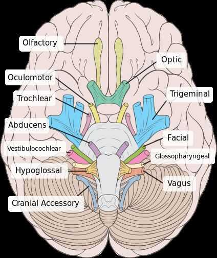
The oculomotor nerve is one of 12 sets of cranial nerves. Many of these nerves are part of the autonomic nervous system. The autonomic nervous system supplies (innervates) organs, like your eyes.
What nerve controls the movement of the eye?
The oculomotor nerve is the third cranial nerve. It controls four of the six muscles that enable eye movement. Conditions affecting cranial nerve III include third nerve palsy. It is often a complication of medical issues such as uncontrolled diabetes or a brain aneurysm. Third nerve palsy might impact your vision and the appearance of one or both of your eyes. Many people make a full recovery, although it can take around two months.
Which cranial nerve is responsible for eye movements?
The oculomotor nerve is the third cranial nerve (CN III). It enables eye movements, such as focusing on an object that’s in motion. Cranial nerve III also makes it possible to move your eyes up, down and side to side.
Which organ controls your ability to raise your upper eyelid?
Levator palpebrae superioris, which controls your ability to raise your upper eyelid.
Which reflex adjusts eye positioning when your head is moving?
Vestibule-ocular reflex, which adjust s eye positioning when your head is moving.
What causes oculomotor dysfunction?
The causes of Oculomotor Dysfunction range from slow development to disease of the central nervous system. An eye exam easily diagnoses this condition.
What happens if your oculomotor muscles are ineffective?
If one’s oculomotor muscles are ineffective, he may have difficulty reading–he may easily lose his place or repeat sentences. People with this condition may also have difficulty with balance, depth perception, sports, or hand eye coordination. Virtually every task requires good control of eye movements.
What is the oculomotor nerve?
The oculomotor nerve is the third cranial nerve (CNIII), and one instance in which the name is a clear indication of the function of the nerve (Oculo = pertaining to the eye, motor = producing movement). Simply from the name then, it is easy to know that the oculomotor nerve will innervate muscles that move the eye itself or components of the eye.
How to test for oculomotor nerve involvement?
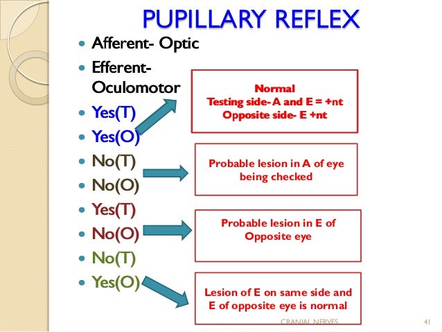
However, also testing the pupillary light reflex will give a more complete picture of oculomotor nerve involvement. This is normally accomplished by shining a bright light into one eye and watching for pupil constriction in the eye being stimulated with light, and also the contralateral eye. Information that is coming into the brain from the eyes travels via the optic nerve (CNII). This information is sent to both the right and left Edinger-Westphal (EW) nuclei, regardless of which eye is being tested. If the practitioner shines a light in one eye and both pupils constrict, this indicates that both the optic and oculomotor nerves are intact. If the light is shone in one eye and neither pupil constricts, an absence of both the direct and consensual light reflexes, this indicates a lesion to the optic nerve in that eye. In this case the information doesn’t reach the EW nucleus to be passed to the oculomotor nerve in either eye.
What nerve innervates the eye?
Simply from the name then, it is easy to know that the oculomotor nerve will innervate muscles that move the eye itself or components of the eye. It is the movement producing functions of the nerve that make it a useful indicator of brain injury. To understand the important clinical significance of the oculomotor nerve, ...
Why is it important to test the oculomotor nerve?
Testing the proper functioning of the oculomotor nerve can be quite simple, but it is incredibly important to test both the somatic and visceral motor functioning of the nerve. In addition, testing must be combined with an understanding of the symptoms that would occur from damage to the oculomotor nerve structures within different regions of the brainstem, and also outside of the brainstem.
How does the somatic motor function of the eye work?
As we know, the somatic motor function of the nerve supplies input to the majority of the muscles that move the eye. Often the proper functioning of these muscles is tested by getting the patient to follow an object, such as a pen, with their eyes in an ‘H’ pattern. For oculomotor nerve functioning, the practitioner needs to pay special attention to adduction, elevation, and depression of the eye to see if these movements occur. Also he or she must observe the eyelid to see if it is drooping or not.
What is the visceral motor axon?
Visceral motor function. The visceral motor axons of the oculomotor nerve are part of the autonomic nervous system, specifically the parasympathetic division. They will arise from the Edinger-Westphal nucleus and innervate two separate intrinsic muscles within the eye.
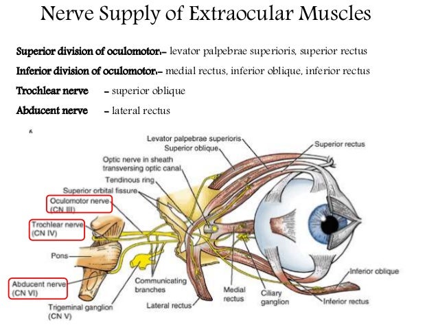
How many extrinsic muscles are there in the eye?
There are seven extrinsic eye muscles (muscles that lay outside of the eye itself) that move the superior eyelid and the eyeball. Five of them are innervated by the oculomotor nerve and will be discussed in detail below.
What is the oculomotor nerve?
The oculomotor nerve is the third of 12 pairs of cranial nerves in the brain. This nerve is responsible for eyeball and eyelid movement.
Which nerve controls the eye?
The oculomotor nerve involves two separate components, each of which has a distinct function. The somatic motor component supplies four extraocular muscles in the eye and the upper eyelid’s levator palpebrae superioris with motor (movement) fibers. It controls the muscles that allow for visual tracking and fixation by the eye.
What is accommodation in the eye?
Accommodation is the ability of the eye to keep an object in focus as the object’s distance from the eye changes. Pupillary light reflexes are automatic changes in dilation (size) of the pupil, which regulate the amount of light that enters the eye, making sure the light is enough to see but not too bright.
Which motor component controls parasympathetic innervation?
The visceral motor component controls parasympathetic innervation (nerves related to involuntary actions) of the ciliary muscles and constrictor papillae, aiding in accommodation and pupillary light reflexes. Accommodation is the ability of the eye to keep an object in focus as the object’s distance from the eye changes. Pupillary light reflexes are automatic changes in dilation (size) of the pupil, which regulate the amount of light that enters the eye, making sure the light is enough to see but not too bright.
Can oculomotor nerves be paralyzed?
The oculomotor nerve can become paralyzed in a condition known as oculomotor nerve palsy. This condition can result from multiple sclerosis or other demyelinating diseases, direct trauma, space-occupying lesions (such as brain cancer), microvascular disease (such as diabetes), or spontaneous subarachnoid hemorrhage (bleeding into the space between two of the membranes that cover the brain). A berry aneurysm is a type of subarachnoid hemorrhage.
Where does the oculomotor nerve start?
The oculomotor nerve begins at the brainstem, which is a structure low in the back of your brain that connects the brain to the spinal column. In the brainstem, two clusters of neurons called nuclei give rise to the oculomotor nerve.
What causes oculomotor palsy?
It's caused by compression of the nerve at the junction of the posterior communicating artery and the internal carotid artery. Symptoms of congenital oculomotor palsy include: A pupil that's "fixed" (doesn't change size in response to light) on the same side as the compression.
What nerves are involved in eye movement?
Anatomy. Function. Associated Conditions. Treatment. The oculomotor nerve enables most of your eye movements, some aspects of vision, and raising the eyelid. It's the third cranial nerve and works with cranial nerves four ( trochlear) and five ( trigeminal) to coordinate eye movement. The oculomotor nerve contains both motor ...
How long does it take to recover from oculomotor palsy?
1 If this approach hasn't lead to much improvement after six months, surgery may be considered.
Which nerve is responsible for the movement of the eye?
Motor function means movement, and the oculomotor nerve is responsible for much of the movement associated with your eyes.
Which muscle keeps the eyelid open?
The sympathetic fibers from the internal carotid plexus that travel with the oculomotor nerve provide motor function to the superior tarsal muscle, which keeps the eyelid open once the levator palpabrae superioris raises it.
Can oculomotor nerves be damaged?
The oculo motor nerve can be damaged or paralyzed in numerous ways. This is called acquired oculomotor palsy and is different from congenital oculomotor palsy, which was discussed above.
What is oculomotor dysfunction?
Oculomotor Dysfunction is a common vision problem and occurs in people of all ages, both children and adults. Oculomotor Dysfunction affects reading, sports, balance, depth perception as well as most visually related tasks. Oculomotor Dysfunction is not a condition that is “out grown”. Instead, over time, an individual develops compensatory ...
How long does it take to treat oculomotor dysfunction?
Oculomotor Dysfunction complicated by accommodatative or convergence deficits typically require an additional 6 to 12 hours of in-office therapy.
What is vision therapy?
Vision therapy involves using lenses, prisms, and specific eye and brain activities designed to improve fixation and saccadic eye movements; integrate oculomotor skills with vergence and accommodative systems; integrate oculomotor skills with information processing.
Why are oculomotor dyslexia and dyslexia confused?
Oculomotor Dysfunction and Dyslexia are often times confused because the symptoms can look very similar. Typical symptoms of Oculomotor Dysfunction include a reluctance or avoidance of reading, poor reading comprehension and frequently rereading the same word or sentence. Often times, the individual will use their fingers or a reading strip ...
What is a CVA stroke?
Oculomotor Dysfunction associated with acquired brain injury or a cerebrovascular accident (CVA), also referred to as stroke, or other types of systemic conditions require additional hours of in-office therapy.
What is OMD in medical terms?
Oculomotor Dysfunction (OMD) is also known as Ocular Motility Dysfunction and is characterized by a deficiency in one or more of the following visual skills:
What type of test does an optometrist use?
An optometrist, such as Dr. Ryan Johnson, residency trained in neuro-optometry, binocular vision and vision therapy will use qualitative chairside testing and observation as well as quantitative (measurable) standardized tests.
What Is Oculomotor Dysfunction?

Eye Movements Affected
- There are six muscles around each eye and these muscles work together with your brain to accurately control your eye movements. If a patient’s oculomotor muscles are impaired, several essential eye functions may be affected including: 1. Convergence/Divergence– the ability to move the eyes inward or outward to focus on objects near or far 2. Saccades– the ability to jum…
Diagnosis
- Oculomotor Dysfunction should be diagnosed and treated by an optometrist who has education and experience in neuro-optometry and vision therapy. A comprehensive eye exam including a binocular vision assessment can help to identify and diagnose Oculomotor Dysfunction as well as any other underlying conditions that may be affecting you or your child’s vision.
Treatment
- Treatment for Oculomotor Dysfunction often includes a form of vision therapy involving specific neuro-optometry activities designed to improve fixation, strengthen your visual muscles, saccadic and pursuit eye movements, as well as improve information processing skills. Our board-certified optometrists and vision therapists promise a personalized approach with one-on-one treatment…