What is a ray amputation of the hand?
In a finger example, ray amputations are the removal of an entire finger along with the corresponding metacarpal bones in the hand. They are same-day surgeries with the patient going home with a bulky soft dressing. The recoveries can vary, but light use of the hand is almost immediate.
What is first ray amputation?
We defined the partial first ray amputation as occurring distal to the first metatarsocuneiform joint, including the phalanges of the hallux, with primary closure at the time of surgery.
What is Ray surgery?
Ray resection for localized necrosis, infection, and osteomyelitis is an accepted procedure allowing removal of the diseased toe and metatarsal. The traditional approach involves a rather lengthy incision and dissection that can compromise the vascular supply to the remaining forefoot.
What is a third ray amputation?
Ray amputation The removal of a single metatarsal in the middle of the foot (ie, the second, third, or fourth metatarsal) results in a V-shaped wedge, which again maintains good function.
How do you do a ray amputation?
The technique of central ray amputation involves the use of a circumferential incision at the midproximal phalanx in conjunction with a dorsal longitudinal incision. The dorsal incision is extended through the extensor. The periosteum is scored at the level of the metacarpal base.
What is 5th ray amputation?
Traditional Partial Fifth Ray Amputation The traditional approach to partial fifth ray amputation involves a standard tennis racket incision consisting of dorsal and plantar soft tissue flaps with midline proximal extension along the lateral border of the foot (Fig. 18.13).
How is a toe amputated?
You will get medicine to help you relax and numb your foot. Then your doctor will make a cut (incision) to remove your toe. If you have healthy skin to cover the wound and have no signs of infection, the doctor will then try to close the wound.
How long does it take to recover from Gamma Knife surgery?
After the procedure, a patient will typically spend 3-5 days recovering in the hospital before being released to return home. Brain tumor recovery following traditional surgery can be relatively lengthy, including activity and work restrictions ranging from 4-8 weeks.
What is finger amputation?
What is amputation? Amputation is the complete removal of an injured or deformed body part. An amputation may be the result of a traumatic injury or may be the result of a planned operation where the finger must be removed.
Why is Ray amputation?
Ray amputation was first described in the early 20th century [3-6], and it was performed on those with proximal interphalangeal joint dysfunction or loss of proximal phalangeal skeleton; it was also used for the treatment of traumatic hand injuries, infections, and tumors [7].
What are the types of amputations?
Common types of amputation involve:Above-knee amputation, removing part of the thigh, knee, shin, foot and toes.Below-knee amputation, removing the lower leg, foot and toes.Arm amputation.Hand amputation.Finger amputation.Foot amputation, removing part of the foot.Toe amputation.
What is a partial Ray amputation of foot?
Partial first-ray resections are used to help salvage the foot and maintain bipedal ambulation. Losing the first metatarsophalangeal joint has biomechanical consequences that lead to further foot deformities and result in more proximal amputations of the ipsilateral limb, such as a transmetatarsal amputation.
What is the first ray of the foot?
The first ray is the segment of the foot composed of the first metatarsal and first cuneiform bones. The location of this joint is important as it intersects the transverse and medial longitudinal arches. This segment serves as a critical element in the structural integrity of the foot.
Where is the 1st metatarsal?
The first metatarsal bone is the bone in the foot just behind the big toe. The first metatarsal bone is the shortest of the metatarsal bones and by far the thickest and strongest of them.
What is Syme amputation?
Syme amputation (SA) is a term used to describe an amputation at the level of the ankle joint in which the heel pad is preserved. It is performed for a number of indications in a pediatric population. SA is purported to hold the advantage of allowing weight bearing without a prosthesis.
What is left hallux amputation?
What Is a Hallux Amputation? A hallux amputation is the partial or total removal of a person's big toe. Typically, you'd undergo a hallux amputation for one of several reasons. For example, you might have undergone trauma or injury or your toe might be infected.
Why do we need to amputation?
The amputation can be performed so that it takes several looks to notice that a digit is missing. The middle digits are a little tougher, but the gap created can be closed so that the cosmetics and functionality are positive. Also, even for people who work with their hands, you're back at work fairly quickly.
What is the procedure called when you remove a finger?
In situations where we are removing digits such as fingers, hand surgeons perform an operation called 'ray amputations '. In a finger example, ray amputations are the removal of an entire finger along with the corresponding metacarpal bones in the hand.
Is it bad to exercise after amputation?
The exercise can help reduce any swelling present as well. Cosmetically, it's not too bad either. Especially when the amputation involves a border digit such as the pointer finger or the pinky.
Can you get back to life after amputation?
While initially thought of as a scary and life-altering incident, amputations of the digits are actually quite common and people get back to living their life at just about the same speed as before. Your body and mind are capable of working together so that you'll develop new, slightly different movements to compensate for the new biological logistics.
Is ray amputation scary?
Ray Amputations. "Amputation" is a frightening word. Many people find the instant images presented in their minds as unpleasant and uncomfortable. When it is a medical treatment possibility for you… it is even more fearful.
What is digit amputation?
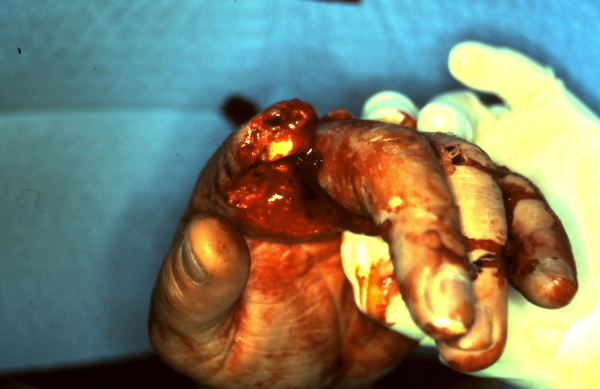
Digit amputations are most commonly performed for bite wounds to the digit or for traumatic degloving, shearing, or crush injuries. Initial management following an injury should be conservative, as many injuries will resolve with treatment with NSAIDs and antibiotics, and marmosets can function well with osteoarthritis and ankylosis of the interphalangeal joints. Digital wounds with severe soft tissue injury and cellulitis, however, may require amputation. Prior to surgery, local anesthetic is applied within the joint at the site of amputation and in the soft tissues. The level of amputation is determined by the extent of the injury and health of the soft tissue, as these will determine if closure of the wound can be achieved. Due to the small size of the digits, dissection of the deeper tissues (flexor and extensor tendons) is limited. For amputation, a circumferential skin incision should be made at a point distal to the joint to allow for disarticulation of the interphalangeal joint via transection of the flexor and extensor tendons, as well as collateral ligaments and joint capsule. Digital arteries and veins can be difficult to identify and ligate, so hemostasis is generally managed by compression and cautery. Closure of the surgical site can include an interrupted subcutaneous suture with simple interrupted skin sutures. A light bandage can be applied over the surgical site but must allow for use of the remaining digits for ambulation around the cage. The authors recommend a small bandage of a nonadherent dressing (Telfa, Coviden, Mansfield, MA, USA) trimmed to cover only the immediate surgical site followed by an adherent dressing retention sheet (Hypafix, BSN Medical, Charlotte, NC, USA or Tegaderm, 3M Health care, St. Paul, MN, USA). Analgesia should include an NSAID to help reduce inflammation at the surgical site.
What is the treatment for sharp amputation?
For sharp amputations where the removed digit is in a clean and fresh state, immediate re-implantation is considered the treatment of choice, which involves microsurgical anastomosis of nerves, blood vessels, bone, and tendon.
What is lumbrical plus digit?
Lumbrical plus digit (paradoxical extension) occurs when the interphalangeal (IP) joints of the injured finger extend when the patient attempts to actively perform a composite fist. When an amputation occurs in which the FDP integrity is disrupted, the FDP muscle retracts, thus pulling the lumbrical origin proximally, which increases tension on its insertion at the radial lateral band. Thus, with FDP contraction, force is transmitted through the tightened lumbrical to the extensor mechanism, causing an extension force of the IP joints. 11,18,19
How many members of Boryoku Dan have had their fingers cut off?
It was found that 194 of 741 (or 26%) members of Boryoku-Dan and 95 of 355 (or 27%) members of Boryoku-Josho-Sha have had their fingers cut off. Digital amputation is usually done at the middle phalanx of the left little finger.
Why are digit prostheses not suitable for pediatric patients?
These prosthetic devices are often associated with complications, such as erosion, infection, and inflammation. Moreover, prosthetic devices are not suitable for pediatric or adolescent patients, as their tissues grow with age. Therefore, most patients decide not to wear the prosthesis, due to the lack of gain in function and its associated complications . Despite the rapid technological advances in medicine, treatment options for digit reconstruction are severely limited. Currently available modalities are involved with a prolonged treatment period, multiple stage surgeries, a lengthy rehabilitation process for a questionable restoration of normal function, and prostheses and surgery-associated complications.
Where is revision finger amputation done?
In general, revision finger amputations are done through the bony shaft, rather than at joint level. Knowing the anatomy of the fingers is important for maintaining attachments of flexor and extensor tendons if possible, as well as contouring bone appropriately for the revision stump (Fig. 3.2A and B ).
Why are tendon resected?
Tendons are resected away from the skin closure to prevent postoperative tendon imbalance or quadrigia. Nerves are resected back from the closure site to make neuroma formation or tip hypersensitivity less likely. It is important to maintain the integrity and mobility of the proximal joints to optimize hand function.
What is a ray resection?
Ray resection, which was pioneered by Bunnell in the 1920s, was initially performed as a salvage procedure for dysfunction of the proximal interphalangeal joint. Successful ray resection with or without an adjacent ray transfer can be useful for treating vascular insufficiency, tumors, infection, trauma, recurrent Dupuytren contracture, ...
What is the name of the journal that studies ray resections of the fingers?
Ray Resections of the Fingers: Indications, Techniques, and... : JAAOS - Journal of the American Academy of Orthopaedic Surgeons
What is the best incision for a central ray resection?
A racquet or V-shaped incision can be used for the border digits ( Figures 2 and 3 ). A web space–preserving incision pioneered by Plasschaert and Hage 25 may be used for a central ray resection, with good results reported ( Figure 4 ). Most authors agree with extending the proximal aspect of the incision using a Brunner zig-zag incision to prevent the development of contractures. 11,15,25 Resection of the central digits is unique in that it may result in difficulty with small objects falling through the persistent gap 3,6,8,11,14-16,18-26 ( Figure 5 ). Thus, central digits are treated with resection and soft-tissue imbrication, bony transposition, carpal wedge resections, or total metacarpal resections to close palmar gaps ( Figure 6 ). Border resections are performed without transposition. Following single ray resection of the fingers, grip and pinch strength can be expected to be 70% to 85% of the strength of the uninvolved hand. 1,2,4,7-11,14-16,18-22,24 Patients are generally satisfied with the outcomes of ray resection, given the improved dexterity and good functional and cosmetic outcomes.
What are the indications for a ray resection?
The one absolute indication for ray resection is ischemic necrosis involving the metacarpal. 1 Severe dysfunction of the proximal interphalangeal joint and amputations at the level of the proximal phalanx are also classic indications for ray resection. 1-6 Some authors argue that maintenance of the metacarpal head and the transverse arch is important for grip stability and strength 7 and advocate that ray resection should be reserved for extreme cases. Amputations performed distal to the proximal interphalangeal joint function well without ray resection. However, a stiff obstructive finger, regardless of length, may result in decreased function and dexterity of the remainder of the hand and, in some cases, a painful, repeatedly traumatized useless digit. 1,8,9 The advantages of ray resection are gap elimination, removal of a cumbersome or painful digit, and better cosmesis in most cases. 2-6,10-22,23 The disadvantages of the procedure include decreased grip and pinch strength, decreased palm width, and an abnormal finger count. 5,7,17,23 Many authors have described the importance of hand width and grip strength to laborers and have recommended against ray resections in this population, 9,14,23 but others have reported good results even in these patients. 6 The primary contraindication is any psychological barrier to amputation because resection may lead to emotional distress.
How to repair the deep transverse intermetacarpal ligament?
Repair of the deep transverse intermetacarpal ligament is performed after bony stabilization. During resection of the long finger, the adductor pollicis origin may be transferred to the transposed index finger to preserve adduction of the thumb. 11,14,18 This is commonly performed by subperiosteal elevation of the origin of the adductor off the long finger and placement of a braided suture to tag the origin. The adductor is then transferred to the transposed index finger with soft-tissue imbrication, 14 through drill holes, 11 or with a suture anchor or anchors. No consensus exists on whether the transfer of the adductor pollicis is necessary. 8 Carroll 8 suggested that subperiosteal elevation without repair allows scarring of the adductor into a functional position.
How strong is the finger after a ray resection?
As with ray resection of border fingers, strength of the central fingers after ray resection has been heavily studied. A decrease in grip, key pinch, and three-point pinch strength of at least 20% to 30% ( even as much as 50% in some cases) can be expected after central ray resection, with or without transposition. 2,3,8-11,14
How long does it take for a finger to be immobilized after border ray resection?
The patient’s finger is immobilized in a bulky soft dressing or protective splint, with the hand placed in the intrinsic-plus position. A drain may be placed at the discretion of the surgeon. The splint is removed 2 to 7 days postoperatively, and a wound check is performed. Active ROM is encouraged to decrease postoperative stiffness. In border ray resection, the patient may be allowed to progress to unrestricted motion after the wound has healed. 1,5,7,13,23
What is a partial first ray amputation?
A partial first ray amputation, an amputation at any level of the hallux or first metatarsal, is a common limb salvage procedure in many of these diabet ic patients. Many patients opt to have a partial first ray resection rather than more a radical surgery because of the many social and psychological factors that accompany an amputation. Unfortunately, amputations at this level can significantly alter the biomechanics of the foot. This in turn may lead to secondary skin breakdown due to increased pressure on areas of the foot that are not intended to bear these loads. As practitioners, it is our responsibility to provide patients with appropriate information, and the risks with each option, allowing them to make appropriate decisions. This article reviews the mechanical impact of the first ray, and the success rates of resections at various levels throughout the first ray.
What is the first ray of the foot?
The first ray is one component of the medial column of the foot and therefore a primary load bearer during gait. The first ray consists of the hallux, the first metatarsal, and the medial cuneiform. It functions in the frontal and longitudinal planes following subtalar joint (STJ) motion. As the STJ pronates, the first ray dorsiflexes and inverts. Inversely, as the STJ supinates, the first ray plantarflexes and everts. These motions allow the first ray to act as a shock absorber or rigid support structure pending its motion.3 The medial column is often referred to as a beam and truss structure. During the early stance phase of gait, the pronated STJ allows the first ray to dorsilfex while it comes into contact with the ground. This motion aids in dispersion of shock from heel strike. During heel strike, the first ray is acting as a beam resisting tensile forces. A lack of dorsiflexion leads to increased pressure under the 1st metatarsal head. In addition, a lack of dorsiflexion of the first ray prevents internal tibial rotation and calcaneal eversion, further preventing the proximal limb from aiding in shock absorption.3
What is the principle of central ray amputation?
The principles of a central ray amputation include removal of the injured finger at the metacarpal base, correcting the rotational deformity, closing the space between the 2 adjacent unamputated fingers, and achieving a satisfactory appearance of the hand.This illustration depicts 1 of 2 techniques that have been described regarding central ray amputation. The procedure involves the transfer of the index finger ray onto the third metacarpal base for the middle finger and the small finger to the ring metacarpal base. The disadvantages of the ray transfer procedure are the requirement for postoperative immobilization and the risk of nonunion.
How is central ray amputation done?
The technique of central ray amputation involves the use of a circumferential incision at the midproximal phalanx in conjunction with a dorsal longitudinal incision (see the images below). The dorsal incision is extended through the extensor. The periosteum is scored at the level of the metacarpal base. The metacarpal is transected at its base. Then, the hand is supinated, and the flexor is divided. The neurovascular bundles are divided proximally to avoid neuroma formation at the skin incision. The deep transverse metacarpal ligaments are identified on either side of the volar plate of the involved finger at the MCP joint.
What is the metacarpal transected at?
With a central ray amputation, the metacarpal is transected at its base. The hand is then supinated and the flexor is divided.
What is the dorsal longitudinal incision for index ray amputation?
In performing an index ray amputation, a dorsal longitudinal incision over the index metacarpal is used in conjunction with a circumferential skin incision at the midproximal phalangeal level. The skin is intentionally left long distally to avoid deficiency that could result in a web-space contracture. (See the image below.)
What is the dorsal incision?
The dorsal incision is extended through the extensor. The periosteum is scored at the level of the metacarpal base. In performing a central ray amputation, the dorsal incision is performed in a tennis racket configuration. The volar incision is completed in the shape of a wedge to facilitate closure without a dog ear.
How is finger ray amputation performed?
The procedure of small finger ray amputation is performed through a tennis racquet incision.
What happens when you amputate your middle finger?
When the middle and ring fingers are amputated at a level near the MCP level, small objects fall through this area, which is created by the gap of the missing digit. Patients describe difficulty in retrieving change from their pockets. This can be corrected with a ray amputation. However, the loss in grip strength and pronation strength has to be considered before ray amputations are performed for these central digits.