What is the action potential of a motor neuron?
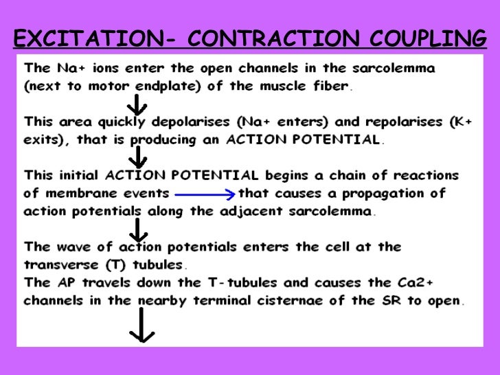
Action Potential An area of muscle fiber membrane that is in close association with the axon terminal of the motor neuron, contain receptors for acetylcholine Motor end plate An area that contains many synaptic vesicles filled with acetylcholine
What is released from the axon terminals of the motor neuron?
Acetylcholine is released by axon terminals of the motor neuron. Action potentials travel the length of the axons of motor neurons to the axon terminals. These motor neurons __________.
What triggers action potential in the axon?
1. electrical potential is set up (resting potential). 2. allowed to suddenly discharge (action potential). 3. triggers action potential further along the axon. Branchlike parts of a neuron that detect information from other neurons. site in neuron where information from thousands of other neurons is collected and integrated.
What happens during the action potential phase?
During the Action Potential. When an impulse is sent out from a cell body, the sodium channels open and the positive sodium cells surge into the cell. Once the cell reaches a certain threshold, an action potential will fire, sending the electrical signal down the axon.

What is the action potential of a motor neuron?
They process synaptic inputs leading up to the production of an action potential in the motor neuron. The action potential propagates down the single axon toward the muscle cell where it makes a junction, variously called the neuromuscular junction, motor end plate, or myoneural junction.
What do the motor neurons release?
Motor neurons release the neurotransmitter acetylcholine at a synapse called the neuromuscular junction. When the acetylcholine binds to acetylcholine receptors on the muscle fiber, an action potential is propagated along the muscle fiber in both directions (see Chapter 4 of Section I for review).
Which substance is released by motor neurons to stimulate a contraction?
When the nervous system signal reaches the neuromuscular junction a chemical message is released by the motor neuron. The chemical message, a neurotransmitter called acetylcholine, binds to receptors on the outside of the muscle fiber. That starts a chemical reaction within the muscle.
When an action potential from a motor neuron arrives?
When an action potential from a motor neuron arrives at the neuromuscular junction (NMJ), a series of events occurs that leads to muscle contraction.
What neurotransmitter is released at the neuromuscular junction?
Acetylcholine (ACh)Acetylcholine (ACh) is the principal neurotransmitter at the vertebrate neuromuscular junction (NMJ), however since the discovery that motoneurons and presynaptic terminals of rodent endplates from the hindlimb muscles extensor digitorum longus (EDL) and soleus are positive for glutamate labelling [1,2], it has been ...
What neurotransmitters are released by the sympathetic nervous system?
Both the sympathetic and parasympathetic nerves release neurotransmitters, primarily norepinephrine and epinephrine for the sympathetic nervous system, and acetylcholine for the parasympathetic nervous system.
Which substance is released by motor neurons to stimulate a contraction quizlet?
A nerve action potential that is initiated in the cell body of a spinal motor neuron propagates out the ventral roots and eventually invades the synaptic terminals of the motor neurons. As a result of the action potential, the chemical transmitter acetylcholine (ACh) is released into the synaptic cleft.
What happens when an action potential reaches motor end plates?
When an action potential reaches the axon terminal of a motor neuron, vesicles carrying neurotransmitters (mostly acetylcholine) are exocytosed and the contents are released into the neuromuscular junction. These neurotransmitters bind to receptors on the postsynaptic membrane and lead to its depolarization.
When muscles contract what is released quizlet?
Muscle contraction occurs when these filaments slide over one another in a series of repetitive events. When an action potential reaches a neuromuscular junction, it causes acetylcholine to be released into this synapse.
What happens when a motor neuron releases acetylcholine quizlet?
Acetylcholine released by the motor neuron at the neuromuscular junction changes the permeability of the cell membrane at the motor end plate. The permeability change allows the influx of positive charge, which triggers an action potential.
What happens when an action potential reaches the neuromuscular junction?
When an action potential reaches a neuromuscular junction, it causes acetylcholine to be released into this synapse. The acetylcholine binds to the nicotinic receptors concentrated on the motor end plate, a specialized area of the muscle fibre's post-synaptic membrane.
When a signal travels from a motor neuron?
An interneuron is a neuron that carries nerve impulses from one neuron to another. A motor neuron sends an impulse to a muscle or gland, and the muscle or gland then reacts in response. Nerve impulses begin in a dendrite, move toward the cell body, and then move down the axon.
What is the purpose of the motor neuron quizlet?
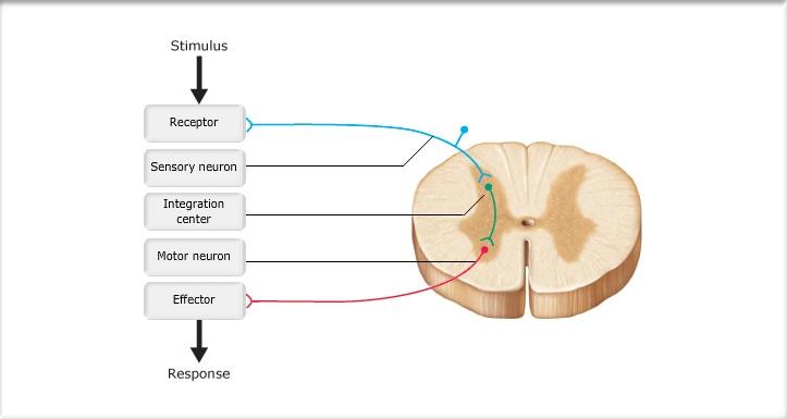
-Motor neurons are responsible for carrying a signal from the central nervous system (CNS) to an effector cell, which then carries out the desired response.
What is the function of the motor nerves?
You have two main types of nerves: Sensory nerves carry signals to your brain to help you touch, taste, smell and see. Motor nerves carry signals to your muscles or glands to help you move and function.
What do motor neurons do to muscles?
Motor neurons (also referred to as efferent neurons) are the nerve cells responsible for carrying signals away from the central nervous system towards muscles to cause movement. They release neurotransmitters to trigger responses leading to muscle movement.
What is the motor function of the nervous system?
0:001:11The motor functions of the nervous system - YouTubeYouTubeStart of suggested clipEnd of suggested clipThe nervous system orders the body's muscles to contract. We can deliberately order the skeletalMoreThe nervous system orders the body's muscles to contract. We can deliberately order the skeletal muscles to contract. Which enables us to perform movements. These voluntary movements are commanded by
What is the initial increase of the membrane potential to the value of the threshold potential?
Hypopolarization is the initial increase of the membrane potential to the value of the threshold potential. The threshold potential opens voltage-gated sodium channels and causes a large influx of sodium ions. This phase is called the depolarization. During depolarization, the inside of the cell becomes more and more electropositive, until the potential gets closer the electrochemical equilibrium for sodium of +61 mV. This phase of extreme positivity is the overshoot phase.
How does action potential work?
So, an action potential is generated when a stimulus changes the membrane potential to the values of threshold potential . The threshold potential is usually around -50 to -55 mV. It is important to know that the action potential behaves upon the all-or-none law. This means that any subthreshold stimulus will cause nothing, while threshold and suprathreshold stimuli produce a full response of the excitable cell.
What are the two types of synapses?
Each synapse consists of the: 1 Presynaptic membrane – membrane of the terminal button of the nerve fiber 2 Postsynaptic membrane – membrane of the target cell 3 Synaptic cleft – a gap between the presynaptic and postsynaptic membranes
What happens to the sodium permeability after an overshoot?
After the overshoot, the sodium permeability suddenly decreases due to the closing of its channels. The overshoot value of the cell potential opens voltage-gated potassium channels, which causes a large potassium efflux, decreasing the cell’s electropositivity.
Why does myelin increase the speed of propagation?
The propagation is also faster if an axon is myelinated. Myelin increases the propagation speed because it increases the thickness of the fiber. In addition, myelin enables saltatory conduction of the action potential, since only the Ranvier nodes depolarize, and myelin nodes are jumped over.
What causes action potential?
From the aspect of ions, an action potential is caused by temporary changes in membrane permeability for diffusible ions. These changes cause ion channels to open and the ions to decrease their concentration gradients. The value of threshold potential depends on the membrane permeability, intra- and extracellular concentration of ions, and the properties of the cell membrane.
Does action potential always propagate forward?
We need to emphasize that the action potential always propagates forward, never backwards. This is due to the refractoriness of the parts of the membrane that were already depolarized, so that the only possible direction of propagation is forward. Because of this, an action potential always propagates from the neuronal body, through the axon to the target tissue.
What neuron is responsible for muscle contraction?
Gamma motor neurons respond to stretch receptors of the skeletal muscle, also known as muscle spindles. Although known as a motor neuron, gamma motor neurons do not cause any motor function directly. Instead, they are thought to be activated alongside the alphas to fine-tune the muscle contraction. Special visceral efferent neurons (also known as ...
How does the axon work?
The axon works to transmit information it receives down its body to the dendrites at the end of the neuron. Motor neurons are known as multipolar neurons in terms of their structure. This means that they have a single axon and multiple dendrites. Motor neurons are the most common structure for neurons.
Which neuron innervates the head and neck?
Special visceral efferent neurons (also known as branchial motor neurons) are responsible for innervate the muscles of the head and neck.
Which type of neuron innervates extrafusal muscle fibers?
Beta motor neuron s are not as well categorized as alpha motor neurons, but are understood to also innervate extrafusal muscle fibers, as well as intrafusal fibers, which serve as specialized sensory organs and are innervated by both motor and sensory fibers.
What are the two types of motor neurons?
There are two types of motor neurons: 1 Lower motor neurons – these are neurons which travel from the spinal cord to the muscles of the body. 2 Upper motor neurons – these are neurons which travel between the brain and the spinal cord.
What causes a lower motor neuron to be damaged?
If the lower motor neurons are damaged, this could be as a result of infections such as Lyme disease, trauma to the peripheral nerves or viruses that can attack the cells . Some of the symptoms of damage to lower motor neurons include muscle paralysis and muscle weakness.
Why do motor neuron diseases occur?
Motor neuron diseases come because of damage to the motor neurons. These diseases tend to affect muscle control and can also affect speaking, eating, breathing, and walking as a result.
How do action potentials work?
The most conspicuous of these voltage changes are action potentials, which are the signals that neurons use to transmit information along long axons. Each action potential lasts slightly less than a millisecond (1 ms) at a particular location along the axon and travels along the axon at a speed that varies from less than 1 m/s to nearly 100m/s depending on the girth of the axon. Action potentials may be recorded from outside an axon by using fine wires as electrodes, as shown in Fig. 2.5a. Although the voltage change between the inside and outside of the neuron is about a tenth of a volt, the signal that the electrodes outside the cell pick up is much smaller, so considerable amplification is needed to display and measure the action potential. Each action potential is conducted rapidly along the axon and passes the two electrodes in succession. As it passes the first electrode, the latter will become positive with respect to the second electrode, and as the action potential passes the second electrode, the situation is rapidly reversed. Consequently, the output signal that the amplifier delivers has an S-shaped waveform when displayed on the oscilloscope (Fig. 2.5b). On a slower time scale, action potentials appear as stick-like departures from the baseline, and because of this appearance they are commonly called spikes.
What is Figure 2.6?
Figure 2.6 Activity in a neuron recorded with an intracellular electrode. In this method (a), one electrode is used to inject current pulses of different magnitudes into the neuron and a second is used to record the voltage, or potential, across the membrane. In (b), the typical appearance on an oscilloscope of responses by a neuron to pulses of current is shown.
What is the effect of a reduction in electrical potential across the cell membrane?
A reduction in electrical potential across the membrane, bringing the intracellular voltage closer to the extracellular, is called a depolarisation. Most depolarising events are excitatory because they increase the likelihood that the neuron will generate an action potential. An increase in the membrane potential from the resting value is called a hyperpolarisation, and the effect of this is inhibitory as it counteracts any depolarisation . Much of the process of integration involves
What happens when a spike occurs?
When a spike occurs at one location, it depolarises the membrane for some distance away. This depolarisation acts as the stimulus for new spikes, but, because of the refractory period, only in the length of axon that has not recently produced a spike. A spike is therefore a stereotyped event, in which the membrane potential swings rapidly between resting potential to about 40 mV positive and back. The amplitudes of spikes in extracellular recordings appear to vary from axon to axon, but this is because the size of the extracellular signals picked up by the electrodes depends on the diameters of the axons and how far away the axons are from the electrodes.
How do neurons make decisions?
Neurons are specialised in their physiology to receive, sort out and pass on information. The signals that neurons deal with involve small changes in the electrical voltage between the inside and outside of the cell, and integration is the process by which these voltage changes are combined together to determine the neuron's output signal. This is essentially how neurons make decisions.
How to record spikes in an axon?
A more precise method of recording a spike at one location along an axon is to use an intracellular electrode . This consists of a glass capillary tube that is drawn out to a very fine point and filled with a conducting salt solution such as potassium acetate . The electrode is connected to an amplifier by way of a silver wire placed into the electrode (Fig. 2.6a). When the tip of the electrode is inserted through the membrane of the cell, the salt solution is in electrical contact with the inside of the cell, and the signal recorded measures the difference in electrical potential, or voltage, between the
How do neuron batteries keep up?
Nevertheless, the neuron needs to keep its batteries topped up by maintaining a difference in concentrations of sodium and potassium between its inside and outside. It does this by means of pumps, proteins in the membrane that consume metabolic energy to transport ions against concentration gradients.
What is an Action Potential?
So, what is an action potential? Many definitions tend to be quite complicated, especially when you are learning about action potentials for the first time. To completely unravel the mystery, we should first look at the meaning of the term.
What causes the cell membrane to move?
In the cell membrane, charged atoms called ions cause the equivalent of motion – they cause action. When a neuron is not firing and when a cell membrane is not allowing large amounts of certain products (we will talk about these later) to enter or leave the cell, that cell has resting potential. When electrical activity is stimulated, the potential stops resting because external forces create electrical movement – an action potential.
What are the phases of cardiac action potential?
It is at the cardiac action potential that many cardiovascular drugs have an effect. The image below shows the cardiac action potential graph (you will soon see that it differs from the neuron action potential graph), and also where different heart medications take effect. The cardiac action potential graph has four phases: 1 Phase Four: diastole and pacemaker potential 2 Phase Zero: depolarization (sodium and calcium ion influx) 3 Phase One: slow repolarization – an extremely short phase of sodium ion gates closing and potassium gates opening 4 Phase Two: slow repolarization – influx of calcium ions to aid with muscle contraction 5 Phase Three: rapid repolarization
Why do ion channels open in the muscle cell?
Muscle membrane ion channels open in reaction to the detection of acetylcholine by muscle receptors. Sodium ions flow into the cell; when the threshold is reached the muscle cell opens its calcium ion stores (Ca 2+ ). It is calcium that enables muscle fibers to contract. When the action potential ends, the sodium ion channels close and the muscle cell can relax.
How is membrane potential measured?
A membrane potential describes how an electrical charge is spread across the membrane. It is measured in millivolts (mV). This is most commonly measured by looking at the charge on the outside of the cell (the side where the extracellular fluid is) and comparing this with the charge on the inside of the cell (the cytosol or intracellular fluid ). To keep calculations as simple as possible it is supposed that the outer side has zero mV.
Which ions are used to make a positive charge?
In the human body – and in the nervous systems of most mammals – the primary ions are sodium and potassium (both with a single positive charge), calcium with two positive charges (Ca 2+ ), and chloride with one negative charge (Cl – ). Chloride is an anion that is used to slow down action potential firing rates in some nerve pathways.
Where is potential energy stored?
This is a continuous energy field more usually associated with the field of physics. In biology, potentials are found at the inner and outer edges of cell membranes. Potential energy is stored energy, that is why it is continuous. When a ball is still, it has potential energy. When a neuron is not firing, it has potential energy. Instead of saying a cell – or rather its membrane – has potential energy, we say is has a resting potential.
What is the end of the axon?
at end of axons, small nodules that release chemical signals from the neuron into the synapse.
What is passive conduction?
passive conduction (diffusion of ions within the intracellular fluid) will ensure that adjacent membrane depolarises; sufficient depolarisation triggers a new action potential.
What is fatty material made up of?
fatty material made up of glial cells, insulates some axons for faster movement of electrical impulses along the axon.
What does depolarization do to the cell membrane?
depolarise (decrease negative charge inside the cell) the cell membrane, increasing the fire likelihood.
How is neurotransmission terminated?
5. neurotransmission is terminated by reuptake, enzyme deactivation or autoreception.
Can ions leak out of an axon?
ions cannot leak out of an insulated axon. so it takes less energy to maintain the resting potential.
Can signals go backwards?
signals can never go backwards, keeps signals separate.
This problem has been solved!
1. How is an action potential passed from the motor neuron to a muscle fiber? Arrange the labels in the order in which they would occur, beginning at the presynaptic membrane. ( Note: ACh = acetylcholine ).
Expert Answer
1. Answer : Step 1 :Signal from the motor neuron arrives at the nerve terminal of the presynaptic cell. Step 2 :Calcium moves into the nerve terminal of the presynaptic cell. Step 3 :Ach is rele view the full answer
