
The spinal cord is a long, thin, tubular bundle of nerves that is an extension of the central nervous system from the brain and is enclosed in and protected by the bony vertebral column. The main function of the spinal cord is transmission of neural inputs between the periphery and the brain.
What is the function of the spinal cord in psychology?
Within the spinal cord, there are 30 segments, each belonging to one of four sections:
- Cervical – These are 8 segments which transmit signals from or to areas of the head, neck, shoulders, arms and hands.
- Thoracic – These are 12 segments which transmit signals from or to areas of the arms, chest and abdominal areas.
- Lumbar – These are 5 segments which transmit signals from or to areas of the legs, feet and some pelvic organs.
What is function of spinal cord?
The existence of intramedullary signal intensity and signal location, as well as the effacement of the spinal cord compression, on the images helps us to diagnose patients with SCI.
What is the meaning of spinal cord?
Whole-body vibration (WBV) is a modality that offers an accessible approach for a generalized activation of sensory afferents. WBV activates presynaptic inhibitory mechanisms that modulate segmental reflex excitability [ 13] and has been shown to reduce spasticity in persons with chronic SCI [ 14, 15 ].
What is the purpose of the spinal cord?
What tissues and fluids make up the spinal cord?
- Dura mater. The outer layer that protects your spinal cord from injury.
- Arachnoid mater. The middle layer between the epidural and subarachnoid space.
- Pia mater. The inner layer that covers your spinal cord. What are the epidural and arachnoid spaces? The epidural space is between the dura mater and arachnoid mater.

What does spinal cord mean in psychology?
the part of the central nervous system that extends from the lower end of the medulla oblongata, at the base of the brain, through a canal in the center of the spine as far as the lumbar region.
What is the simple definition of spinal cord?
(SPY-nul kord) A column of nerve tissue that runs from the base of the skull down the center of the back. It is covered by three thin layers of protective tissue called membranes. The spinal cord and membranes are surrounded by the vertebrae (back bones).
What is the spinal cord in the brain?
The spinal cord is a thick column of nerves surrounded by vertebrae that runs from the brain stem to the lumbar region of the spine. Like the brain, the spinal cord has both grey and white matter. The spinal cord sends information between the brain and most of the body through the spinal nerves.
How does spinal cord affect behavior?
Emotional and behavioral problems may develop or worsen after a SCI. There is often a period of adjustment after a spinal cord injury. Sometimes feelings of sadness or anxiety may develop. In some cases, clinical depression may develop.
How important is spinal cord?
The spinal cord controls various parts of the body and plays an important role when it comes to bladder control. The spinal cord forms a vital link between the brain and the rest of the body and is part of the central nervous system. Together with the brain it controls bodily functions, including movement and behavior.
What is spinal cord Class 10?
The spinal cord is a part of the central nervous system. It is a long pipe-like structure arising from the medulla oblongata, part of the brain consisting of a collection of nerve fibres, running through the vertebral column of the backbone.
Where is the spinal cord?
The spinal cord lies inside the spinal column, which is made up of 33 bones called vertebrae. Five vertebrae are fused together to form the sacrum (part of the pelvis), and four small vertebrae are fused together to form the coccyx (tailbone).
What is the difference between spinal cord and brain?
The brain and spinal cord are the two major components of the nervous system....Difference between Brain and Spinal CordBrainSpinal CordThe brain is housed inside the cranium – a bony covering that protects the brain from mechanical shocks and injuriesThe spinal cord is enclosed inside the vertebral column.Injury4 more rows
What are the functions of spine?
Your spine, or backbone, is your body's central support structure. It connects different parts of your musculoskeletal system. Your spine helps you sit, stand, walk, twist and bend. Back injuries, spinal cord conditions and other problems can damage the spine and cause back pain.
What happens when spinal cord is damaged?
Emergency signs and symptoms of a spinal cord injury after an accident include: Extreme back pain or pressure in your neck, head or back. Weakness, incoordination or paralysis in any part of your body. Numbness, tingling or loss of sensation in your hands, fingers, feet or toes.
Can spinal injury cause mental issues?
In a new study, published in Mayo Clinic Proceedings, researchers from Michigan Medicine find adults with spinal cord injury are at a higher risk of developing mental health disorders, including depression and anxiety, compared to adults without the condition.
What happens if brain and spinal cord are non functional?
Paralysis occurs when communication between the brain and spinal cord fails. This can result from injury to neurons in the brain (a stroke), or in the spinal cord. Trauma to the spinal cord affects only the areas below the level of injury.
Where is the spinal cord?
The spinal cord lies inside the spinal column, which is made up of 33 bones called vertebrae. Five vertebrae are fused together to form the sacrum (part of the pelvis), and four small vertebrae are fused together to form the coccyx (tailbone).
What is a sentence for spinal cord?
The 47-year-old suffers from a spinal cord injury that affects her nervous system and has chronic pain. The numb feeling is caused by damage to your spinal cord and nerves. He was soon involved in research on the nervous system and the spinal cord at an army institute.
Which is the part of spinal cord?
The spinal cord comprises three parts: the cervical (neck), thoracic (chest), and lumbar (lower back) regions. Three layers of tissue protect the spinal cord: the dura mater, arachnoid mater, and pia mater.
How many bones are in the spinal cord?
Vertebrae: The spine has 33 stacked vertebrae (small bones) that form the spinal canal. The spinal canal is a tunnel that houses the spinal cord and nerves, protecting them from injury. Most vertebrae move to allow for a range of motion.
What is a spinal cord stroke?
Spinal cord stroke is a rare disorder characterized by acute paraplegia and sensory level and bladder/bowel paralysis due to spinal cord infarction. The stroke may develop in cases of profound hypotension with preexisting vascular disease affecting the arteries supplying the spinal cord, such as occurs in diabetes and vasculitides (central nervous system vasculitis, lupus, and salmonellosis have been reported). It has also been described as a complication of spinal cord arterial-venous malformations. However, the most common cause of spinal cord stroke is thoracoabdominal aortic aneurysms (TAAA) that involve the intercostal artery, which gives rise to the ‘Magna’ artery (anterior spinal) of Adamkiewicz. TAAA are usually due to arteriosclerosis or associated with different Marfan syndrome subtypes. Surgical repair or dissection of TAAA is the main predisposing factor for spinal cord stroke, although rarely, embolism to the cord arising from the TAAA may occur.
What is the pathogenesis of spinal cord occlusion?
The pathogenesis of the occlusion has been linked to pressurized injection or herniation of disc material into vertebral body sinusoids during axial stress, e.g., during heavy lifting ( Feigin et al., 1965; Srigley et al., 1981; Toro et al., 1994; McLean et al., 1995; Tosi et al., 1996; Fahey et al., 1998 ).
What is an embolic obstruction of the spinal cord?
Embolic obstruction of spinal blood vessels is not commonly encountered. It is usually due to atheromatous or fibrocartilaginous embolism and may also be seen in cases of decompression sickness (see below). Atheromatous embolism, which occurs strictly on the arterial side of the circulation, may produce infarcts of varying size, depending on the caliber of the vessels affected ( Périer et al., 1960; Hughes and Brownell, 1966; Jellinger, 1967; Wolman and Bradshaw, 1968; Fieschi et al., 1970; Herrick and Mills, 1971; Laguna and Cravioto, 1973; Slavin et al., 1975; Rodriguez-Baeza et al., 1991 ). It may also be observed in the absence of any damage to the spinal cord parenchyma ( Soloway and Aronson, 1964 ). Finding atheromatous emboli in any given case may require an exhaustive search through many sections; typically, only a small proportion of thrombosed vessels show evidence of cholesterol cleft formation ( Hashizume et al., 1997 ).
What is the thrombosis of the anterior spinal artery?
Thrombosis of the anterior spinal artery causes distinctive clinical syndrome characterized by paraplegia or quadriplegia with sparing of posterior column sensation. Thrombosis may be seen in patients with systemic vasculitis, opium addicts, women on oral contraceptives, and patients with hypercoagulable states. Rothman and Nelson (1980) describe a case of spinal cord infarction due to occlusion of branches of the anterior spinal artery in the patient with sickle-cell disease ( Fig. 29.5 ).
How many spinal cords are there?
The spinal cord has 31 segments of paired spinal nerves and ends with a thin, fibrous thread called filum terminale, in the coccyx. The 31 segments are divided into regions: (1) the top 8 segments comprise the cervical region such that C1–8 nerves exit the spinal cord in the cervical region or in the neck; (2) the next 12 segments are part of the thoracic region such that T1–12 nerves exit the spinal cord in the thoracic region or the chest; (3) the next 5 segments make up the sacral region such that L1–5 nerves exit the spinal cord in the lumbar region or the lower back; (4) the S1–5 nerves exit the spinal cord in the sacral region or near the pelvis; and finally, (5) the last segment consists of the coccygeal nerves which exit the spinal cord near the coccyx, or tailbone. The main purpose of the spinal cord is to transmit and receive information between the brain and the rest of the body (muscles and glands). The spinal cord also has some of its own neural circuits that are responsible for reflexes and rhythmic movements, such as locomotion and respiration.
What causes decompression sickness?
Decompression sickness develops when gas bubbles form within the circulation in association with excessively rapid decompression in divers or others who have been subjected to increased atmospheric pressure. Neurologic signs and symptoms, which are often referable to the cervical or upper thoracic spinal cord, are due to the appearance of multiple small foci of perivascular necrosis, particularly within the white matter ( Haymaker and Davison, 1950; Haymaker and Johnston, 1955; Haymaker, 1957; Palmer et al., 1981, 1987; Calder et al., 1989 ). The genesis of these lesions is unclear. Some believe that epidural venous obstruction is primarily responsible ( Haymaker and Johnston, 1955; Hallenbeck et al., 1975 ), while others ( Ries et al., 1999) believe that the lesions are due to paradoxic gas bubble embolism through a patent foramen ovale ( Wilmshurst et al., 1989 ).
Which spinal cord is most vulnerable to ischemia?
The thoracolumbar territory is the most vulnerable spinal cord area to ischemia because there is only one major feeder to the anterior spinal artery in this region.
What is the white matter of the spinal cord?
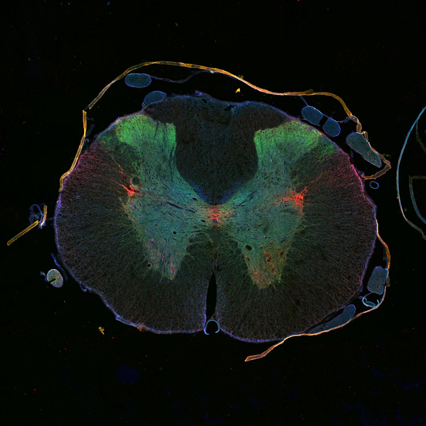
White Matter & Grey Matter. The spinal cord is split into grey matter (which is in the shape of a butterfly) and white matter (which is the material surrounding the grey). The white matter is made up of nerve fibers, called axons, which run up and down the length of the cord. Each group of axons carries a specific type of information it needs ...
What is the spinal cord?
The spinal cord is a complex cylinder of nerves that starts at the base of your brain and runs down the vertebral canal to the backbone. It is part of the body’s collection of nerves, called the central nervous system, along with the brain. In each of the spinal cord’s many segments lives a pair of roots that are made up of nerve fibers. These roots are referred to as the dorsal (which is towards the back) and the ventral (which is away from the back) roots. We depend on the spinal column to be the main support of our body. It allows us to stand upright, bend, and twist while protecting the spinal cord from injury. If the spinal cord is injured, it often causes issues like:
What are the functions of the spinal cord?
The spinal cord’s major functions include: 1 Electrochemical communication. Electrical currents travel up and down the spinal cord and across nerves, sending signals which allow different segments of the body to communicate with the brain. 2 Walking. While a person walks, a collection of muscle groups in the legs are constantly contracting and relaxing. The action of taking step after step may seem incredibly simple to us since we have been doing it all of our lives, but there are actually a lot of factors that have to be coordinated properly to allow this to happen. This central pattern generators in the spinal cord are made up of neurons which send signals to the muscles in the legs, making them relax or contract, and produce the alternating movements which occur when a person walks. 3 Reflexes. Reflexes are involuntary responses resulting from stimuli involving the brain, spinal cord, and nerves of the peripheral nervous system.
How many pairs of spinal nerves are there?
A nerve is an organ shaped like a small cord that is made up of several axons that are bound together. There are 31 pairs of spinal nerves
How long will it take technology to reverse spinal cord damage?
Research is progressing quickly, and in just 5 years we could have the means to reverse the most severe of spinal cord injuries.
How does electrical current travel?
Electrical currents travel up and down the spinal cord and across nerves, sending signals which allow different segments of the body to communicate with the brain . Walking. While a person walks, a collection of muscle groups in the legs are constantly contracting and relaxing.
What are involuntary responses resulting from stimuli involving the brain, spinal cord, and nerves of?
Reflexes . Reflexes are involuntary responses resulting from stimuli involving the brain, spinal cord, and nerves of the peripheral nervous system.
How are the two hemispheres connected?
The two hemispheres are connected by a thick band of neural fibers known as the corpus callosum, consisting of about 200 million axons. The corpus callosum allows the two hemispheres to communicate with each other and allows for information being processed on one side of the brain to be shared with the other side.
Which part of the brain contains the cerebral cortex?
The two hemispheres of the cerebral cortex are part of the forebrain ( [link] ), which is the largest part of the brain. The forebrain contains the cerebral cortex and a number of other structures that lie beneath the cortex (called subcortical structures): thalamus, hypothalamus, pituitary gland, and the limbic system (collection of structures). The cerebral cortex, which is the outer surface of the brain, is associated with higher level processes such as consciousness, thought, emotion, reasoning, language, and memory. Each cerebral hemisphere can be subdivided into four lobes, each associated with different functions.
Which lobe of the brain is responsible for processing information from the body's senses?
The brain’s parietal lobe is located immediately behind the frontal lobe, and is involved in processing information from the body’s senses. It contains the somatosensory cortex, which is essential for processing sensory information from across the body, such as touch, temperature, and pain. The somatosensory cortex is organized topographically, which means that spatial relationships that exist in the body are maintained on the surface of the somatosensory cortex ( [link] ). For example, the portion of the cortex that processes sensory information from the hand is adjacent to the portion that processes information from the wrist.
Where is the reticular formation located?
The midbrain is comprised of structures located deep within the brain, between the forebrain and the hindbrain. The reticular formation is centered in the midbrain , but it actually extends up into the forebrain and down into the hindbrain. The reticular formation is important in regulating the sleep/wake cycle, arousal, alertness, and motor activity.
What is the surface of the brain called?
The surface of the brain, known as the cerebral cortex, is very uneven, characterized by a distinctive pattern of folds or bumps, known as gyri (singular: gyrus), and grooves, known as sulci (singular: sulcus), shown in [link]. These gyri and sulci form important landmarks that allow us to separate the brain into functional centers. The most prominent sulcus, known as the longitudinal fissure, is the deep groove that separates the brain into two halves or hemispheres: the left hemisphere and the right hemisphere.
How does the spinal cord respond to sensory signals?
Withdrawal from heat and knee jerk are two examples. When a sensory message meets certain parameters, the spinal cord initiates an automatic reflex. The signal passes from the sensory nerve to a simple processing center, which initiates a motor command. Seconds are saved, because messages don’t have to go the brain, be processed, and get sent back. In matters of survival, the spinal reflexes allow the body to react extraordinarily fast.
What happens when the spinal cord is damaged?
When the spinal cord is damaged in a particular segment, all lower segments are cut off from the brain, causing paralysis. Therefore, the lower on the spine damage is, the fewer functions an injured individual loses.
How are the two hemispheres connected?
The two hemispheres are connected by a thick band of neural fibres known as the corpus callosum, consisting of about 200 million axons. The corpus callosum allows the two hemispheres to communicate with each other and allows for information being processed on one side of the brain to be shared with the other side.
Which part of the brain is the largest?
The two hemispheres of the cerebral cortex are part of the forebrain ( Figure BB.12 ), which is the largest part of the brain. The forebrain contains the cerebral cortex and a number of other structures that lie beneath the cortex (called subcortical structures): thalamus, hypothalamus, pituitary gland, and the limbic system (a collection of structures). The cerebral cortex, which is the outer surface of the brain, is associated with higher level processes such as consciousness, thought, emotion, reasoning, language, and memory. Each cerebral hemisphere can be subdivided into four lobes, each associated with different functions.
Where is the reticular formation located?
The midbrain is comprised of structures located deep within the brain, between the forebrain and the hindbrain. The reticular formation is centred in the midbrain , but it actually extends up into the forebrain and down into the hindbrain. The reticular formation is important in regulating the sleep/wake cycle, arousal, alertness, and motor activity.
How do we know the functions of the brain?
Much of what we know about the functions of different areas of the brain comes from studying changes in the behaviour and ability of individuals who have suffered damage to the brain. For example, researchers study the behavioural changes caused by strokes to learn about the functions of specific brain areas. A stroke, caused by an interruption of blood flow to a region in the brain, causes a loss of brain function in the affected region. The damage can be in a small area, and, if it is, this gives researchers the opportunity to link any resulting behavioural changes to a specific area. The types of deficits displayed after a stroke will be largely dependent on where in the brain the damage occurred.
What happens when a sensory nerve meets a certain parameter?
When a sensory message meets certain parameters, the spinal cord initiates an automatic reflex. The signal passes from the sensory nerve to a simple processing centre, which initiates a motor command. Seconds are saved, because messages don’t have to go the brain, be processed, and get sent back.
Why did Schiavo's eyes move?
Why did Schiavo’s eyes sometimes move, and why did she groan? Although the parts of her brain that control thought, voluntary movement, and feeling were completely damaged, her brainstem was still intact. Her medulla and pons maintained her breathing and caused involuntary movements of her eyes and the occasional groans.
What nerves send messages to the brain?
Sensory nerves bring messages in; motor nerves send messages out to the muscles and organs. Messages travel to and from the brain through every segment. Some sensory messages are immediately acted on by the spinal cord, without any input from the brain. Withdrawal from a hot object and the knee jerk are two examples.
How does the spinal cord respond to sensory signals?
Withdrawal from heat and knee jerk are two examples. When a sensory message meets certain parameters, the spinal cord initiates an automatic reflex. The signal passes from the sensory nerve to a simple processing center, which initiates a motor command. Seconds are saved, because messages don’t have to go the brain, be processed, and get sent back. In matters of survival, the spinal reflexes allow the body to react extraordinarily fast.
Where does the spinal cord end?
In the opposite direction, the spinal cord ends just below the ribs —contrary to what we might expect, it does not extend all the way to the base of the spine.
Which part of the brain contains the cerebral cortex?
The two hemispheres of the cerebral cortex are part of the forebrain ( [link] ), which is the largest part of the brain. The forebrain contains the cerebral cortex and a number of other structures that lie beneath the cortex (called subcortical structures): thalamus, hypothalamus, pituitary gland, and the limbic system (collection of structures). The cerebral cortex, which is the outer surface of the brain, is associated with higher level processes such as consciousness, thought, emotion, reasoning, language, and memory. Each cerebral hemisphere can be subdivided into four lobes, each associated with different functions.
What is the surface of the brain called?
The surface of the brain, known as the cerebral cortex, is very uneven, characterized by a distinctive pattern of folds or bumps, known as gyri (singular: gyrus), and grooves, known as sulci (singular: sulcus), shown in [link]. These gyri and sulci form important landmarks that allow us to separate the brain into functional centers. The most prominent sulcus, known as the longitudinal fissure, is the deep groove that separates the brain into two halves or hemispheres: the left hemisphere and the right hemisphere.
What happens when the spinal cord is damaged?
When the spinal cord is damaged in a particular segment, all lower segments are cut off from the brain, causing paralysis. Therefore, the lower on the spine damage is, the fewer functions an injured individual loses.
How many segments are there in the spinal cord?
The spinal cord is functionally organized in 30 segments, corresponding with the vertebrae. Each segment is connected to a specific part of the body through the peripheral nervous system. Nerves branch out from the spine at each vertebra. Sensory nerves bring messages in; motor nerves send messages out to the muscles and organs. Messages travel to and from the brain through every segment.
What connects the brain to the outside world?
It can be said that the spinal cord is what connects the brain to the outside world. Because of it, the brain can act. The spinal cord is like a relay station, but a very smart one. It not only routes messages to and from the brain, but it also has its own system of automatic processes, called reflexes.
The Spinal Cord
It can be said that the spinal cord is what connects the brain to the outside world. Because of it, the brain can act. The spinal cord is like a relay station, but a very smart one. It not only routes messages to and from the brain, but it also has its own system of automatic processes, called reflexes.
The Two Hemispheres
The surface of the brain, known as the cerebral cortex, is very uneven, characterized by a distinctive pattern of folds or bumps, known as (singular: gyrus), and grooves, known as (singular: sulcus), shown in Figure 4.15. These gyri and sulci form important landmarks that allow us to separate the brain into functional centers.
Forebrain Structures
The two hemispheres of the cerebral cortex are part of the (Figure 4.17), which is the largest part of the brain. The forebrain contains the cerebral cortex and a number of other structures that lie beneath the cortex (called subcortical structures): thalamus, hypothalamus, pituitary gland, and the limbic system (a collection of structures). The
Midbrain and Hindbrain Structures
The is comprised of structures located deep within the brain, between the and the hindbrain. The reticular formation is centered in the midbrain, but it actually extends up into the forebrain and down into the hindbrain. The reticular formation is important in regulating the sleep/wake cycle, arousal, alertness, and motor activity.
Brain Imaging
You have learned how brain injury can provide information about the functions of different parts of the brain. Increasingly, however, we are able to obtain that information using brain imaging techniques on individuals who have not suffered brain injury.
What is the spinal cord?
Spinal Cord. The spinal cord is a part of the central nervous system. It is a long pipe-like structure arising from the medulla oblongata, part of the brain consisting of a collection of nerve fibres, running through the vertebral column of the backbone. It is segmented with a pair of roots ...
Why is it important to understand the physiology of the spinal cord?
Understanding the physiology of the spinal cord helps in detecting and determining the various methods to deal with diseases and damage related to the spinal cord. Also Read: Peripheral Nervous System.
What is the structure of the spinal cord?
Structure Of Spinal Cord. The Spinal cord runs through a hollow case from the skull enclosed within the vertebral column. Spinal nerves arise from different regions of the vertebral column and are named accordingly, the regions are – Neck, chest, pelvic and abdominal. Cross-section of spinal cord displays grey matter shaped like a butterfly ...
How many spinal nerves are there?
Several spinal nerves emerge out of each segment of the spinal cord. There are 8 pairs of cervical, 5 lumbar, 12 thoracics, 5 sacral and 1 co ccygeal pair of spinal nerves
How long is the spinal cord?
In adults, the spinal cord is usually 40cm long and 2cm wide. It forms a vital link between the brain and the body.
What is the white matter of the brain?
The white matter consists of a collection of axons permitting communication between different layers of CNS. A tract is a collection of axons and carries specialized information. Ascending tracts and descending tracts send and transmit signals from the brain respectively to various nerve cells across the body.
What is the subarachnoid space?
Subarachnoid space lies between the arachnoid mater and pia mater. It is filled with cerebrospinal fluid.
The Major Functions of The Spinal Cord
- The spinal cord’s major functions include: 1. Electrochemical communication. Electrical currents travel up and down the spinal cord and across nerves, sending signals which allow different segments of the body to communicate with the brain. 2. Walking.While a person walks, a collection of muscle groups in the legs are constantly contracting and rel...
The Structure of The Spinal Cord
- Theoverall structureof the spinal cord is enclosed by the protection of the vertebral column. The spinal nerves are located in the spaces between the arches of the vertebrae. Spinal nerves are divided into these separate regions: 1. Cervical (neck) 2. Thoracic (chest) 3. Lumbar (abdominal) 4. Sacral (pelvic) 5. Coccygeal (tailbone)
Spinal Cord Injury
- A spinal cord injury (SCI) is when a part of the cord or the nerves located at the base of the spine are damaged. This can have a major effect on the body’s sensory, motor, and reflex capabilities if the brain is unable to send information past the location of the injury." The closer the injury is to the brain, the more expansive the damage. As you can probably imagine, an SCI can alter a pers…