What is systemic venous system in human body?
Systemic venous system starts out as the network of venules that coalesce to form progressively larger diameter veins – all draining blood from the tissues into the right atrium. In the lower extremities, deep system consists of the anterior and posterior tibial veins and peroneal vein.
What is venous blood?
Venous blood is deoxygenated blood which travels from the peripheral vessels, through the venous system into the right atrium of the heart.
What is systemic circulation in the heart?
Systemic Circulation The systemic circulation is the part of the vascular system that carries blood from the left ventricle to organs and tissues of the body. From:Encyclopedia of Heart Diseases, 2006
What is the difference between capillaries and venous system?
Capillaries are the smallest blood vessels in your body. Oxygen passes through the walls of your capillaries to your tissues. Carbon dioxide can also move into your capillaries from the tissue before entering your veins. The venous system refers to the network of veins that work to deliver deoxygenated blood back to your heart.

What is the systemic venous system?
In human anatomy, the systemic venous system refers to veins that drain into the right atrium without passing through two vascular beds (i.e. they originate from a set of capillaries and do not pass through a second set of capillaries before reaching the right side of the heart.
What is the meaning of venous blood?
Blood that has passed through the capillaries of various tissues other than the lungs, is found in the veins, in the right chambers of the heart, and in pulmonary arteries, and is usually dark red as a result of a lower content of oxygen.
Where is the systemic vein?
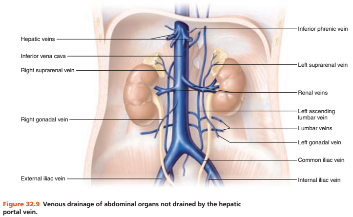
The systemic veins may be arranged into three groups: (1) The veins of the heart. (2) The veins of the upper extremities, head, neck, and thorax, which end in the superior vena cava. (3) The veins of the lower extremities, abdomen, and pelvis, which end in the inferior vena cava. —Most of the veins of the heart (Fig.
What is the function of systemic veins?
Systemic circulation carries oxygenated blood from the left ventricle, through the arteries, to the capillaries in the tissues of the body. From the tissue capillaries, the deoxygenated blood returns through a system of veins to the right atrium of the heart.
What is the difference between arterial blood and venous blood?
The main difference between arterial and venous blood is that arterial blood is oxygenated whereas venous blood is deoxygenated. The arterial blood is bright red in color and the venous blood is blackish red in color.
What contains venous blood?
Venous blood is deoxygenated blood which travels from the peripheral blood vessels, through the venous system into the right atrium of the heart.
What are the two major systemic veins?
The inferior vena cava. The common iliac vein of the pelvis.
What are the 3 types of veins?
What are the different types of veins?Deep veins are located within muscle tissue. ... Superficial veins are closer to the skin's surface. ... Pulmonary veins transport blood that's been filled with oxygen by the lungs to the heart.More items...•
What are the major veins of the systemic circulation?
cephalic vein ß lateral arm and forearm.basilic vein ß medial arm and forearm.brachial vein (deep vein)radial vein.ulnar vein.
What are the 4 major veins?
The major blood vessels connected to your heart are the aorta, the superior vena cava, the inferior vena cava, the pulmonary artery (which takes oxygen-poor blood from the heart to the lungs where it is oxygenated), the pulmonary veins (which bring oxygen-rich blood from the lungs to the heart), and the coronary ...
Do systemic veins have valves?
Lymphatics are thin-walled vessels that carry a protein-rich fluid from the periphery back to the central circulation. As in the systemic venous system, unidirectional flow is assisted by valves throughout lymphatics of all calibers.
Where does systemic circulation begin?
The heart pumps oxygenated blood out of the left ventricle and into the aorta to begin systemic circulation. After the blood has supplied cells throughout the body with oxygen and nutrients, it returns deoxygenated blood to the right atrium of the heart.
What does venous mean in medical terms?
Definition of venous 1 : of, relating to, or full of veins a venous thrombosis a venous rock. 2 of blood : having passed through the capillaries and given up oxygen for the tissues and become charged with carbon dioxide.
What Colour is venous blood?
dark redVenous blood is dark red and not blue.
What is the venous?
The venous system refers to the network of veins that work to deliver deoxygenated blood back to your heart.
What is the meaning of deoxygenated blood?
Deoxygenated blood refers to the blood that has a low oxygen saturation relative to blood leaving the lungs. The oxygenated blood is also called arterial blood. The deoxygenated blood is also called venous blood. The oxygen concentration of oxygenated blood is high.
How to determine the blood flow rate of a portosystemic vessel?
The physiological basis for the opening and growth of native portosystemic collateral vessels relies on a simple principle of fluid mechanics based on Ohm's law: the blood flow rate between two points in a vessel is proportional to the driving pressure (i.e., the pressure gradient) between these two points. This relationship is defined by the equation: Δ P = Q × R, in which Δ P is the pressure gradient, Q is the magnitude of blood flow, and R is the vascular resistance opposing that flow, which in portal hypertension represents the sum of the serial resistances of the portal vein and the hepatic vascular bed, and of the parallel resistance of the collaterals. Under normal conditions, the pressure difference between the portal and systemic circulation or portal pressure gradient (most commonly evaluated in clinical practice by its equivalent, the hepatic venous pressure gradient [HVPG]) is quite similar at both ends of the preexisting collateral vessels ( Bosch et al., 2009 ). Consequently, these vessels are “dormant,” have structure but do not carry detectable blood flow. They are constricted but not obstructed, and therefore can be opened and become functional following a hemodynamically relevant obstruction of the portal venous system and consequent elevated portal pressure.
What is a varice in the splanchnic system?
Varices are dilated vessels, linking the portal and systemic venous systems, which return blood from the splanchnic circulation to the systemic circulation, by passing the liver, decompressing the portal venous system and reducing portal pressure. Varices can be large, and haemorrhage, sometimes catastrophic, occurs from any of these vessels when intravascular pressures reach a threshold. Lower oesophageal or gastric varices, which are thin-walled and submucosal, are most prone to haemorrhage because intravascular pressures rise beyond that threshold with everyday physiological processes.
What causes a gas embolism?
VGE and AGE can result from nonsurgical procedures including pulmonary overexpansion during mechanical ventilation 30 and during hemodialysis. 31,32 VGE caused by a computed tomography (CT) injector has been reported, but in the majority of cases the air does not arterialize. 33,34 Penetrating chest injuries can allow air to enter the circulation leading to VGE and AGE. 35,36 Massive cerebral air embolism may occur after entrance of air into the circulatory system via ruptured pulmonary vessels during cardiopulmonary resuscitation. 37 Cerebral and coronary gas embolism have occurred from inhalation of pressurized helium 38,39 and ingestion of hydrogen peroxide. 40
How does a thrombus form?
Decreased or turbulent flow within veins may instigate thrombus formation. This can be caused by extrinsic compression by a mass lesion or by prolonged positional obstruction of the veins such as in long-distance travel. 1,2 In addition, prolonged immobility particularly of the lower extremities results in reduced extrinsic pump function that is normally performed by muscular movements. This results in decreased venous flow, edema, and potential thrombus formation in the affected extremity. Turbulent flow can also contribute to thrombus formation. This can be seen related to foreign bodies such as catheters that may cause thrombosis through chronic inflammation of the vein wall and direct triggering of the coagulation pathways along the surface of the catheter. Central venous catheters are a significant risk factor for thrombus formation with up to 4% of patients with central venous catheters developing catheter-related thrombus. 3 The majority of upper extremity DVTs are associated with catheters. 4
Why do I have to have an air embolus to die?
Symptoms are immediate. When air embolus leads to death, it is generally due to obstruction of blood flow in the pulmonary artery outflow tract. The autopsy pathologist, therefore, must carefully incise the main pulmonary artery under water to document the release of air bubbles from the vessel.
Where is the supracardiac vein located?
Type I: supracardiac (50%). The four pulmonary veins drain into a venous collector located behind the left atrium, from which a vertical vein runs up to reach the innominate vein, and this terminates in the right superior vena cava and right atrium. It has been reported that the vertical vein may be obstructed, but this is rarely severe in the perinatal period.
Where does the pulmonary venous confluence drain into the RA?
Intracardiac TAPVR. Here, the pulmonary venous confluence drains directly into the RA via the coronary sinus.
What are the conditions that affect the venous system?
Many conditions can affect your venous system. Some of the most common ones include: Deep vein thrombosis (DVT). A blood clot forms in a deep vein, usually in your leg. This clot can potentially travel to your lungs, causing pulmonary embolism. Superficial thrombophlebitis.
What is the blood vessel that returns deoxygenated blood from your organs back to your heart?
Veins are a type of blood vessel that return deoxygenated blood from your organs back to your heart. These are different from your arteries, which deliver oxygenated blood from your heart to the rest of your body.
What is the outer layer of the vein wall?
The walls of your veins are made up of three different layers: Tunica externa. This is the outer layer of the vein wall, and it’s also the thickest. It’s mostly made up of connective tissue. The tunica externa also contains tiny blood vessels called vasa vasorum that supply blood to the walls of your veins. Tunica media.
Why do my veins swell?
Superficial veins near the surface of the skin visibly swell. This happens when one-way valves break down or vein walls weaken, allowing blood to flow backward. Chronic venous insufficiency. Blood collects in the superficial and deep veins of your legs due to improper functioning of one-way valves.
Why are veins unique?
They’re unique because they carry oxygenated blood. All other veins carry only deoxygenated blood. Systemic veins. The systemic circuit carries deoxygenated blood from the rest of the body back to your heart, where it then enters the pulmonary circuit for oxygen. Most veins are systemic veins.
How to stop veins from forming overtime?
High blood pressure can weaken your veins overtime due to added pressure. Avoid long periods of standing or sitting. Try to change positions regularly throughout the day. When sitting down, avoid crossing your legs for long periods of time or regularly switch positions so one leg isn’t on top for a long period of time.
What are the two circuits of blood circulation?
Your body circulates blood on two different tracks called the systemic circuit and the pulmonary circuit . Veins are based on the circuit they’re found in: Pulmonary veins. The pulmonary circuit carries deoxygenated blood from your heart to your lungs.
Why does blood flow from the portal vein to the systemic vein?
Whenever a macroscopic venous connection is present between the portal vein (or one of its tributaries) and a systemic vein, the blood will flow from the portal vein to the systemic vein, because the pressure in the portal vein is higher (8 to 10 mm Hg) than that in the systemic veins (0-5 mm Hg). 8
What are varices in the splanchnic system?
Varices are dilated vessels, linking the portal and systemic venous systems, which return blood from the splanchnic circulation to the systemic circulation , bypassing the liver, decompressing the portal venous system and reducing portal pressure. Varices can be large, and haemorrhage, sometimes catastrophic, occurs from any of these vessels when intravascular pressures reach a threshold. Lower oesophageal or gastric varices, which are thin-walled and submucosal, are most prone to haemorrhage because intravascular pressures rise beyond that threshold with everyday physiological processes.
How does central venous pressure affect ventricular end diastolic pressure?
Central venous blood pressures vary with the horse's weight and exert a significant influence on ventricular end-diastolic pressure because the right ventricle is in direct continuity with the systemic venous system and right atrium.86,87 Changes in central venous blood pressure cause corresponding alterations in the right ventricular end-diastolic pressure. The atrial pressure wave consists of two or three positive waves ( a wave from atrial contraction, sometimes c wave following tricuspid valve closure, and v wave from atrial filling during ventricular systole) and two negative waves or “descents” ( x' descent from downward displacement of the tricuspid valve annulus during ventricular systole and y descent from emptying of the atrial blood into the ventricle after opening of the tricuspid valve; see Figure 3-9 ). The pulsations vary with the phase of ventilation and are dramatically influenced by positive-pressure ventilation during anesthesia (see Chapter 17 ).
Which veins drain blood from the head?
It, in turn, becomes subclavian vein once it dives under the clavicle. Finally, subclavian veins on each side joined by the jugular veins drain blood from the head. On the left, brachiocephalic vein is formed, which traverses the chest under the manubrium. On the right, the confluence of subclavian and jugular veins forms the origin of the superior vena cava. Superior vena cava drains in to the right atrium at its superior aspect.
Why do I have to have an air embolus to die?
Symptoms are immediate. When air embolus leads to death, it is generally due to obstruction of blood flow in the pulmonary artery outflow tract. The autopsy pathologist, therefore, must carefully incise the main pulmonary artery under water to document the release of air bubbles from the vessel.
What is an amniotic fluid embolus?
Amniotic fluid embolus in the setting of labor and delivery can be a devastating event, associated with ARDS and disseminated intravascular coagulation ( DIC). The diagnosis is made clinically or at autopsy due to the presence of amniotic epithelium or “squames” in thrombosed vessels ( Fig. 8.12 ). Other causes of thromboembolic disease include fragments of catheters or injected foreign materials.
Which system drains blood from the tissues into the right atrium?
Systemic venous system starts out as the network of venules that coalesce to form progressively larger diameter veins – all draining blood from the tissues into the right atrium.
What is the difference between arterial blood and venous blood?
The difference in the oxygen content of arterial blood and venous blood is known as the arteriovenous oxygen difference. Most medical laboratory tests are conducted on venous blood, with the exception of arterial blood gas tests. Venous blood is obtained for lab work by venipuncture (also called phlebotomy), or by finger prick for small quantities.
Why do veins turn blue?
The blue appearance of surface veins is caused mostly by the scattering of blue light away from the outside of venous tissue if the vein is at 0.5 mm deep or more. Veins and arteries appear similar when skin is removed and are seen directly.
Why is deoxygenated blood darker than normal blood?
Deoxygenated blood is darker due to the difference in shape of the red blood cell when oxygen binds to haemoglobin in the blood cell (oxygenated) versus does not bind to it (deoxygenated). Human blood is never blue.
What is deoxygenated blood?
Deoxygenated blood. Concentrated blood after oxygenation. Venous blood is deoxygenated blood which travels from the peripheral blood vessels, through the venous system into the right atrium of the heart.
Where is deoxygenated blood pumped to?
Deoxygenated blood is then pumped by the right ventricle to the lungs via the pulmonary artery which is divided in two branches, left and right to the left and right lungs respectively. Blood is oxygenated in the lungs and returns to the left atrium through the pulmonary veins .
Is venous blood colder than arterial blood?
Venous blood is typically colder than arterial blood, and has a lower oxygen content and pH. It also has lower concentrations of glucose and other nutrients, and has higher concentrations of urea and other waste products. The difference in the oxygen content of arterial blood and venous blood is known as the arteriovenous oxygen difference.
Where is venous blood drained?
Venous blood from orbits, inferior aspects of parietal and frontal lobes are drained into cavernous sinus. (1,2) Traditionally, acid-base status is assessed with an arterial blood gas (ABG); however, venous blood samples are frequently taken for other reasons.
What is the blood that passes through the capillaries of the lungs?
ve·nous blood. Blood that has passed through capillaries of various tissues, except the lungs, and is found in veins, right chambers of heart, and pulmonary arteries; usually dark red due to lower oxygen content.
What does it mean when blood is red?
( vē'nŭs blŭd) That which has passed through the capillaries of various tissues, except the lungs, and found in the veins, the right chambers of the heart, and the pulmonary arteries; usually dark red as a result of a lower oxygen content. Medical Dictionary for the Health Professions and Nursing © Farlex 2012.
Why is blood red?
blood which has passed through the capillaries of various tissues, except the lungs, and is found in the veins, the right chambers of the heart, and the pulmonary arteries; it is usually dark red as a result of a lower content of oxygen.
Can venous blood be drained into a capillary tube?
Venous blood should not be collected into a capillary tube or a micro-tube. Much of it is absorbed by tissue, bone and venous blood, but these amounts do not change dramatically over short periods of time. Venous blood from orbits, inferior aspects of parietal and frontal lobes are drained into cavernous sinus.
Is there a correlation between pulpal blood and venous blood?
The finidngs of this investigation showed that there is high correlation of the pulpal blood group with venous blood. So the blood group of this population can determined from dental pulp.
Where is venous blood found?
Venous Blood: Venous blood is the blood that has passed through various blood capillaries of various organs except for the lungs, and is found in veins, right chambers of the heart, and pulmonary artery.
What is the color of blood in the venous end?
Figure 2: Formation of Venous Blood. Since blood in the venous end is deoxygenated, the color of the blood is blackish red. This deoxygenated blood moves through the venules to the veins. Ultimately, all deoxygenated blood from the body comes to the right atrium of the heart through superior and inferior vena cava.
What is the difference between a pulmonary vein and an arterial vein?
The main difference between arterial and venous blood is that arterial blood is oxygenated whereas venous blood is deoxygenated. The arterial blood is bright red in color and the venous blood is blackish red in color. However, pulmonary artery and pulmonary vein are two exceptions to this; pulmonary artery carries deoxygenated blood away from the heart while pulmonary vein carries oxygenated blood towards the heart.
What is the blood that flows from the heart to the metabolizing tissues throughout the body?
The arterial blood is rich in oxygen and other nutrients such as glucose, amino acids, and vitamins. It flows from the heart to the metabolizing tissues throughout the body to supply oxygen and nutrients to the cells.
How is arterial blood collected?
Arterial Blood: The arterial blood is collected by the direct puncture of an artery.
Which blood type is rich in oxygen and nutrients such as glucose, amino acids, and vitamins?
Arterial Blood : The arterial blood is rich in oxygen and nutrients such as glucose, amino acids, and vitamins. Venous Blood: The venous blood is rich in HCO 3 and metabolic wastes such as urea.
Which blood vessels flow in the heart?
Flow. Arterial Blood: Arterial blood flows in lungs, left chambers of the heart and in arteries. Venous Blood: Venous blood flows in the right chamber of the heart and in veins.
What is systemic blood flow?
Systemic blood flow is a complex dynamic variable with rapid fluctuations caused by changes in the functional activity and metabolic demand of the different organs. Doppler echocardiography offers direct and indirect measures of systemic and organ blood flow. 22,23
What is the systemic circulation?
The systemic circulation is the part of the vascular system that carries blood from the left ventricle to organs and tissues of the body. As outlined above, the aorta is the major artery of the systemic circulation. It extends down the length of the chest and abdomen and reaches the pelvis dividing into two branches, the iliac arteries (see Fig. 10 ). Veins collect blood from the capillaries of tissues and organs (see Fig. 11 ). The large veins drain into the superior and inferior vena cavae that return blood to the right atrium.
What is the function of bronchial arteries?
In addition, bronchial arteries provide nutritive flow to the lower trachea, airway nerves, and lymph nodes.22,23 The drainage of bronchial vessels into the pulmonary circulation and the large veins has a complex arrangement ( eFig. 6-1 ). Interconnections have been demonstrated between bronchial vessels and precapillary, capillary, and postcapillary vessels of the pulmonary circulation. 24 Despite the fact that the normal adult lung remains viable without the bronchial circulation (as well as in the absence of innervation), as is the case in the transplanted lung, bronchial blood flow is critical in the lung development in the fetus and contributes to gas exchange in the presence of congenital cardiac anomalies. There is a striking increase in the size and number of bronchial arteries (due to angiogenesis) in lung disorders such as pulmonary fibrosis, lung carcinoma, and disorders characterized by pulmonary vascular occlusion. 25-27 Neovascularization of the systemic circulation into the lung after pulmonary artery obstruction is now well recognized in multiple species. 22,26
How does the vasodilator effect work?
The vasodilator effect is mediated via release of acetylcholine with hyperpolarisation of the vascular smooth muscle. Long-term neural regulation of the circulation is modulated by humoral and other factors. Angiotensin II is an important facilitator of sympathetic transmission.
How to calculate blood flow velocity?
Blood flow by Doppler echocardiography is calculated as the product of the displacement of the velocity profile , called the velocity-time integral (VTI) of blood flow velocity; the cross-sectional area of the vessel at the site of the measurement; and the heart rate. VTI is measured by tracing the pulsed Doppler velocity signal and the cross-sectional area by measuring the diameter (D) and calculating the area as π × radius (D/2) 2. 24 Assuming a circular vessel with a constant cross-sectional area, blood flow (volume per time, usually mL/min) is calculated as VTI (for one heartbeat) × π × (D/2) 2 × heart rate. 25,26 A prerequisite for this calculation is a laminar parabolic flow profile representative of long, straight blood vessels under steady flow conditions. Blood flow in neonates is usually indexed by weight (mL/kg/min). In obtaining the VTI, it is important to minimize the angle of insonation and to assess the diameter with the ultrasound beam perpendicular to the vessel at the true (maximal) diameter. An inappropriate angle will underestimate the VTI and overestimate the diameter. Owing to the squaring of the radius in the formula, inaccurate measures of the diameter may have considerable influence. It is recommended to average the measurements from a minimum of five cardiac cycles in order to minimize measurement error. 27 Flow measurements are hampered by significant intra- and interobserver variability. 28
Which system controls blood vessels?
The autonomic nervous system represents the efferent component of the neural control of the circulation. Up to three types of fibres may innervate blood vessels: sympathetic vasoconstrictor fibres, sympathetic vasodilator fibres, and parasympathetic vasodilator fibres. As the size of vessel decreases, the density of autonomic innervation increases. The small arteries and arterioles are therefore the most richly innervated arteries.
What type of receptors are stimulated by volume distention of the atriums and fire during ventricular s?
Type B barorecept ors are stretch receptors stimulated by volume distention of the atriums and fire during ventricular systole. The afferents are via unmyelinated vagal fibres. Atrial distention decreases sympathetic activity. Receptors which respond to stretch and contractility are also present in the ventricles.
Where do varicose veins appear?
Usually appearing in the legs, varicose veins may also occur in the anus, where they are known as hemorrhoids. While not a serious health risk, varicose veins can be eliminated for cosmetic reasons or if they cause discomfort.
How to treat varicose veins?
Doctors at Johns Hopkins recommend the following for treating varicose veins: Elevated feet. Raise the foot of your bed from two to four inches with blocks to aid circulation at night. Avoid scratching itchy skin above varicose veins, as this may cause ulceration or profuse bleeding.
What is the inflammation of the veins in the legs?
Thrombophlebitis is the inflammation of a vein (usually in an extremity, especially one of the legs) that occurs in response to a blood clot in the vessel. When it occurs in a vein near the surface of the skin, it is known as superficial thrombophlebitis, a minor disorder commonly identified by a red, tender vein.
What causes blood to flow backwards?
Damaged vein walls hinder the circulatory system, allowing blood to collect and flow in a retrograde (backward) fashion when the muscles relax. This creates an unusually high pressure buildup in the veins. This buildup causes further stretching and twisting of the veins, increased swelling, more valve incompetence, sluggish blood flow and potential blood clot formation. Eventually, this condition can lead to various disorders known as venous disease.
What to do if your veins are swollen?
Call your doctor if you have a painful, swollen vein that does not disappear in a few days, or if you have unexplained swelling in an arm or leg.
How many people in the US have varicose veins?
Approximately 15 percent of the United States population is affected by varicose veins, which generally do not pose great health risk. However, thrombophlebitis can be much more serious, even life-threatening, affecting millions of people each year.
What is the name of the thin-walled structures inside of which a set of valves keeps blood in the?
Venous Disease Overview. Veins are thin-walled structures inside of which a set of valves keeps blood in the body flowing in one direction. The heart pumps oxygen-rich blood to the body’s tissues through thicker-walled arteries; the veins return that blood to the heart. Veins located close to the surface of the skin are called superficial veins ...