Explore
Your gallbladder is a small, pear-shaped organ in your upper right abdomen. Your gallbladder stores and releases bile to help your digestive system break down fats. The most common issue you may develop with your gallbladder is gallstones. Gallstones are …
What is the anatomical position of the gallbladder?
· The gallbladder is essentially a pear-shaped cul-de-sac that communicates with the common hepatic ducts via the cystic duct. In vivo, the sac is actually grey-blue in appearance (and not green as depicted in the texts).
What are signs that something is wrong with your gallbladder?
The gallbladder is a pear shaped organ that is about 7 to 10 centimeters long (3 to 4 inches) and 2 to 3 centimeters wide (about 1 inch). It has the ability to hold about 50 milliliters of bile which can be emptied via the cystic duct (gallbladder duct) into the common bile duct. From here, the bile will empty into the lumen of the duodenum.
What is the physiology of the gallbladder?
Your gallbladder is a four-inch, pear-shaped organ. It's positioned under your liver in the upper-right section of your abdomen. The gallbladder stores bile, a combination of fluids, fat, and cholesterol. Bile helps break down fat from food in your intestine.
Is the gallbladder the same thing as bladder?
The gallbladder is a small pouch that sits just under the liver. The gallbladder stores bile produced by the liver. After meals, the gallbladder is empty and flat, like a …
What is the gross anatomy of the gallbladder?
The part of the gallbladder projecting beyond the undersurface of the liver is called the fundus; fundus continues into the main body of the gallbladder, which lies in a fossa on the undersurface of the liver. The body of the gallbladder narrows into an infundibulum, which leads through the neck to the cystic duct.
What are the main functions of the gallbladder?
The gallbladder is a pear-shaped organ located on the undersurface of the liver. The gallbladder functions as a concentrating reservoir for bile, which it delivers to the duodenum in response to meals.
What happens when you have your gallbladder removed?
Living without a gallbladder You can lead a perfectly normal life without a gallbladder. Your liver will still make enough bile to digest your food, but instead of being stored in the gallbladder, it drips continuously into your digestive system.
What are the most common gallbladder problems?
Cholecystitis is the most common type of gallbladder disease. It presents itself as either an acute or chronic inflammation of the gallbladder. Acute cholecystitis is generally caused by gallstones. But it may also be the result of tumors or various other illnesses.
How do you know if your having gallbladder problems?
SymptomsSudden and rapidly intensifying pain in the upper right portion of your abdomen.Sudden and rapidly intensifying pain in the center of your abdomen, just below your breastbone.Back pain between your shoulder blades.Pain in your right shoulder.Nausea or vomiting.
How do you cleanse your gallbladder?
In most cases, a gallbladder cleanse involves eating or drinking a combination of olive oil, herbs and some type of fruit juice over several hours. Proponents claim that gallbladder cleansing helps break up gallstones and stimulates the gallbladder to release them in stool.
What foods should you avoid if you have no gallbladder?
To side-step this gastrointestinal discomfort, avoid eating high-fat or spicy foods, including:French fries and potato chips.High-fat meats, such as bologna, sausage and ground beef.High-fat dairy, such as cheese, ice cream and whole milk.Pizza.Lard and butter.Creamy soups and sauces.Meat gravies.Chocolate.More items...•
Is a gallbladder surgery a major surgery?
Gallbladder removal surgery is known as a cholecystectomy. This isn't a surgery that most doctors will rush into. While it's a common surgery, it's still major surgery with some serious risks and complications. However, in most cases, you'll go home the same day as you've had the surgery.
Do you gain weight after gallbladder removal?
People who undergo gallbladder removal surgery will often experience changes in their body weight ahead of and following this procedure. Many people will lose weight initially but may see an increase in their BMI in the long term. It is usually possible to manage these weight changes with diet and exercise.
How do you know if your gallbladder needs to be removed?
Some symptoms that may indicate the need for gallbladder removal include: sharp pain in the right upper portion of your abdomen that can radiate to the middle of your abdomen, right shoulder, or back. fever....Why open gallbladder removal is donebloating.nausea.vomiting.further pain.
What can be mistaken for gallbladder problems?
Also known as the “stomach flu,” gastroenteritis may be mistaken for a gallbladder issue. Symptoms such as nausea, vomiting, watery diarrhea, and cramping are hallmarks of the stomach flu. Kidney stones. Kidney stones can cause sharp pains in your abdomen, side, and back.
When should a gallbladder be removed?
You may need gallbladder surgery if you have pain or other symptoms caused by gallstones — small stones that can form in the gallbladder. They can block the flow of bile and irritate the gallbladder. Common symptoms of gallbladder problems include: Indigestion, with bloating, heartburn, and gas.
Anatomy of The Gallbladder
The gallbladder is a pear shaped organ that is about 7 to 10 centimeters long (3 to 4 inches) and 2 to 3 centimeters wide (about 1 inch). It has th...
Location of The Gallbladder
The gallbladder sits in the shallow gallbladder fossa on the visceral surface of the liver. Here the hepatic surface of the gallbladder is connecte...
Functions of The Gallbladder
The primary functions of the gallbladder relates to the storage and secretion of bile. 1. The gallbladder stores bile. It is also able to concentra...
Where is the gallbladder located?
Your gallbladder is located in the upper right part of your abdomen (belly). It sits just under your liver.
What is the inflammation of the gallbladder?
Cholecystitis: Cholecystitis is inflammation of your gallbladder. It can occur when a gallstone blocks bile from exiting your gallbladder. Cholecystitis causes fever and pain and usually requires surgery.
What causes gallbladder problems?
Several conditions can cause problems in your gallbladder. The most common condition is gallstones. Gallstones are typically harmless but can sometimes lead to disease states. Gallbladder issues include:
What happens to the gallbladder when you eat?
Before you start eating, your gallbladder is full of bile. When you start eating, your gallbladder receives signals to contract and squeeze the stored bile through the biliary tract. The bile eventually finds its way to your largest bile duct, the common bile duct. Bile passes through the common bile duct into the duodenum, the first part of your small intestine, where it mixes with food waiting to be digested. After you eat, your gallbladder is empty and resembles a deflated balloon, waiting to be filled up again.
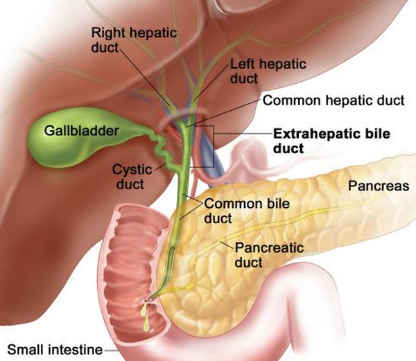
What is the name of the system that carries bile from the liver?
Your gallbladder is connected to other parts of your digestive system through a series of bile ducts called the biliary tract. The biliary tract (sometimes called biliary system or biliary tree) is a pipe-like system that carries bile from your liver to your small intestine.
What is the organ that stores and releases bile?
Your gallbladder is a small, pear-shaped organ located under your liver that stores and releases bile. Bile is the fluid your liver produces that helps digest fats in the food you eat.
What is gallstone pancreatitis?
Gallstone pancreatitis: Gallstone pancreatitis is inflammation of your pancreas. It occurs when a gallstone travels down the common bile duct and blocks the pancreatic duct at a common point just before draining into the small intestine.
Which part of the liver is embedded in the gallbladder?
This is the portion of the sac that is either embedded in, or in contact with the gallbladder fossa of the liver. The pars descendens (second part) of the duodenum, as well as the hepatic flexure and proximal transverse colon, are posteriorly related to the gallbladder.
What is the structure of the gallbladder and biliary tree?
This structure, known as the hepatic diverticulum, gives rise to the gallbladder and associated biliary duct; as well as the liver.
What is the splanchnic mesoderm that grows between the heart and the midgut?
Therefore, an organized layer of splanchnic mesoderm known as the septum transversum grows between the heart and the midgut. As the hepatic diverticulum grows, it divides unequally. The larger cranial bud, commits to becoming the liver primordium and the extrahepatic biliary tree.
How does the biliary tree work?
Once the pressure within the biliary tree reaches 10 mm H2O of bile, there is relaxation of the sphincter of Oddi. Therefore, bile can be released from both the gallbladder and directly from the liver via the biliary tree. However, during fasting states, the absence of cholecystokinin results in contraction of the sphincter of Oddi. Increased pressure in the biliary tree results in diversion of bile into the gallbladder where it is stored and concentrated.The endothelial membrane of the gallbladder is equipped with numerous ion channels that actively absorb sodium, chloride and bicarbonate ions. The water molecules subsequently follow the osmotic gradient generated by the ion shift, resulting in concentration of bile. Finally, the gallbladder also produces about 15 – 20 mL of mucus throughout the course of each day.
How much mucus does the gallbladder produce?
Finally, the gallbladder also produces about 15 – 20 mL of mucus throughout the course of each day. To master the anatomy of the liver, take a look at the following resources: Gallbladder Explore study unit. Overview of the liver Explore study unit.
Why do you need surgery for gallbladder?
Surgery is usually advised in order to avoid a gallbladder infection that could potentially spread, among other risks.
Which organ has fibrous ducts?
The extrahepatic bile ducts extend into the mesenchyme of the septum transversum and gives rise to the characteristic fibrous appearance of the liver. Gallbladder (axial view) Within the liver, the endometrial cells that overlap each other to form the hepatocytes also give rise to the intrahepatic biliary system.
Where is the gallbladder located?
Location of the Gallbladder. The gallbladder sits in the shallow gallbladder fossa on the visceral surface of the liver. Here the hepatic surface of the gallbladder is connected by connective tissue to the capsule surrounding the liver.
What are the layers of the gallbladder?
The wall of the gallbladder has several layers including the epithelium (inner), lamina propria, muscularis, perimuscular and serosa (outer).
What is the role of the gallbladder in the body?
Sitting under the liver, the gallbladder controls the expulsion of bile into the duodenum plays an important role in the digestion of fats.
How much bile can a gallbladder hold?
It has the ability to hold about 50 milliliters of bile which can be emptied via the cystic duct (gallbladder duct) into the common bile duct. From here, the bile will empty into the lumen of the duodenum. This is explained in detail under Bile Duct Anatomy.
What is the secretion of bile in the gallbladder?
The gallbladder secretes bile by muscular contractions of its wall in response to both neural and hormonal factors stimulated by food, especially fatty foods, in the duodenum.
How long is the cystic duct?
The cystic duct, which is about 3 to 4 centimeters long (about 1 to 2 inches), carries bile into the common bile duct.
What is the gallbladder?
The gallbladder is a small pouch that sits just under the liver. The gallbladder stores bile produced by the liver. After meals, the gallbladder is empty and flat, like a deflated balloon. Before a meal, the gallbladder may be full of bile and about the size of a small pear.
Where is the gallbladder located?
The gallbladder is a small pouch that sits just under the liver. The gallbladder stores bile produced by the liver. After meals, the gallbladder is empty and flat, like a deflated balloon. Before a meal, the gallbladder may be full of bile and about the size of a small pear.
What is the best test for gallstones?
Ultrasound is an excellent test for gallstones and to check the gallbladder wall. HIDA scan(cholescintigraphy): In this nuclear medicine test, radioactive dye is injected intravenously and is secreted into the bile. Cholecystitis is likely if the scan shows bile doesn’t make it from the liver into the gallbladder.
What is gallstone pancreatitis?
Gallstone pancreatitis: An impacted gallstone blocks the ducts that drain the pancreas. Inflammation of the pancreas results, a serious condition. Gallbladder Tests. Abdominal ultrasound: a noninvasive test in which a probe on the skin bounces high-frequency sound waves off structures in the belly.
How do you dissolve gallstones?
Contact solvent dissolution: A needle is inserted through the skin into the gallbladder, and chemicals are injected that dissolve gallstones. This technique is rarely used.
Can an X-ray detect gallstones?
Abdominal X-ray: Although they may be used to look for other problems in the abdomen, X-rays generally cannot diagnose gallbladder disease. However, X-rays may be able to detect gallstones. Gallbladder Treatments.
Does the gallbladder help digest fat?
In response to signals, the gallbladder squeezes stored bile into the small intestine through a series of tubes called ducts. Bile helps digest fats, but the gallbladder itself is not essential. Removing the gallbladder in an otherwise healthy individual typically causes no observable problems with health or digestion yet there may be a small risk of diarrhea and fat malabsorption.
Where is the gallbladder located?
The gallbladder is a hollow organ that sits beneath the liver and stores bile that is made in the liver.
How big is the gallbladder?
Anatomy of the Gallbladder. The gallbladder is a hollow organ that sits beneath the liver and stores bile made in the liver. In adults, the gallbladder measures approximately eight centimeters (3.1 in) in length and four centimeters (1.6 in) in diameter when fully distended.
What is the muscle that squirts bile into the duct?
The muscularis is a layer of smooth muscular tissue that helps the gallbladder contract and squirt its bile into the bile duct. The perimuscular (meaning around the muscle) is a fibrous connective tissue layer that surrounds the muscularis . The serosa is a smooth membrane that is the outer covering of the gallbladder.
How many sections does the gallbladder have?
The gallbladder is divided into three sections:
Which layer of the gallbladder is closest to the inside of the gallbladder?
The epithelium is a thin sheet of cells that is closest to the inside of the gallbladder. The lamina propria is a thin layer of loose connective tissue, which together with the epithelium , forms the mucosa. The muscularis is a layer of smooth muscular tissue that helps the gallbladder contract and squirt its bile into the bile duct.
What is the outer covering of the gallbladder?
The serosa is a smooth membrane that is the outer covering of the gallbladder.
Which organ is responsible for fat digestion?
gallbladder: In vertebrates, a small organ that aids mainly in fat digestion and concentrates bile produced by the liver.
Where is the gallbladder located?
Your gallbladder is located in the upper right portion of your abdomen. Its function is to store bile that’s produced by the liver.
What is the function of the gallbladder?
The gallbladder is an organ that’s found in your abdomen. Its function is to store bile until it’s needed for digestion. When we eat, the gallbladder contracts, or squeezes, to send bile into your digestive tract.
What percentage of gallbladder polyps are benign?
Gallbladder polyps are growths that project into the inside of the gallbladder. About 95 percent of polyps are benign (noncancerous).
How to treat gallstones in duct?
The condition is treated by removing the gallstone from the duct using an endoscope. Removal of the gallbladder may also be recommended to prevent the condition from happening again.
How do you know if you have a gallbladder problem?
Other indications that you may have a gallbladder issue are digestive symptoms . These can include nausea and vomiting.
What are the symptoms of a gallbladder issue?
One of the most common symptoms of a gallbladder issue is pain. This pain can:
What causes gallbladder cancer?
Gallbladder cancer. Gallbladder cancer is a rare type of cancer. Little is known about what causes it, but risk factors can include things like being female and having gallstones or obesity.
Where is the gallbladder located?
The gallbladder is a pear-shaped organ located in the right upper quadrant of the abdomen. It measures approximately 7 cm to 10 cm in length and 4 cm in width. Even though the organ is small, it is a common cause of abdominal pain due to gallstones, which often require surgical removal of the organ. Anatomically, the gallbladder is located anteriorly on the undersurface of liver segments IV and V. There are many variants of the anatomy of the biliary system making exact knowledge of these anatomic possibilities crucial when performing gallbladder and biliary surgery. The gallbladder has an inferior peritoneal surface and a superior liver surface. It has no capsule however some authors describe an extension of the liver capsule (Glisson's capsule) covering the exposed surface of the body of the gallbladder. The gallbladder fundus is wide, and as it continues into the main body, it narrows in diameter. The gallbladder body tapers into the infundibulum, which then connects to the neck and cystic duct. At the distal portion of the gallbladder and into the cystic duct are spiral valves of Heister. These valves may be responsible to aide gallbladder emptying with neural and hormonal stimulation. In most people, there is an inferior outpouching of the gallbladder infundibulum or neck called Hartmann's Pouch. Occasionally there is a paucity located at the top of the gallbladder fundus. This is called a Phrygian cap and has no pathologic or surgical significance. [1]
What are the layers of the gallbladder?
The other layers are the lamina propria, smooth muscularis, and serosa. Rotitansky-Aschoff sinuses are deep inclusions from the mucosal layer extending into the muscularis layer. [5]
What is the function of the gallbladder?
The function of the gallbladder is to store bile (30 ml to 50 ml), which is released during the digestive and absorptive processes in the intestine. Contraction of the gallbladder with the release of bile into the biliary tree and duodenum is caused by gastric distension and fatty food content. This stimulates the secretion of cholecystokinin (CCK) from inclusion cells of the duodenum which causes contraction of the gallbladder. Disruption of neuro innervation, blockage of the cystic duct from gallstones, or other etiologies can cause symptoms of chronic or acute cholecystitis. Tests such as a nuclear HIDA (hepatobiliary) scan with CCK, or ultrasound are used to diagnose gallbladder disease. The gallbladder is distensible, and when the cystic duct is obstructed, it can enlarge to twice its size. This may result in infection requiring surgical removal. [6]
What is the diameter of the bile duct?
The short common bile duct travels inferiorly in the hepatoduodenal ligament along with the hepatic artery on the right and the portal vein posteriorly. The normal common bile duct diameter varies from 4 mm to 7 mm. Dilation of this duct usually indicates a distal obstruction from a common bile duct stone, benign stricture or neoplasm of the bile duct, pancreatic head, or ampulla Vater.
What arteries do gallbladders get their blood from?
The gallbladder receives most of its blood supply from the cystic artery. The cystic artery is a branch of the right hepatic artery which arises from the common hepatic artery. Anatomic variants of this vascular supply are also frequently encountered. The common bile duct receives blood from the proper hepatic, the right gastric, the gastroduodenal, and the posterior superior pancreaticoduodenal arteries. These small vessels must be preserved during surgery to ensure adequate perfusion of the cystic and common bile ducts. Disruption of these vessels will increase rates of duct ischemia and leaks. There is no formal cystic vein. Venous drainage is by direct emptying into the gallbladder bed of the liver by short venules from the gallbladder into the liver parenchyma. Larger venous sinuses of the liver can also be encountered during cholecystectomy, and these can be problematic when trying to control bleeding. Subserosal and submucosal lymphatics drain the gallbladder to the cystic lymph node of Lund or node of Calot located in the Calot triangle. Cancer of the gallbladder often bypasses this lymph node and spreads directly to nodes located in the porta hepatis. [3]
Which nerves innervate the gallbladder?
The gallbladder and cystic duct receive innervation from the following three nerves: 1) the right phrenic nerve conveys sensory information, 2) the hepatic branch of the right vagus nerve provides parasympathetic innervation, and 3) the celiac plexus provides sympathetic information. Gastric surgeries such as resections or bariatric procedures, or vagotomies done for peptic ulcer disease will de-innervate the gallbladder and cause dysfunctioning of the organ. This will, in turn, lead to the formation of gallstones and cholecystitis. Many times when such surgeries are performed, prophylactic cholecystectomies are done simultaneously to prevent cholecystitis. [4]
Which muscle prevents reflux of the bile duct?
The distal end of the common bile duct is formed by the union of the pancreatic duct to form the common channel known as the ampulla of Vater. The ampulla of Vater is surrounded by a smooth muscle called the sphincter of Oddi, which prevents the reflux of duodenal secretions into the bile duct.
Which ducts secrete bile?
Hepatocytes synthesize and secrete bile via the right and left hepatic ducts. These ducts fuse into a single common hepatic duct in the lateral part of the porta hepatis. The neck of the gallbladder funnels off into the short cystic duct. This duct combines with the common hepatic duct to form the common bile duct.
Which duct is responsible for the flow of bile and pancreatic juice?
The common bile duct unites with the pancreatic duct to form the ampulla of Vater (hepatopancreatic ampulla), which opens into the duodenum on the major duodenal papilla. The flow of bile and pancreatic juice is controlled by the sphincter of Oddi. Anatomy of the biliary system and gallbladder location: Anterior view.
What are the two organs that help with digestion?
The liver and gallbladder are the two accessory organs of the gastrointestinal tract, which carry out a multifunctional role that aids digestion and homeostasis. The liver consists of several lobes and receives its blood supply mainly from the hepatic portal vein. This organ also detoxifies the body, so take good care of it because it is your best friend while celebrating after your exams!
What are the microscopic structures of the liver parenchyma?
The microscopic anatomy of the liver parenchyma is represented by the hepatic lobules. They consist of cords of hepatocytes surrounding a central vein . Sinusoids and portal triads are also part of the hepatic lobules.
What are the two surfaces of the liver?
The liver has two surfaces; diaphragmatic and visceral. The surfaces show several fissures, which together with the ligaments divide the liver into four lobes: 1 Left and right lobes, separated by the falciform ligament 2 Caudate and quadrate lobes, delimited by the fissures of the visceral surface
How many lobes does the liver have?
The liver has two surfaces; diaphragmatic and visceral. The surfaces show several fissures, which together with the ligaments divide the liver into four lobes: Left and right lobes, separated by the falciform ligament. Caudate and quadrate lobes, delimited by the fissures of the visceral surface.
What veins are responsible for the venous drainage of the liver?
The hepatic veins (right, middle, left) are responsible for the venous drainage of the liver. They flow into the inferior vena cava . Do you want to find out more blood supply details, including the ‘danger zones’ during transplant surgery? Click below for more anatomy!
