B-form DNA
- B-DNA is the Watson–Crick form of the double helix that most people are familiar with.
- They proposed two strands of DNA — each in a right‑hand helix — wound around the same axis. ...
- The two strands of the duplex are antiparallel and plectonemically coiled. ...
What is the difference in the a form B form and Z form of DNA?
The key difference between B DNA and Z DNA is that the B DNA is the commonest form of DNA , which is a right-handed helix while the Z DNA is the long and thin version of B DNA, which is a left-handed helix. James Watson and Francis Crick in 1953 discovered the double helix structure of DNA. Therefore, DNA appears as a twisted ladder.
What is a difference between a DNA B DNA and Z DNA?
Difference Between B DNA and Z DNA Definition. B-DNA refers to the typical form of double helix DNA in which the chains twist up and to the right around the front of the axis of the helix. Formation. ... Helix Sense. ... Major and Minor Grooves. ... Bases and the Backbone. ... The orientation of Sugar Residues. ... Repeating Unit. ... Residues per Turn. ... The Angle of Twist per Repeating Unit. ... Rise per Residue. ... More items...
What is the basic structure of DNA?
The molecular structure of DNA
- DNA molecules are polymers. Polymers are large molecules that are built up by repeatedly linking together smaller molecules, called monomers.
- DNA monomers are called nucleotides. ...
- There are four nucleotide monomers. ...
- The sugar and acid in all four monomers are the same. ...
What are the basic subunits of DNA?
What Are the Subunits of DNA?
- Subunits of DNA. Nucleotides are the subunits of DNA. ...
- Arrangements of Subunits. The four nucleotides join with each other and form what is famously known as the DNA ladder. ...
- Human DNA. The Human Genome Project determined the sequence of the three billion bases present in human DNA. ...
- An Interesting Fact. ...
Where is B form DNA found?
DNA is usually found in the B form under physiological conditions. Sometimes kinks are found in the B helix at transcriptional control regions. These kinks can either be intrinsic to the DNA sequence or caused by transcription factor binding.
What is B and Z form of DNA?
Important Differences between B DNA and Z DNA Commonly occurring structural conformations of DNA are – A-DNA, B-DNA and Z-DNA. The key difference between form B DNA and Z DNA is that the B-DNA is right-handed, while the Z-DNA is left-handed.
Is DNA A form or B form?
A-DNA is thought to be one of three biologically active double helical structures along with B-DNA and Z-DNA. It is a right-handed double helix fairly similar to the more common B-DNA form, but with a shorter, more compact helical structure whose base pairs are not perpendicular to the helix-axis as in B-DNA.
Is human DNA in B form?
A‑form nucleic acids and Z‑DNA The most common form, present in most DNA at neutral pH and physiological salt concentrations, is B-form. That is the classic, right-handed double helical structure we have been discussing.
What are the A and B forms of DNA?
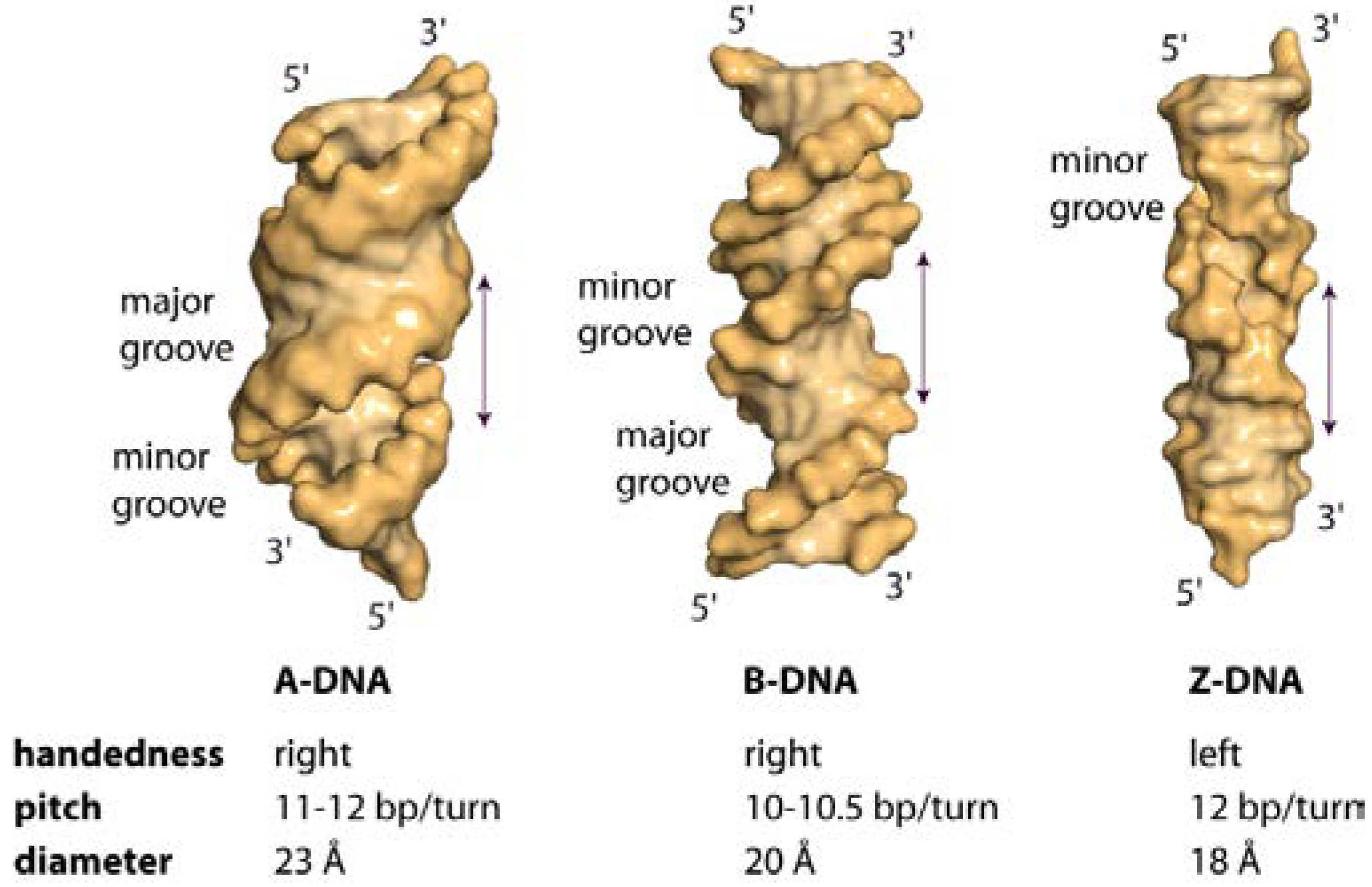
A-form DNA. A-DNA is a right-handed double helix made up of deoxyribonucleotides. ... B-form DNA. B-form DNA is a right-handed double helix, which was discovered by Watson and Crick based on the X-ray diffraction patterns. ... Z-form DNA. Z-form DNA is a left-handed double helix.
What are 3 types of DNA?
There are three different DNA types:A-DNA: It is a right-handed double helix similar to the B-DNA form. ... B-DNA: This is the most common DNA conformation and is a right-handed helix. ... Z-DNA: Z-DNA is a left-handed DNA where the double helix winds to the left in a zig-zag pattern.
Why is B form DNA most stable?
It is known that the stability of the double helical structure of B-DNA is supplied by the hydrogen bonds as proposed by Watson and Crick3 and by the stacking interactions. However, the relative importance of both stabilizing interactions as well as how they interfere with each other is largely unknown.
What are the 4 types of DNA?
There are four nucleotides, or bases, in DNA: adenine (A), cytosine (C), guanine (G), and thymine (T).
How can A B type DNA be transformed to its A form?
The proposed mechanism for the B- to A-DNA transition is that direct electrostatic interactions between mobile metal ions and phosphate groups across the major groove lead to DNA bending, then to sugar repuckering, and therefore to the transition.
Is B-DNA left or right-handed?
right-handedNormal B-DNA, as first described by Watson and Crick, is a right-handed helix. GC-rich DNA can also exist in a form known as Z-DNA, which forms a left-handed helix.
What type of DNA is human?
Cells have two types of DNA – mitochondrial DNA and autosomal DNA. Nuclear DNA (autosomal DNA) is enveloped into 22 pairs of chromosomes. In every pair of autosomes, one has inherited, one set is derived from the father and the other from the mother.
Is B-DNA right-handed?
Deoxyribonucleic acid (DNA hereafter) is a double-stranded macromolecule used by living organisms to carry their genetic information. The right-handed helix conformation called B-DNA is the dominant form in vivo. The diameter of the helix is 20 Å and one turn consists of 10 bp.
What is the function of Z-DNA?
Z-DNA is thought to play a role in the regulation of gene expression; Z-DNA is also thought to be involved in DNA processing events and/or genetic instability. For example, Z-DNA-forming sequences have the potential to enhance the frequencies of recombination, deletion, and translocation events in cellular systems.
What are the 4 types of DNA?
There are four nucleotides, or bases, in DNA: adenine (A), cytosine (C), guanine (G), and thymine (T).
What are the two different types of DNA?
There are two types of DNA in the cell – autosomal DNA and mitochondrial DNA. Autosomal DNA (also called nuclear DNA) is packaged into 22 paired chromosomes. In each pair of autosomes, one was inherited from the mother and one was inherited from the father.
Is B-DNA right-handed?
Deoxyribonucleic acid (DNA hereafter) is a double-stranded macromolecule used by living organisms to carry their genetic information. The right-handed helix conformation called B-DNA is the dominant form in vivo. The diameter of the helix is 20 Å and one turn consists of 10 bp.
How long is the residence time in the minor groove of the B-DNA sequence?
For comparison, the residence time in the minor groove of the regular B-DNA sequence d (CGCG A TCGCG) is 0.3 ns at 10°, shorter than in the case of the B'-DNA sequences, but not by a large factor. As for the major groove residence times, they are comparable for the three sequences, and a few times shorter than those of minor groove water. At − 8° the value is 0.36 ns, or even more if there is rotation of water, a phenomenon that may be considered more likely in the major than in the minor groove.
Which DNA conformation inhibits cleavage?
In contrast to the usual recognition sequence in B-DNA, the left-handed Z-DNA conformation within the recognition sequence or at a distance of four or less base pairs inhibits the cleavage by Eco RI (and Hha I and Bam HI as well) ( 39 ). Note that certain restriction enzymes, e.g., Mbo I (GATC), show enhanced cleavage activity at a B–Z junction.
What Does the Future Hold for the Study of Nucleic Acid Modifications in the Brain?
Since Watson and Crick proposed the model of the right-handed double helix, which was later called B-DNA, the field has come to appreciate that this particular conformational state is but one possible shape. In fact, when the crystal structure of DNA was revealed, the field was amazed to discover that DNA could also assume a zig-zag confirmation with a left-handed double helix, which was completely opposite to that proposed 25 years earlier ( Wang et al., 1979 ). It is evident that this interesting conformational state of DNA, or Z-DNA as it became known, together with at least 20 other possible DNA states such as the G-quadruplex, which interacts with DNA helicases and other epigenetic regulators, have very important biological functions in the cell ( Murat & Balasubramanian, 2014; Rich & Zhang, 2003 ). It is now known that the structural properties of DNA can influence its ability to recognize and interact with transcription factors and other protein partners that, in turn, drive gene expression as well as coordinating the organization and integrity of the genome ( Parker et al., 2009; Rohs et al., 2009; Rohs, West, et al., 2009; H. Zhou et al., 2015; K.I. Zhou et al., 2015; and reviewed in Harteis & Schneider, 2014 ).
What are the structures of trinucleotide repeats?
Most trinucleotide repeats show non-B DNA structures. CAG/CTG repeats are known to form hairpins with slipped strands, which may play an important role in the mechanism of repeat-size instability ( Pearson et al., 1998, 2003 ). GAA repeats at the FRDA locus have been shown to form the “sticky DNA” structure ( Sakamoto et al., 1999 ). In contrast, in vitro studies provided evidence that ATTCT repeats take unpaired structures, in which two strands are separated ( Potaman et al., 2003 ). Two-dimension (2D) agarose gel electrophoresis of plasmid topoisomers containing (ATTCT)11–46 repeats suggested that the repeats form uncoiled DNA under superhelical tension. Atomic force microscopy confirmed unpairing of the two strands in these ATTCT repeats. Furthermore, chloroacetylaldehyde bound the repeat region of the supercoiled plasmid, indicating accessibility of this reagent to the uncoiled region ( Potaman et al., 2003 ). Although the unpaired DNA structure may induce chromosome fragility and DNA methylation, our cytogenetic study, in collaboration with Dr. Lisa G. Shaffer of Washington State University showed no evidence of chromosome-22 fragility in leukocytes obtained from SCA10 patients (unpublished data). Our Southern blot analysis of genomic SCA10 DNA digested with methylation-dependent and -independent enzymes showed no evidence of aberrant methylation in the SCA10 region (data not shown).
What is sticky DNA?
A new type of DNA structure that implies intramolecular triplex formation was shown to be adopted by lengths of GAA as found in FRDA. This structure was called “sticky DNA” and was first demonstrated in plasmids containing long tracts of GAA [ 187 ]. Sticky DNA was discovered as an anomalously retarded band in agarose gels in which linearized plasmids containing GAA repeats were separated [ 188 ]. Such a slow-migrating band was shown to have a number of physicochemical properties that are typical of intramolcular R·R·Y triplexes. In particular, the retarded band appeared only if the plasmid was negatively supercoiled prior to linearization, and it was sensitive to divalent ion concentration and temperature as is typical for R·R·Y triplexes. The possible intermolecular nature of the structure was suggested by the correlation between its abundance and plasmid DNA concentration. This was proven by electron microscopy analysis, which revealed bimolecular complexes formed by joining two plasmids through the region containing the GAA repeat. An excellent correlation was found between the lengths of GAA and the formation of this novel conformation: FRDA patients have 66 or more repeats; sticky DNA was found only for repeats longer than 59 units. In vitro transcription studies of (GAA) n repeats (where n = 9–150) using 17 or SP6 RNA polymerase showed that, when a gel-isolated sticky DNA template was transcribed, the amount of full-length RNA synthesized was significantly reduced compared with the transcription of the linear template. Surprisingly, transcriptional inhibition was observed not only for the sticky DNA template, but also for another DNA molecule used as an internal control in an orientation-independent manner. The molecular mechanism of transcriptional inhibition by sticky DNA was sequestration of the RNA polymerases by direct binding to the complex DNA structure [ 189 ]. A (GAAGGA)65 sequence, also found in intron 1 of the frataxin gene, does not form sticky DNA, nor does it inhibit transcription in vivo and in vitro or associate with the FRDA disease state [ 190 ]. This finding suggests that interruptions in the GAA sequence may destabilize its structure and facilitate transcription. A systematic analysis of the effects of introducing interruptions into a (GAA·TTC) 150 repeat by substituting an increasing number of A's with G's has confirmed that the sticky DNA/triplex structure is progressively destabilized and it fails to form when the sequence becomes (GAAGGA) 75. As the tendency to form a sticky DNA/triplex structure decreases, less and less inhibition of transcription is observed in vivo and in vitro [ 191 ].
What is the topology of DNA?
Figure 3.1. DNA topology. (A) By definition a topologically closed DNA requires constrained DNA ends (gray bars). The relaxed state of the DNA double helix is shown in the center. When DNA is under torsional force (indicated by circular arrows), this will manifest as a change in Tw and/or formation of Wr. Untwisted DNA is negatively supercoiled (left), overtwisted DNA is positively supercoiled (right). (B) Twisting of DNA by protein complexes tracking along the double helix and restrictions to the twist diffusion are establishing quasi-topological boundaries that confine dynamic DNA supercoiling at particular genomic regions.
What is the link number difference of DNA?
DNA with a nonzero linking number difference (ΔLk=Lk − Lk 0) is called supercoiled DNA. Since DNA behaves as an elastic rod, any deviation of linking number from the reference Lk 0 value imparts torsional stress upon the DNA. Thus, supercoiling occurs when torsional stress introduces changes in the helical repeat of the DNA and/or induces formation of a coiled helical structure. The linking number difference normalized to the length of DNA in units of unstressed helical turns (supercoiling density, σ) is often used to describe the DNA topological state: σ =ΔLk/Lk 0. If the outcome of DNA torsional stress is an under-Twisted double helix, then DNA is negatively supercoiled. In positively supercoiled DNA, the double helix is over-Twisted ( Fig. 3.1A ). To relieve torsional stress in a topologically closed DNA, strand-breakage is required. Relaxation of DNA supercoiling is performed by a special class of enzymes named DNA topoisomerases. Topoisomerases temporarily cleave one or both DNA strands altering the linking number from Lk to Lk 0.
What is the B form of DNA?
B-form DNA. B-DNA is the Watson–Crick form of the double helix that most people are familiar with. They proposed two strands of DNA — each in a right‑hand helix — wound around the same axis. The two strands are held together by H‑bonding between the bases (in anti-conformation). The two strands of the duplex are antiparallel ...
What is the difference between A-form and B-form DNA?
It is in the C2′ endoconformation for B-form, whereas it is in the C3′ endoconformation in A-form. A second major difference between A-form and B-form nucleic acid is the placement of base-pairs within the duplex.
Why do different forms of DNA exist?
In nearly all cells, from simple bacteria through complex eukaryotes, the DNA must be compacted by more than a thousand fold in order even to fit inside the cell or nucleus.
How do bases fit in the double helical model?
Bases fit in the double helical model if pyrimidine on one strand is always paired with purine on the other. From Chargaff’s rules, the two strands will pair A with T and G with C. This pairs a keto base with an amino base, a purine with a pyrimidine. Two H‑bonds can form between A and T, and three can form between G and C.
What is Z DNA?
Z-DNA is a radically different duplex structure, with the two strands coiling in left-handed helices and a pronounced zig-zag (hence the name) pattern in the phosphodiester backbone.
What are the two strands of a duplex?
The two strands of the duplex are antiparallel and plectonemically coiled . The nucleotides arrayed in a 5′ to 3′ orientation on one strand align with complementary nucleotides in the 3′ to 5′ orientation of the opposite strand.
What is the duplex in Z DNA?
The duplex in Z-DNA has to accommodate the distortion of this G nucleotide in the synconformation. The cytosine in the adjacent nucleotide of Z-DNA is in the “normal” C2′ endo, anticonformation.
What are the different forms of DNA?
Different forms of DNA like B, A, C, D, E and Z have revealed after the X-ray diffraction analysis of DNA crystals at atomic resolution.
What is DNA based on?
DNA (hereditary material of the cell) consists of a long polynucleotide chain and shows structural diversity by changing its structural configuration, based on various factors like: The hydration level. Salt concentration.
What is the sugar pucker in Z-type DNA?
In Z-form also, there is zig-zag plectonemic coiling. It possesses antiparallel strands like B-DNA. In Z-type, the sugar pucker or deoxyribose ring is present at the “C 3 ” endoform for the purine bases and the “C 2 ” endoform for the pyrimidine bases.
What is the Z-DNA conformation?
The coiling of the Z-form is in a left-handed and zig-zag pattern. The conformation of Z-DNA is long and thin compared to the B-DNA. It shows an asymmetric structural configuration, which gives rise to the zig-zag helical structure with no internal space.
How many degrees does C-DNA rotate?
Both the strands of C-DNA are antiparallel to each other, unlike Z-DNA. C-DNA has a helix diameter of 19.0 Å. The rotation per base pair is 38.6 degrees, with a base pair tilt of 7.8 degrees. The size and shape of the C-DNA are smaller than the B-DNA and A-DNA.
How does Z-DNA form?
The Z-DNA forms by the alternating stretching of purines and pyrimidines bases. The Z-form helix’s diameter is 18Å, and the base-pair tilt is 7 Å, which is lower than the B and A-form. A single turn in B-form comprises 12 base pairs, which are perpendicular to the helical axis.
How many base pairs does C-DNA have?
The helix of C-DNA is twisted in a right-handed fashion with a helix pitch of 30.97Å. It consists of 9.33 base pairs per turn.
What is the most common form of DNA?
As we are taught in school, the double stranded DNA molecule is a right-handed helix as determined by Watson and Crick using Franklin's x-ray diffraction images [1]. This B-form of DNA has approximately 10 nucleotides per turn of the helix and is the most common form of DNA found in nature.
What is the name of the DNA that is dehydrated?
In addition to B- and Z-DNA, DNA can exist in another form known as A-DNA. A-DNA occurs when DNA is dehydrated, but also in DNA/RNA hybrids and double stranded RNA. A-DNA with the elements colored. A-DNA with the bases colored. A-DNA with the strands colored.
What is Z DNA?
Z-DNA occurs in nature, but is most frequently used by marketing departments to [incorrectly] create company logos. Z-DNA with the elements colored. Z-DNA with the bases colored. Z-DNA with the stands colored. In addition to B- and Z-DNA, DNA can exist in another form known as A-DNA.
When is DNA day?
April 25th (4/25) is national DNA day. Digital World Biology ™ celebrates by sharing some of our favorite DNA structures. We created these photos with Molecule World ™ Molecule World is a tools for exploring molecular and chemical structures on an iPhone or iPad.
Can you download DNA structures on Molecule World?
Go to our new DNA Exploration Collection. Download the file named DNA_exploration.mwc and open it in Molecule World on an iPad. Alternatively, you can download the structures below and open them in Molecule World on an iPhone.
Can you see DNA on iPhone?
Update: These same structures can also be viewed on the iPhone with Molecule World for iPhone. Visit the iTu nes app store to download either app and explore these DNA structures on your phone or iPad.
Is DNA right handed or left handed?
Classic structure with the strands colored. However, we've heard it said that the first crystal structure of DNA was not right-hand ed (pers. communication S. Elgin, Washington University). Instead, the high salt and GC base-pairs, used to form the DNA crystals caused the helix to twist in a left-handed way, ...
Which is better, B-DNA or Crick?
Important structural features of B-DNA are given below: Ø Majority of the DNA in a cell is in B-DNA conformation. Ø B-DNA is a right handed helix.
How tall is each turn on helix in B-DNA?
Ø Each turn on helix in B-DNA possess a helical height of 34 Å.
What is the helical turn per base pair in Z-DNA?
Ø The helical turn per base pair in Z-DNA is 9⁰ for pyrimidine – purine step and 51⁰ for purine – pyrimidine step.
How many base pairs are tilted in A-DNA?
Ø Individual base pairs in A-DNA are 20⁰ tilted with respect to the helical axis.
Which bond conformation is anti- for pyrimidines and syn- for purines?
Ø The glycosidic bond conformation is anti- for pyrimidines and syn- for purines.
What are the different types of conformations that DNA can adopt?
The various types of conformations that the DNA can adopt depend on different factors such as: 1. Hydration level.
What is the most abundant type of DNA?
Among these three types, the most abundant type of DNA is B-DNA, commonly known as Watson-Crick Model of DNA double helix. The present post describes the structural features of A, B and Z forms of DNA in a comparative manner. We will also discuss the similarities and differences between A-DNA, B-DNA and Z-DNA.
What is DNA made of?
DNA stands for deoxyribonucleic acid. It’s made up of units of biological building blocks called nucleotides. DNA is a vitally important molecule for not only humans, but for most other organisms as well. DNA contains our hereditary material and our genes — it’s what makes us unique.
What are the components of DNA?
The DNA molecule is made up of nucleotides. Each nucleotide contains three different components — a sugar, a phosphate group, and a nitrogen base. The sugar in DNA is called 2’-deoxyribose. These sugar molecules alternate with the phosphate groups, making up the “backbone” of the DNA strand.
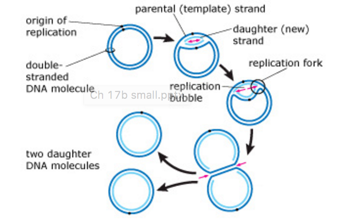
How does DNA get split?
In order to achieve this, your DNA must undergo a process called replication. When this occurs, the two DNA strands split apart. Then, specialized cellular proteins use each strand as a template to make a new DNA strand. When replication is completed, there are two double-stranded DNA molecules.
What part of DNA is responsible for aging?
Another part of DNA that may be involved in aging are telomeres. Telomeres are stretches of repetitive DNA sequences that are found at the ends of your chromosomes. They help to protect DNA from damage, but they also shorten with each round of DNA replication.
Why do cells read the code 3 bases at a time?
Your cells read this code three bases at a time in order to generate proteins that are essential for growth and survival. The DNA sequence that houses the information to make a protein is called a gene. Each group of three bases corresponds to specific amino acids, which are the building blocks of proteins.
Why is DNA damaged?
In fact, it’s estimated that tens of thousands of DNA damage events occur every day in each of our cells. Damage can occur due to things like errors in DNA replication, free radicals, and exposure to UV radiation. But never fear!
Where is DNA found in eukaryotic cells?
In a eukaryotic cell, DNA is within the nucleus. A small amount of DNA is also found in organelles called mitochondria, which are the powerhouses of the cell.

Why Do Different Forms of DNA Exist?
B-Form DNA
- B-DNA is the Watson–Crick form of the double helix that most people are familiar with.
- They proposed two strands of DNA — each in a right‑hand helix — wound around the same axis. The two strands are held together by H‑bonding between the bases (in anti-conformation).
- The two strands of the duplex are antiparallel and plectonemically coiled. The nucleotides arr…
- B-DNA is the Watson–Crick form of the double helix that most people are familiar with.
- They proposed two strands of DNA — each in a right‑hand helix — wound around the same axis. The two strands are held together by H‑bonding between the bases (in anti-conformation).
- The two strands of the duplex are antiparallel and plectonemically coiled. The nucleotides arrayed in a 5′ to 3′ orientation on one strand align with complementary nucleotides in the 3′ to 5′ orien...
- Bases fit in the double helical model if pyrimidine on one strand is always paired with purine on the other. From Chargaff’s rules, the two strands will pair A with T and G with C. This pairs a ket...
A-Form DNA
- The major difference between A-form and B-form nucleic acid is in the confirmation of the deoxyribose sugar ring. It is in the C2′ endoconformation for B-form, whereas it is in the C3′ endoconforma...
- A second major difference between A-form and B-form nucleic acid is the placement of base-pairs within the duplex. In B-form, the base-pairs are almost centered over the helical axis bu…
- The major difference between A-form and B-form nucleic acid is in the confirmation of the deoxyribose sugar ring. It is in the C2′ endoconformation for B-form, whereas it is in the C3′ endoconforma...
- A second major difference between A-form and B-form nucleic acid is the placement of base-pairs within the duplex. In B-form, the base-pairs are almost centered over the helical axis but in A-form,...
- Right-handed helix
- 11 bp per turn; 0.26 nm axial rise; 28o helix pitch; 20obase-pair tilt
Z-Form DNA
- Z-DNA is a radically different duplex structure, with the two strands coiling in left-handed helices and a pronounced zig-zag (hence the name) pattern in the phosphodiester backbone.
- Z-DNA can form when the DNA is in an alternating purine-pyrimidine sequence such as GCGCGC, and indeed the G and C nucleotides are in different conformations, leading to the zig-zag pattern.
- The big difference is at the G nucleotide.
- It has the sugar in the C3′ endoconformation (like A-form nucleic acid, and in contrast to B-form DNA) and the guanine base is in the synconformation.
Conditions Favoring A-Form, B-Form, and Z-Form of DNA
- Whether a DNA sequence will be in the A-, B-or Z-DNA conformation depends on at least three conditions.
- The first is the ionic or hydration environment, which can facilitate conversion between different helical forms.
- A-DNA is favored by low hydration, whereas Z-DNA can be favored by high salt.
- The second condition is the DNA sequence: A-DNA is favored by certain stretches of purines (or pyrimidines), whereas Z-DNA can be most readily formed by alternating purine-pyrimidine steps.
Other Rare Forms of DNA
- C-DNA 1. Formed at 66% relative humidity and in presence of Li+ and Mg2+ ions. 2. Right-handed with the axial rise of 3.32A° per base pair 3. 33 base pairs per turn 4. Helical pitch 3.32A°×9.33°A=30.97A°. 5. Base pair rotation=38.58°. 6. Has a diameter of 19 A°, smaller than that of A-&B- DNA. 7. The tilt of base is 7.8° D-DNA 1. Rare variant with 8 base pairs per helical tu…
References
- https://www.researchgate.net/publication/10837288_A_glossary_of_DNA_structures_from_A…
- http://people.bu.edu/mfk/restricted566/dnastructure.pdf
- Alberts, B., Johnson, A., Lewis, J., Raff, M., Roberts, K., & Walter, P. (2002). Molecular biology of the cell. New York: Garland Science.
- http://www.newworldencyclopedia.org/entry/Deoxyribose