Which blood vessels transport blood towards the heart?
- These are blood vessels that take oxygen-poor blood back to the heart.
- Veins become larger and larger as they get closer to the heart.
- The superior vena cava is the large vein that brings blood from the head and arms to the heart, and the inferior vena cava brings blood from the abdomen and ...
What blood vessel carries blood away from the heart?
- Arteries begin with the aorta, the large artery leaving the heart.
- They carry oxygen-rich blood away from the heart to all of the body’s tissues.
- They branch several times, becoming smaller and smaller as they carry blood further from the heart.
How does blood flow through the heart step by step?
- Capillaries separate oxygenated blood from deoxygenated blood and arteries from veins
- After passing through the capillaries, blood is deoxygenated and needs to head back to the heart to be pumped to the lungs to pick up oxygen
- The Superior Vena Cava is the vein that gets deoxygenated blood from the upper body and returns it to the heart
What arteries supply blood to the heart?
What are the main arteries?
- Aorta. The largest artery in the body that connects directly to the left ventricle.
- Head and neck arteries (carotid arteries)
- Stem arteries (lower aorta, coronary and subclavian)

What are the blood supplies to the heart?
Coronary arteries supply blood to the heart muscle. Like all other tissues in the body, the heart muscle needs oxygen-rich blood to function. Also, oxygen-depleted blood must be carried away. The coronary arteries wrap around the outside of the heart.
What veins supply the heart with blood?
Pulmonary veins - bring oxygen-rich blood back to the heart from the lungs. Right coronary artery (RCA) - supplies blood to the right atrium, right ventricle, bottom portion of the left ventricle and back of the septum.
Does the heart have its own blood supply?
Your heart muscle needs its own supply of blood because, like the rest of your body, it needs oxygen and other nutrients to stay healthy. For this reason, your heart pumps oxygen-rich blood to its own muscle through your coronary arteries. Keep blood flowing efficiently.
Where does the heart's blood supply come from?
Your coronary arteries supply blood to your heart. These arteries branch off from the aorta so that oxygen-rich blood is delivered to your heart as well as the rest of your body.
What are the 4 main blood vessels in the heart?
The major blood vessels connected to your heart are the aorta, the superior vena cava, the inferior vena cava, the pulmonary artery (which takes oxygen-poor blood from the heart to the lungs where it is oxygenated), the pulmonary veins (which bring oxygen-rich blood from the lungs to the heart), and the coronary ...
Which artery supplies the most blood to the heart?
The aorta (the main blood supplier to the body) branches off into two main coronary blood vessels (also called arteries). These coronary arteries branch off into smaller arteries, which supply oxygen-rich blood to the entire heart muscle. The right coronary artery supplies blood mainly to the right side of the heart.
What are the 3 main arteries of the heart?
The coronary arteries are also called the epicardial arteries because they run along the outer surface of the heart on the epicardium; the main ones are the left coronary artery and the right coronary artery. The left coronary artery divides into the left anterior descending and the left circumflex arteries.
What is the nerve supply to the heart?
The nervous supply to the heart is autonomic, consisting of both sympathetic and parasympathetic parts. The sympathetic fibres arise from the pressor centre, while the parasympathetic fibres arise in the depressor centre.
What are the 7 steps of blood flow through the heart?
Blood flows through the heart in the following order: 1) body –> 2) inferior/superior vena cava –> 3) right atrium –> 4) tricuspid valve –> 5) right ventricle –> 6) pulmonary arteries –> 7) lungs –> 8) pulmonary veins –> 9) left atrium –> 10) mitral or bicuspid valve –> 11) left ventricle –> 12) aortic valve –> 13) ...
What is the main artery in the heart?
the aortaThe largest artery is the aorta, the main high-pressure pipeline connected to the heart's left ventricle. The aorta branches into a network of smaller arteries that extend throughout the body. The arteries' smaller branches are called arterioles and capillaries.
How many arteries feed the heart?
two coronary arteriesThere are two coronary arteries, each containing several branches: Right coronary artery (RCA): The RCA supplies blood to your right atrium and right ventricle (where deoxygenated blood goes before heading to the lungs).
How the blood flows through the heart step by step?
0:232:12Blood Flow through the Heart in 2 MINUTES - YouTubeYouTubeStart of suggested clipEnd of suggested clipThe inferior vena cava receives blood from the trunk viscera. And lower limbs. Both the superior andMoreThe inferior vena cava receives blood from the trunk viscera. And lower limbs. Both the superior and inferior vena cava end up in the right atrium. One of the four chambers of the heart. The heart not
What are the 3 main veins of the heart?
The inferior vena cava. pulmonary trunk artery. Approximately two inches superior to the base of the heart, this vessel branches into the left and right pulmonary arteries, which transport blood into the lungs. The pulmonary trunk artery.
What are the 4 major veins?
Veins can be categorized into four main types: pulmonary, systemic, superficial, and deep veins.
How many veins connect to the heart?
Answer and Explanation: There are around 10 veins that are found in or connected to the heart. The two largest veins that bring blood from the body to the heart are the superior vena cava and inferior vena cava.
What are the 3 main vessels of the heart?
There are three kinds of blood vessels: arteries, veins, and capillaries. Each of these plays a very specific role in the circulation process. Arteries carry oxygenated blood away from the heart.
Which artery supplies blood to the heart?
Now the question arises which artery supplies blood to the heart. The answer to this question is coronary arteries. The blood supply of the heart is mediated by the coronary artery; these arteries are divided into two main classes: the left coronary arteries and the right coronary arteries. This complete circulation including the venous supply of the heart is known as coronary circulation.
What is the function of the heart?
The heart is a major organ of the human body which is responsible for the circulation of the blood. Blood circulation in the body ensures that every cell of the organ receives nutrients and oxygen supply to perform its metabolism. To understand the blood supply of the heart it is important to understand the location of the heart, anatomy of the heart, the vasculature of the nerve which answers questions like which artery supplies blood to the heart. This article dwells with the intricacies of blood supply of heart anatomy and functioning, which arteries supply blood to heart, the circulation system of the heart known as coronary circulation.
What are the layers of the heart?
The heart wall can be defined as the muscular structure of the heart. The heart wall has 4 layers pericardium, epicardium, myocardium, and endocardium. The pericardium is the outermost layer of the heart wall, it is the layer in which arteries supply blood to the heart. It has a fibrous sac-like structure from the outside and the inner surface is generally parietal. Epicardium is the layer just below the pericardium, between both these layers there is a presence of the cavity; this cavity is known as the pericardial cavity. It is a fluid filled cavity. This layer, the epicardium, is also known as the visceral layer of the pericardium. The myocardium is the layer that is responsible for the contraction and relaxation of the atrium and ventricle, thus ensuring the blood supply of the heart. It has a modification known as an intercalated disk, which is specialized to the cardiac muscle only, it ensures the connection between the left and right ventricle and similarly left and right atrium. It is important to note that valves lack this intercalated disk. The blood vessels responsible for the blood supply of heart protrudes into this layer.
Which valve allows blood to come into the ventricular section of the heart?
Ans: They are the atrioventricular valve that allows the blood to come into the ventricular section of the heart leading to ventricular relaxation and atrial contraction.
Where is the heart located?
The heart is present in the thoracic cavity, which is posterior to the sternum, inferior to the diaphragm. It is located in between the lungs. The heart is made up of myocardium muscle, this muscle has exceptionally high thickness thus the heart can not work with the normal vasculature of the circulation. The blood supply of the heart is also known as coronary circulation and the artery that supplies blood to the heart is known as the coronary artery. The heart is the four-chambered organ. It is divided into two atria and two ventricles.
How many valves are there in the heart?
There are four major valves present in the heart they are called, atrioventricular valve and the semilunar valve. Each of these valves is prest in pair thus forming 4 valves.
What is the function of the cardiovascular system?
human nervous system: The cardiovascular system. The function of the cardiovascular system is to maintain an adequate supply of oxygen to all tissues of the body. In order to maintain this... The right and left coronary arteries originate from the right and left aortic sinuses (the sinuses of Valsalva), which are bulges at the origin ...
Why does the heart not depend on the blood in its own chambers?
Because of the watertight lining of the heart (the endocardium) and the thickness of the myocardium, the heart cannot depend on the blood contained in its own chambers for oxygen and nourishment. It possesses a vascular system of its own, called the coronary arterial system.
Which artery runs posteriorly along the atrioventricular groove?
There are many branches of the left anterior descending artery; the first and usually the largest septal branch is important because of its prominent role in supplying blood to the septum. The left circumflex artery leaves the left main coronary artery to run posteriorly along the atrioventricular groove.
How does electrical impulse affect cardiac contraction?
Voltage-sensitive protein channels on the surface of the sarcolemma, the membrane that surrounds the muscle fibre, support the flow of current as it relates to the flow of specific ions (ion-specific channels). These voltage-sensitive channels open and close as a function of the voltage that is sensed on the outer side and inner side (referred to as being “across the membrane,” or transmembrane) of the sarcolemma, between which a difference in electrical potential exists. An electrical potential gradient is created by an excess of negative ions immediately inside the sarcolemma and an equal excess of positive ions on the outside of the sarcolemma (a stage known as the resting potential). When a nerve impulse stimulates ion channels to open, positive ions flow into the cell and cause depolarization, which leads to muscle cell contraction.
Why is repolarization important for the heart?
When the heart is repolarized, the sarcoplasmic reticulum reabsorbs the excess calcium, and the cellular calcium concentration returns to its formerly low level, letting the heart muscle relax. Reabsorption of cellular calcium by the sarcoplasmic reticulum is important because it prevents the development of muscle tension.
Where is the ostium located?
The ostium, or opening, of the right coronary artery is in the right aortic sinus and that of the left coronary artery is in the left aortic sinus, just above the aortic valve ring. There is also a non-coronary sinus of Valsalva, which lies to the left and posteriorly at the origin of the ascending aorta. The left coronary arterial system is more ...
How does the heart beat?
Regular beating of the heart is achieved as a result of the inherent rhythmicity of cardiac muscle; no nerves are located within the heart itself, and no outside regulatory mechanisms are necessary to stimulate the muscle to contract rhythmically. That these rhythmic contractions originate in the cardiac muscle can be substantiated by observing cardiac development in the embryo ( see above ); cardiac pulsations begin before adequate development of nerve fibres. In addition, it can be demonstrated in the laboratory that even fragments of cardiac muscle in tissue culture continue to contract rhythmically. Furthermore, there is no gradation in degree of contraction of the muscle fibres of the heart, as would be expected if they were primarily under nervous control.
What Is Lad And Rca Disease
LAD: left anterior descending coronary artery LCx: left circumflex coronary artery RCA: right coronary artery SCA: single coronary artery. This case demonstrates a rare congenital coronary artery anomaly found incidentally with a presentation of atypical angina and three-vessel coronary artery disease .
Improving Health With Current Research
Learn about the following ways the NHLBI continues to translate current research and science into improved health for people who have heart conditions. Research on this topic is part of the NHLBI’s broader commitment to advancing heart and vascular disease scientific discovery.
Anatomy Of The Heart And Blood Vessels
The heart is a muscular pump that pushes blood through blood vessels around the body. The heart beats continuously, pumping the equivalent of more than 14,000 litres of blood every day through five main types of blood vessels: arteries, arterioles, capillaries, venules and veins.
About Blood Supply Of Heart
The heart is a major organ of the human body which is responsible for the circulation of the blood. Blood circulation in the body ensures that every cell of the organ receives nutrients and oxygen supply to perform its metabolism.
How The Heart Works
The heart is a large, muscular organ that pumps blood filled with oxygen and nutrients through the blood vessels to the body tissues. It’s made up of:
How Do The Heart And Blood Vessels Work
The heart works by following a sequence of electrical signals that cause the muscles in the chambers of the heart to contract in a certain order. If these electrical signals change, the heart may not pump as well as it should.
Diagnosis And Treatment Of Coronary Artery Disease
Fig 1.6 A coronary angiogram. Two critical narrowings have been labelled.
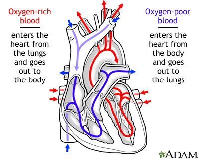
Which side of the heart is the blood entering?
Right Side. Blood enters the heart through two large veins, the inferior and superior vena cava, emptying oxygen-poor blood from the body into the right atrium.
How does blood flow through the right ventricle?
Blood flows from your right atrium into your right ventricle through the open tricuspid valve. When the ventricles are full, the tricuspid valve shuts. This prevents blood from flowing backward into the atria while the ventricles contract (squeeze).
How does blood flow through your lungs?
Once blood travels through the pulmonic valve, it enters your lungs. This is called the pulmonary circulation. From your pulmonic valve, blood travels to the pulmonary artery to tiny capillary vessels in the lungs. Here, oxygen travels from the tiny air sacs in the lungs, through the walls of the capillaries, into the blood. At the same time, carbon dioxide, a waste product of metabolism, passes from the blood into the air sacs. Carbon dioxide leaves the body when you exhale. Once the blood is purified and oxygenated, it travels back to the left atrium through the pulmonary veins.
Where does blood travel through the pulmonary artery?
Once blood travels through the pulmonic valve, it enters your lungs. This is called the pulmonary circulation. From your pulmonic valve, blood travels to the pulmonary artery to tiny capillary vessels in the lungs. Here, oxygen travels from the tiny air sacs in the lungs , through the walls of the capillaries, into the blood.
Which vein empties oxygen rich blood from the lungs into the left atrium?
The pulmonary vein empties oxygen-rich blood, from the lungs into the left atrium.
Which side of the heart works together?
The right and left sides of the heart work together
Where does oxygen travel?
Here, oxygen travels from the tiny air sacs in the lungs, through the walls of the capillaries, into the blood. At the same time, carbon dioxide, a waste product of metabolism, passes from the blood into the air sacs. Carbon dioxide leaves the body when you exhale.
What Are The Different Types Of Arteries
Arteries are the blood vessels that carry blood away from the heart. The different types of arteries include:
Venous Drainage Of The Brain
Circulation to the brain is both critical and complex. Many smaller veins of the brain stem and the superficial veins of the cerebrum lead to larger vessels referred to as intracranial sinuses.
Classification & Structure Of Blood Vessels
Blood vessels are the channels or conduits through which blood is distributed to body tissues. The vessels make up two closed systems of tubes that begin and end at the heart. One system, the pulmonary vessels, transports blood from the right ventricle to the lungs and back to the left atrium.
What Do The Heart And Blood Vessels Do
The heart’s main function is to pump blood around the body. Blood carries nutrients and waste products and is vital to life. One of the essential nutrients found in blood is oxygen.
Anatomy Of The Heart And Blood Vessels
The heart is a muscular pump that pushes blood through blood vessels around the body. The heart beats continuously, pumping the equivalent of more than 14,000 litres of blood every day through five main types of blood vessels: arteries, arterioles, capillaries, venules and veins.
What Is Collateral Circulation
Collateral circulation is a network of tiny blood vessels, and, under normal conditions, not open. When the coronary arteries narrow to the point that blood flow to the heart muscle is limited , collateral vessels may enlarge and become active.
Describe Briefly The Cardiac Dominance
Coronary artery dominance: The artery which gives rise to the posterior interventricular artery arises determines the coronary dominance.
Which artery supplies blood to the left side of the heart muscle?
The left main coronary artery supplies blood to the left side of the heart muscle (the left ventricle and left atrium). The left main coronary divides into branches: The left anterior descending artery branches off the left coronary artery and supplies blood to the front of the left side of the heart. The circumflex artery branches ...
Which artery supplies blood to the right ventricle?
This artery supplies blood to the outer side and back of the heart. Right coronary artery (RCA). The right coronary artery supplies blood to the right ventricle, the right atrium, and the SA (sinoatrial) and AV (atrioventricular) nodes, which regulate the heart rhythm.
Why are the coronary arteries important?
Since coronary arteries deliver blood to the heart muscle, any coronary artery disorder or disease can have serious implications by reducing the flow of oxygen and nutrients to the heart muscle. This can lead to a heart attack and possibly death. Atherosclerosis (a buildup of plaque in the inner lining of an artery causing it to narrow or become blocked) is the most common cause of heart disease.
What is the function of the heart muscle?
Like all other tissues in the body, the heart muscle needs oxygen-rich blood to function. Also, oxygen-depleted blood must be carried away. The coronary arteries wrap around the outside of the heart. Small branches dive into the heart muscle to bring it blood.
Which artery divides into smaller branches?
The right coronary artery divides into smaller branches, including the right posterior descending artery and the acute marginal artery. Together with the left anterior descending artery, the right coronary artery helps supply blood to the middle or septum of the heart.