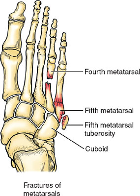
Calcaneal tuberosity fractures involve the posterosuperior aspect of the calcaneus and may involve the insertion of the Achilles tendon. In this report, we describe an unusual presentation of a calcaneal avulsion fracture involving 2 displaced fragments in a male patient who did not seek treatment for 1 month after the original injury.
What is the prevalence of calcaneal tuberosity fractures?
Fractures of the calcaneal tuberosity are relatively uncommon and are seen most frequently in elderly and diabetic patients. These injuries are typically avulsion fractures caused by concentric contraction of the gastrocnemius-soleus muscle complex.
What is the pathophysiology of calcaneal tuberosity?
The plantar fascia inserts on the medial process of the calcaneal tuberosity. It is the most common cause of heel pain, and the typical presentation is sharp pain localized at the anterior aspect of the calcaneus. All content published on Kenhub is reviewed by medical and anatomy experts.
What muscles attach to the calcaneal tuberosity?
There is a prominent medial margin on the medial process of the calcaneal tuberosity which provides attachments for the superficial part of the flexor retinaculum and distally the plantar aponeurosis. Some muscles also attach there including abductor hallucis and flexor digitorum brevis.
What is the inferior surface of the calcaneus?
The inferior surface (plantar surface) of calcaneus is bounded posteriorly by a transverse elevation, the calcaneal tuberosity, which is depressed in the middle and prolonged at either end into a process; the lateral process, small, prominent, and rounded, gives origin to part of the Abductor digiti quinti; the medial process, broader and larger...
What attaches to the calcaneal tuberosity?
The Achilles tendon attaches to the calcaneal tubercle.
What does calcaneal mean?
relating to the heelMedical Definition of calcaneal 1 : relating to the heel. 2 : relating to the calcaneus.
Where is the calcaneal?
heel boneThe calcaneus (heel bone) is the largest of the tarsal bones in the foot. It lies at the back of the foot (hindfoot) below the three bones that make up the ankle joint.
How serious is a calcaneal fracture?
Calcaneal fractures can be serious injuries that may produce lifelong problems. Arthritis, stiffness and pain in the joint frequently develop. Sometimes the fractured bone fails to heal in the proper position.
Can calcaneal spur be cured?
Heel spurs can't be cured. Healthcare providers recommend non-surgical treatments to ease symptoms associated with heel spurs.
How long does it take for a calcaneus fracture to heal?
If your injury is minor, such as a crack in the bone with little muscle damage, you may be able to resume normal activities from 3 to 4 months after surgery. If your fracture is severe, however, it may take from 1 to 2 years before recovery is complete.
How do you treat calcaneus pain?
Dorest and raise your heel when you can.put an ice pack (or bag of frozen peas) in a towel on your heel for up to 20 minutes every 2 to 3 hours.wear wide comfortable shoes with a low heel and soft sole.use soft insoles or heel pads in your shoes.wrap a bandage around your heel and ankle to support it.More items...
How do you fix calcaneus pain?
Treating heel pain resting your heel – avoiding walking long distances and standing for long periods. regular stretching – stretching your calf muscles and plantar fascia. pain relief – using an icepack on the affected heel and taking painkillers, such as non-steroidal anti-inflammatory drugs (NSAIDs)
How do you palpate calcaneal tuberosity?
4:025:28Palpation Of The Foot And Ankle - YouTubeYouTubeStart of suggested clipEnd of suggested clipAnd this is where the plantar fascia originates you'll often have tenderness through here you canMoreAnd this is where the plantar fascia originates you'll often have tenderness through here you can feel through the plantar fascia of the foot as well. Down into the ball of the foot.
Can you walk with a calcaneus fracture?
Symptoms of a heel fracture include pain, swelling and bruising of the heel. Patients usually are unable to walk.
How painful is a calcaneal fracture?
Pain is usually severe enough to require an emergency room visit. If the fracture is caused by a stress fracture, over time, then symptoms may be far more vague. There may be some pain, increasing throughout the day, often described as being dull and achy. Bruising may or may not be present.
Does a calcaneus fracture require surgery?
General Treatment Some, but not all, calcaneus fractures require surgery. The broken bone will take 3-4 months to heal with or without surgery. If surgery is not needed, there will still be a time where movement and weight bearing is limited.
What is the medical term for pertaining to the heel bone?
Calcaneum = Calcaneus bone, a heel bone. Calcanea (Plural) Calcane/al: Pertaining to the heel.
What does plantar mean in medical terms?
the sole of the footMedical Definition of plantar : of, relating to, or typical of the sole of the foot the plantar aspect of the foot.
What is the meaning of carples?
British Dictionary definitions for carpal carpal. / (ˈkɑːpəl) / noun. any bone of the wrist. (as modifier)carpal bones Also: carpale (kɑːˈpeɪlɪ)
What does the prefix dynia mean?
to painDynia definition Forms medical terms relating to pain.
What is calcaneal tuberosity fracture?from pubmed.ncbi.nlm.nih.gov
These injuries are typically avulsion fractures caused by concentric contraction of the gastrocnemius-soleus muscle complex.
How many fractures of the tuberosity of the calcaneus are there?from pubmed.ncbi.nlm.nih.gov
We describe 24 fractures of the tuberosity of the calcaneus in 22 patients. Three were similar to the type of avulsion fracture which has been well-defined but the remainder represent a group which has been unrecognised previously. Using CT and operative findings we have defined the different patter …
What is the calcaneal sulcus?
The calcaneal sulcus provides attachments for the: interosseous, talocalcaneal, cervical ligaments and. medial root of the inferior extensor retinaculum. Calcaneal sulcus (superior view) There is also a non-articular area on the calcaneus, distal to the posterior talar facet which provides an attachment surface for:
Which aspect of the calcaneus features many curves to accommodate the talus and the many different?
Anterior aspect. The front of the calcaneus features many curves to accommodate the talus and the many different tarsal bones, which lead to the metatarsals and phalanges. The back of the calacaneus is not as complex, featuring a tuberosity and a medial process.
What are the two bones that the calcaneus articulates with?
Role with the talus and cuboid. The calcaneus articulates with two bones: the talus and the cuboid. There are three articular surfaces for the talus. The largest is the slightly convex oval surface for the body of the talus.
What is the posterior aspect of the calcaneus?
The posterior aspect is rough and concavo-convex in shape. This convexity supports the fibroadipose tissue (Kager's fat pad) between the calcaneal tendon and the ankle joint.
What is the main band of the inferior extensor retinaculum?
the main band of the inferior extensor retinaculum. the stem of the bifurcate ligament. There is a prominent medial margin on the medial process of the calcaneal tuberosity which provides attachments for the superficial part of the flexor retinaculum and distally the plantar aponeurosis.
Which ligament attaches to the medial margin of the sustentaculum tali?
Flexor retinaculum of the foot (posterior view) The plantar calcaneonavicular ligament attaches to the medial margin of the sustentaculum tali.
Which tali bears the greatest weight per area?
The sustentaclulum tali bears the greatest weight per area and also has a lot of soft tissue structures attaching around it. The lateral wall of the calcaneus is thin and has attachments for the calcaneofibular ligament and the osseus reflection of the peroneal tendons.
What is a calcaneal spur?
A calcaneal spur is also called as a heel spur, it is a bony outgrowth from the calcaneal tuberosity. Heel spurs are typically detected by a radiographic examination which is commonly referred to as an “x-ray”. When a foot bone is exposed to constant stress, calcium deposits build up on the bottom of the calcaneal bone.
What is the best oil for calcaneal spurs?
Flaxseed Oil. Flaxseed oil, also called as linseed oil, contains alpha-linolenic acid, a form of omega-3 fatty acid, which can help to reduce pain and inflammation. It can also lessen morning stiffness associated with calcaneal spurs. Heat ¼ cup of flaxseed oil in a bowl until it is warm.
How to get rid of calcaneal spurs on foot?
Roll it under your foot/heel for 10 to 15 minutes. Do this as soon as you feel or get pain after a day of rigorous activity. 3. Baking Soda. Baking soda is another good natural remedy for calcaneal spurs. The crystals in baking soda will help you to reduce calcium deposits built up on the bottom of the heel bone.
What are the three facets of the calcaneus?
The posterior part of the calcaneus is circular, with three facets (superior, middle and inferior). The superior facet is separated from the calcaneal tendon by the retrocalcaneal bursa. The middle facet provides the attachment site for the calcaneal tendon ( Achilles tendon). The inferior facet curves anteriorly and is continuous with calcaneal tuberosity on the plantar surface. The plantar surface of the calcaneal tuberosity projects forward on the plantar surface as a medial (larger) and lateral (smaller) process and at its most anterior projection is the calcaneal tubercle, where the short plantar ligament attaches.
Which bones articulate with the calcaneus?
Anteriorly, the calcaneus articulates with the cuboid ( calcaneocuboid joint) bones.
Which part of the medial surface supports the head of the talus?
Protruding anteromedially from upper margin of the medial surface is the sustentaculum tali which supports the more posterior part of the head of the talus. At its inferior aspect is a groove accommodating the flexor hallucis longus tendon. Superiorly is a cartilage covered facet (middle talar articular facet) for the corresponding middle facet of the head of talus as part of the subtalar joint, which is inclined anteriorly.
Where are the anterior and posterior facets of the talocalcaneal joint located?
The anterior and posterior facets of the talocalcaneal joint are on the superior surface of the calcaneus. The anterior facet is small and the posterior facet is large, inclined anteriorly and located near the middle of the superior surface. Between these two facets runs a fairly deep sulcus, the calcaneal sulcus, which together with the opposing talar sulcus forms the tarsal sinus (sinus tarsi). The tarsal sinus is a large gap between the anterior ends of the talus and calcaneus on its lateral aspect.
Which ligament is anterior to the sustentaculum tali?
anterior: plantar calcaneonavicular ligament (anterior margin of the sustentaculum tali of the calcaneus)
How to perform ultrasound evaluation of calcaneus?from radiologykey.com
To perform ultrasound evaluation of the calcaneus, the patient is placed in the prone position with the patient’s ankle hanging off the edge of the table. With the patient in the above position, a high-frequency linear ultrasound transducer is placed in a longitudinal plane with the inferior portion of the ultrasound transducer over plantar surface of the foot with the superior end of the transducer on the anterior portion of the calcaneus, and an ultrasound survey scan is taken ( Fig. 17.16 ). The calcaneus, calcaneal spur, and linear plantar fascia are identified at its insertion on the calcaneus ( Fig. 17.17 ). When the insertion of the plantar fascia is identified, it is evaluated for evidence of calcaneal spurs, insertional tendinopathy, and plantar fasciitis ( Figs. 17.18 to 17.20 ).
What is a plantar calcaneal spur?from radiopaedia.org
Plantar calcaneal spur. Plantar calcaneal spurs , or sometimes simply referred to as calcaneal spurs, are a commonly seen finding in radiology practice.
What causes a spur on the calcaneus?from healthhype.com
It is believed that the plantar fascia may cause excessive pulling of the periosteum (outer bone layer) of the calcaneus, friction with neighboring bones or pressure on prone parts of the calcaneus when walking or standing which ultimately results in the development of a spur.
Why do older men have plantar calcaneal spurs?from radiopaedia.org
Epidemiology. Plantar calcaneal spurs tend to usually occur in older men and women and may be related to obesity, osteoarthritis and current or previous heel pain.
What does it mean when your foot is tender?from healthhype.com
A tender protrusion may be at the back of the sole upon firm pressure. Sharp pains when walking on hard surfaces, especially when barefoot. Pain upon carrying a heavy object that may distort the pressure applied on the foot.
Who first discovered the osseous spurring of the plantar aspect of the calcaneus?from radiopaedia.org
Osseous spurring of the plantar aspect of the calcaneus was first documented in 1900 by the German physician P Plettner , who coined the term Kalkaneussporn (calcaneal spur) 7.
Can calcaneal spurs be treated?from healthhype.com
A calcaneal spur is not easily treated and the patient needs to learn to live with the condition by managing it conservatively. Special footwear, resting regularly when walking or running over long distances and professional care by a podiatrist may assist.
