
What is the calcaneocuboid ligament?
The calcaneocuboid joint is a type of saddle joint between the calcaneus and the cuboid bone. There are five ligaments connecting the calcaneus and the cuboid bone, forming parts of the articular capsule : the dorsal calcaneocuboid ligament.
What is the plantar calcaneocuboid joint?
the long plantar ligament. and the plantar calcaneocuboid ligament. The calcaneocuboid joint is conventionally described as among the least mobile joints in the human foot. The articular surfaces of the two bones are relatively flat with some irregular undulations, which seem to suggest movement limited to a single rotation and some translation.
What stabilizes the calcaneocuboid joint?
The calcaneocuboid joint is stabilized by ligaments on its superior and inferior aspects. The plantar calcaneocuboid ligament (short plantar ligament) is a thick band of fibrous tissue that extends from the anterior tubercle of the calcaneus to the posterior part of the cuboid just behind the groove for the fibularis longus tendon 1.
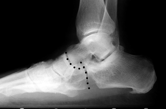
What is calcaneal cuboid joint joint joint arthritis?
CALCANEAL CUBOID JOINT ARTHRITIS. By: Robert H. Sheinberg, D.P.M., D.A.B.F.A.S., F.A.C.F.A.S. Pain on the outside of the foot just in front of the ankle may be due to injury or arthritic changes to the calcaneal cuboid joint.
Where is the calcaneocuboid joint located?
Calcaneocuboid arthritis is a form of osteoarthritis which affects the calcaneocuboid joint, that is located between the cuboid bone and the heel bone. Located on the outside of the foot, in front of the heel bone, the cuboid bone is characterised by its cube shape.
What type of joint is calcaneocuboid joint?
gliding typeJoint, calcaneocuboid: The joint located in the foot between the calcaneus bone and the cuboid bone. It is a gliding type of joint. The ligaments that serve to support and strengthen this joint are called the capsular, dorsal calcaneocuboid, bifurcated, long plantar, and plantar calcaneocuboid ligaments.
What is the function of the calcaneocuboid joint?
Function. The calcaneocuboid joint is conventionally described as among the least mobile joints in the human foot. The articular surfaces of the two bones are relatively flat with some irregular undulations, which seem to suggest movement limited to a single rotation and some translation.
What movements occur at the calcaneocuboid joint?
The three movement patterns identified for the calcaneocuboid joint; a rotation and translation as the result of the inversion-eversion driving motion and a rotation produced by the medial-lateral rotation of the leg on the foot.
Is calcaneocuboid joint intermediate?
True Blue. Response: I think of ankle, subtalar, talo-navicular and calcaneo-cuboid joints as intermediate joints (CPT 20605).
What is the calcaneocuboid joint reinforced by?
Calcaneocuboid ligaments Similar to its neighbour, the calcaneocuboid joint is also directly reinforced by four major ligaments: Laterally by the calcaneocuboid part of the bifurcate ligament (mentioned above) Long plantar ligament. Inferiorly by the plantar calcaneocuboid and the long plantar ligaments.
What bones make up the calcaneocuboid joint?
Calcaneocuboid joint: is formed between the front of the calcaneus and the posterior surface of the cuboid bone. Cuneonavicular joint: is formed between the navicular bone and the three cuneiform bones.
How do you pronounce calcaneocuboid?
0:051:01How To Say Calcaneocuboid - YouTubeYouTubeStart of suggested clipEnd of suggested clipHawking void hawking y aquí voy.MoreHawking void hawking y aquí voy.
What type of joint is between tarsal bones?
The joints between the tarsal bones of the foot are known as the intertarsal joints. The specific intertarsal joints of the foot include the subtalar joint, talocalcaneonavicular joint, calcaneocuboid joint, cuneonavicular joint, cuboideonavicular joint, and the intercuneiform joint.
What is the ankle joint called?
Talocrural jointThe ankle joint, or Talocrural joint, is a large synovial joint. It is a hinge joint that allows plantarflexion and dorsiflexion, moving the foot up and down.
What type of joint is the ankle?
hinged synovial jointThe ankle joint is a hinged synovial joint with primarily up-and-down movement (plantarflexion and dorsiflexion).
What muscles stabilize the ankle?
Peroneals. The peroneal muscles feature two divisions: the peroneus longus and the peroneus brevis muscles. These muscles wrap around the arch of the foot and past the ankle. Combined with the tibialis muscles, the peroneal muscles work to support and stabilize the ankle.
What type of joint is the talocalcaneonavicular joint?
ball and socket jointThe talocalcaneonavicular joint is a ball and socket joint; the rounded head of the talus is received into the concavity formed by the posterior surface of the navicular, the anterior articular surface of the calcaneus, and the upper surface of the plantar calcaneonavicular ligament.
What type of joint is the cuneonavicular joint?
Description. The Cuneonavicular articulation is a joint formed between the navicular bone and the three cuneiform bones. The navicular and cuneiform bones is connected by dorsal and plantar ligament. The dorsal ligaments are three small bundles, one attached to each of the cuneiform bones.
What type of joint is the Tarsometatarsal joint?
Lisfranc jointThe tarsometatarsal joint is an articulation (Lisfranc joint) that consists of the three cuneiforms and the cuboid as they join with the five metatarsals. Transverse ligamentous supports span the base of the metatarsals with the exception of the first and second metatarsals.
What kind of joint is the subtalar joint?
synovial jointThe subtalar (ST) joint is an articulation between two of the tarsal bones in the foot, the talus and calcaneus. The joint is classed structurally as a synovial joint, and functionally as a plane synovial joint.
What causes a change in the pressure distribution to that joint area?
Severe flattening of the arch causes a change in the pressure distribution to that joint area.
Can bone spurs develop in the foot?
With excessive stress to that region, bone spurs can develop in the joint. Trauma to the outside of the foot from a sporting activity or from a motor vehicle accident may predispose the joint to early wear and tear.. Severe flattening of the arch causes a change in the pressure distribution to that joint area.
Can you have surgery on a cuboid?
Rarely is surgery necessary in an isolated calcaneal cuboid joint injury that is arthritic. However, if pain is unremitting, structural realignment of the joint or possibly fusing the joint may provide long-term benefits.
Is joint space narrowing present?
Joint space narrowing may also be present. However, it is often accompanied by arthritic changes in other joints around the midfoot and outer foot region. TREATMENT: Early identification as to the cause and degree of injury are important to provide a good long-term prognosis.
What is the calcaneocuboid joint?
The calcaneocuboid joint is part of the mid-tarsal (Chopart) joint. It is a synovial articulation between the calcaneus and the cuboid bones of the foot.
Which ligaments are connected to the dorsal calcaneocuboid ligament?
The dorsal calcaneocuboid ligament attaches to the anterolateral calcaneus (lateral to the bifurcate ligament) and to the dorsolateral cuboid.
Which ligament is located in the posterior part of the cuboid?
The calcaneocuboid joint is stabilized by ligaments on its superior and inferior aspects. The plantar calcaneocuboid ligament (short plantar ligament) is a thick band of fibrous tissue that extends from the anterior tubercle of the calcaneus to the posterior part of the cuboid just behind the groove for the fibularis longus tendon 1.
Where is the plantar ligament attached?
The superficial fibers of the long plantar ligament continue to the second to fourth metatarsals, bridging over the groove containing the fibularis longus tendon 1.
Can calcaneocuboid joint be dislocated?
The calcaneocuboid joint can be rarely associated with lateral ankle instability and pain if it is dislocated or subluxed 3. It can also be injured in combined plantarflexion-inversion injuries, as is the case with Chopart fracture 4.
Symptoms
Pain occurs when initially standing after sitting or lying down for prolonged period of time
Treatment
Treatment is aimed at reducing mechanical stress on the CC joint during walking.
What is Calcaneocuboid Joint Arthrodesis?
Calcaneocuboid joint arthrodesis involves the fusion of the cuboid and calcaneus bones. They are fused together with metal plates, pins, or screws.
What are some alternatives to a calcaneocuboid joint fusion?
Using custom orthotics, walking aids, or foot braces are also alternatives. Calcaneocuboid joint arthroplasty and foot fusion are surgical alternatives to the procedure.
How long does it take to recover from calcaneocuboid arthritis?
The total recovery time for calcaneocuboid joint arthrodesis is eight to 12 weeks.
What is calcaneocuboid fusion?
Calcaneocuboid fusion treatment is a procedure that stabilizes the ankle joint to restore functions and alleviate pain. Although this surgery offers high success rates with minimal risks, it should still be considered as a final resort only after non-invasive treatment methods (such as medication and physical therapy) have been unsuccessful in relieving pain.
Who will evaluate patients for calcaneocuboid fusion?
Dr. Ebert will carefully evaluate patients to determine if they are ideal candidates for calcaneocuboid fusion before performing surgery.
How long do you stay in the hospital after calcaneocuboid fusion?
Patients can expect to stay in the hospital for a few days following calcaneocuboid fusion treatment. The ankle will be swollen, so the patient will need to rest with the foot elevated. Once swelling goes down and surgical incisions heal, the foot and ankle will be cast.
Can calcaneocuboid fusion be used for foot pain?
Potential candidates for calcaneocuboid fusion treatment include those who are experiencing foot and ankle pain as a result of one of the following conditions:
What is the joint between the ankle and the foot called?
Ankle joint. The ankle joint, also known as the talocrural joint, is a hinge joint that involves the tibia and fibula of the leg and the talus of the foot. The body of the talus sits within a deep recess referred to as the mortise. This mortise is formed by the: Medial malleolus of the tibia.
What are the bones of the foot?
It consists of 28 bones, which can be divided functionally into three groups, referred to as the tarsus, metatarsus and phalanges.
Which nerves innervate the talocalcaneal joint?
The talocalcaneal joint is innervated by branches of the sural, medial plantar and posterior tibial nerves .
How many bands of ligaments are involved in stabilising joints?
There are three bands of ligaments involved in stabilising these joints:
How many ligaments are in the scapula?
This joint is stabilized by a fibrous capsule and four ligaments:
What is the medial collateral ligament?
The medial collateral ligament, also known as the deltoid ligament, is a triangular band that attaches to the medial malleolus proximally and to the calcaneus, talus and navicular bones distally. The ligament consists of four main groups of fibres:
What is the articulating surface of the lateral malleolus?
The talar articulating surface for the tibial medial malleolus is flat and comma shaped, whereas the articulating surface for the lateral malleolus of the fibula is concave and triangular. The articulating surface for the inferior tibia is convex in the parasagittal plane but slightly concave transversely.