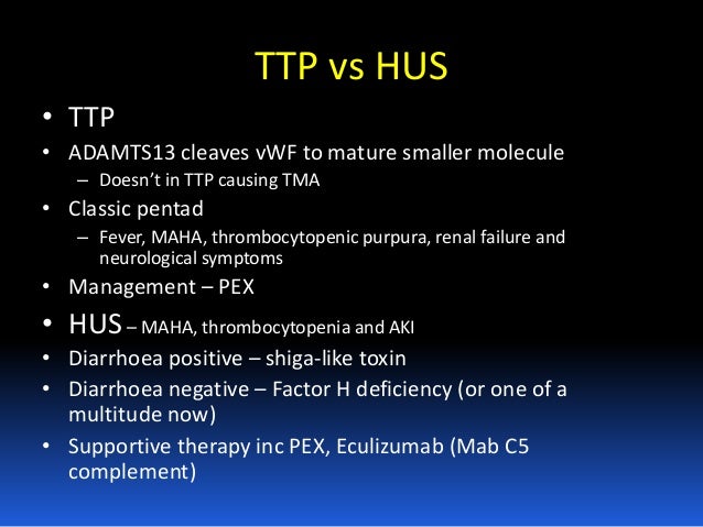
HUS is characterized by thrombocytopenia, anaemia and renal insufficiency, whereas the pentad of signs and symptoms including thrombocytopenia, anaemia, neurologic deficit, renal dysfunction and fever is observed in TTP.
What is the difference between aHUS and TTP?
For both aHUS and TTP, a key characteristic is the tiny clots (thrombotic microangiopathy or TMA) that can form throughout the body, in addition to other common features such as red blood cell destruction, low platelet counts and kidney injury. Patients with TTP additionally experience fever and neurological symptoms.
What is the difference between TTP/Hus and microangiopathy?
The terms TTP/HUS, microangiopathic haemolytic anaemia and thrombotic microangiopathy are sometimes used to avoid an uncertain differential diagnosis. HUS and TTP are characterized by disseminated microthrombi composed of agglutinated platelets, that occlude arterioles and capillaries in the microcirculation.
What is atypical HUS or thrombosis purpura?
Atypical HUS or Thrombotic Thrombocytopenic Purpura (TTP)? For both aHUS and TTP, a key characteristic is the tiny clots (thrombotic microangiopathy or TMA) that can form throughout the body, in addition to other common features such as red blood cell destruction, low platelet counts and kidney injury.
What is the pathophysiology of TTP and Hus?
HUS and TTP are characterized by disseminated microthrombi composed of agglutinated platelets, that occlude arterioles and capillaries in the microcirculation. In HUS the platelet microthrombi are primarily confined to the kidneys, and thus renal failure is the dominant feature.

What is TTP and HUS?
Thrombotic thrombocytopenic purpura (TTP) and hemolytic uremic syndrome (HUS) are multisystemic disorders that are characterized by thrombocytopenia, microangiopathic hemolytic anemia, and ischemic manifestations, resulting from platelet agglutination in the arterial microvasculature.
Does HUS cause TTP?
HUS is related to thrombotic thrombocytopenic purpura Thrombotic Thrombocytopenic Purpura (TTP) Thrombotic thrombocytopenic purpura (TTP) is a serious disorder that involves the formation of small blood clots throughout the body that block the flow of blood to vital organs such as the... read more (TTP), but it occurs ...
What is the difference between HUS and aHUS?
Typical HUS (ie, STEC-HUS) follows a gastrointestinal infection with STEC, whereas aHUS is associated primarily with mutations or autoantibodies leading to dysregulated complement activation.
How do you get TTP HUS?
TTP is caused by a deficiency of a normal blood enzyme that is named ADAMTS13 (you don't need to know why it has this name). To cause TTP, ADAMTS13 must be absent or severely deficient. We usually describe a severe deficiency as less than 10% of the normal levels.
When should you suspect TTP?
TTP should be suspected in all patients with MAHA and thrombocytopenia unless an obvious alternative etiology is present. Although MAHA and thrombocytopenia are the hallmarks of TTP,2,3 end-organ involvement and its severity are extremely variable.
What causes TTP?
What causes TTP? TTP occurs when you do not have the right amount of an enzyme (a type of protein in your blood) called ADAMTS13. This enzyme controls how your blood clots. If you do not have enough ADAMTS13, your body makes too many blood clots.
What is the treatment for HUS?
HUS is generally treated with medical care in the hospital. Fluid volume management is crucial and may include: intravenous (IV) fluids. nutritional supplementation by IV or tube feeding.
Can you survive aHUS?
Atypical hemolytic uremic syndrome (aHUS) is a disease that causes blood clots in small blood vessels in your kidneys and other organs. These clots keep blood from getting to your kidneys, which can lead to serious medical problems, including kidney failure. There's no cure, but treatment can help manage the condition.
How rare is hemolytic uremic syndrome?
Typical hemolytic uremic syndrome (HUS) is an uncommon disease that occurs in 5 to 15 percent of individuals, especially children, who are infected by the Escherichia coli (E. coli) bacterium, usually O157:H7 but also 0104:H4.
How long can you live with TTP?
The most striking evidence for the impact of morbidities following recovery from TTP is decreased survival. Among the 77 patients who survived their initial episode of TTP (1995-2017), 16 (21%) have subsequently died, all before their expected age of death (median difference, 22 years; range 4-55 years).
How is HUS diagnosed?
To confirm a diagnosis of HUS , your doctor is likely to perform a physical exam and recommend lab tests, including:Blood tests. These tests can determine if your red blood cells are damaged. ... Urine test. This test can detect abnormal levels of protein, blood and signs of infection in your urine.Stool sample.
How do you rule out TTP?
Complete Blood Count This test measures the number of red blood cells, white blood cells, and platelets in your blood. For this test, a sample of blood is drawn from a vein, usually in your arm. If you have TTP, you'll have a lower than normal number of platelets and red blood cells (anemia).
What is the difference between TTP and DIC?
Thrombotic thrombocytopenic purpura (TTP) – hemolytic uremic syndrome (HUS) is a thrombotic microangiopathy superficially like DIC, but distinctly different; in contrast to DIC, the mechanism of thrombosis is not via the tissue factor (TF)/factor VIIa pathway. Results of blood coagulation assays in TTP-HUS are normal.
How do you treat TTP HUS?
For patients with inherited TTP (inherited ADAMTS13 deficiency), simple infusion of plasma in sufficient. Most children and adults only need plasma infusion when they have some illness which may trigger an episode of TTP.
How does HUS cause renal failure?
Overview. Hemolytic uremic syndrome (HUS) is a condition that can occur when the small blood vessels in your kidneys become damaged and inflamed. This damage can cause clots to form in the vessels. The clots clog the filtering system in the kidneys and lead to kidney failure, which could be life-threatening.
Is ITP and TTP the same?
Are ITP and TTP the same thing? No, ITP and TTP are not the same thing. Both ITP and TTP are bleeding disorders, but they occur for different reasons and may require different treatments.
What is the difference between HUS and TTP?
HUS and TTP are characterized by disseminated microthrombi composed of agglutinated platelets, that occlude arterioles and capillaries in the microcirculation. In HUS the platelet microthrombi are primarily confined to the kidneys, and thus renal failure is the dominant feature.
What are the therapeutic strategies for D+HUS?
Correction of volume depletion, electrolyte disorder and acidosis are the main therapeutic strategies in D+HUS. Control of blood pressure and early dialysis are essential in patients with renal damage. Infusion of FFP or plasma exchange have shown little if any beneficial effect in D+HUS, but the absence or presence of an unknown humoral factor that may be replaced or removed by plasma therapy, may be considered in D−HUS.
What is the risk factor for TTP?
The majority of patients who develop TTP have no identifiable associated risk factor or underlying disease . TTP and HUS in adults have been associated occasionally with autoimmune phenomena (autoantibodies directed against endothelial cells or platelets, circulating immune complexes), specific platelet agglutinating plasma proteins, release of a calcium‐dependent protease (calpain) or of lysosomal cathepsin‐like cysteine proteases, deficiency of prostacyclin, free radical formation, and abnormal processing of vWF multimers. vWF is a plasma glycoprotein composed of a variable number of 270 kD subunits linked by disulphide bonds. vWF multimers circulating in normal human plasma range in size between 500 and 20 000 kD. Unusually large vWF multimers are secreted from specific storage organelles (Weibel‐Palade bodies) of endothelial cells. These highly polymeric forms of vWF may bind spontaneously to platelets at high levels of shear stress [ 6] and may agglutinate intact circulating platelets. Moake et al. [ 7] observed unusually large vWF multimers in plasma samples from patients with chronic relapsing forms of TTP. They tentatively predicted that in normal individuals a ‘depolymerase’ may be responsible for the conversion of unusually large vWF multimers to smaller polymers normally found in the circulation. This enzyme was proposed to be either a protease or a disulphide bond reductase reducing the size of the unusually large vWF multimers binding spontaneously to platelets thus giving rise to platelet thrombi within the microvasculature [ 7 ]. A specific protease that cleaves purified human vWF in vitro to fragments produced by in vivo proteolysis has been recently purified from normal plasma [ 8, 9 ]. This protease is different from all known proteases, is activated by divalent cations such as Ba 2+ and less by Ca 2+. Cleavage of the vWF subunit at Tyr 842‐Met 843 is strongly enhanced at low ionic strength, in the presence of urea or guanidinium chloride, or under high fluid shear stress. It has been postulated that shear stress of flowing blood modulates the conformation of vWF and enhances its susceptibility to proteolysis [ 10 ].
What is atypical HUS?
Atypical HUS, also denoted as sporadic HUS, is characterized by the absence of antecedent diarrhoea (D−HUS), the tendency to relapse, a generally poor outcome, and often by a positive family history. Hypocomplementaemia and low complement C3 levels have been observed in atypical HUS. Several cases of familial HUS have been reported [ 2 – 5] with deficiency of complement factor H, also denoted as β1 H globulin. Factor H is a cofactor for the factor I‐mediated cleavage of the active form C3b of the complement factor C3. Factor H accelerates the decay of the C3 convertase and inhibits its formation. Low levels or absence of factor H result in increased activation of the alternative complement pathway. In addition to abnormal factor H, abnormalities of other regulators of the alternative pathway may also be implicated in the development of HUS.
Is thrombotic thrombocytopenic purpura the same as HUS?
Haemolytic‐uraemic syndrome (HUS) and thrombotic thrombocytopenic purpura (TTP) are two clinically similar disorders characterized by severe microangiopathic haemolytic anaemia and thrombocytopenia. HUS is characterized by thrombocytopenia, anaemia and renal insufficiency, whereas the pentad of signs and symptoms including thrombocytopenia, anaemia, neurologic deficit, renal dysfunction and fever is observed in TTP. However, about 60% of patients diagnosed with acute TTP lack one or more of these criteria, while about 30% of those receiving a diagnosis of HUS exhibit neurologic symptoms and fever. Thus, the two disorders are often difficult to distinguish. Given the lack of consistent clinical findings, the same disorder might be diagnosed as HUS by a nephrologist while a haematologist might call it TTP. Since both HUS and TTP have been reported to occur in siblings and on different occasions even in the same patient, it sometimes appears that the two disorders are different manifestations of the same pathophysiologic process. The terms TTP/HUS, microangiopathic haemolytic anaemia and thrombotic microangiopathy are sometimes used to avoid an uncertain differential diagnosis. HUS and TTP are characterized by disseminated microthrombi composed of agglutinated platelets, that occlude arterioles and capillaries in the microcirculation. In HUS the platelet microthrombi are primarily confined to the kidneys, and thus renal failure is the dominant feature. In TTP the microvascular occlusions may occur in any tissue and lead to fluctuating ischaemic dysfunction of the respective organs, most frequently of the brain, producing intermittent neurological symptoms. Markers of activated coagulation are only mildly increased in contrast to disseminated intravascular coagulation. No significant amounts of fibrin are formed and rather low amounts of fibrin degradation products are present. Blood clotting times (prothrombin time, activated partial thromboplastin time) are usually normal. The hypotheses concerning the aetiology of HUS and TTP are controversial and suggest that different pathogenetic mechanisms may trigger the disease. Acute episodes of thrombotic microangiopathies have been observed in association with viral and bacterial infections, toxins, pregnancy, HELLP syndrome, bone marrow transplantation, drug (mitomycin, cyclosporin A, ticlopidine) therapy, and cancer, and have been variously referred to as TTP, HUS, TTP/HUS, TTP‐like disease or secondary TTP. It appears that endothelial cell injury is primarily involved in the sequence of events leading to acute episodes of HUS and TTP.
Is vwf deficient in TTP?
Both patients had complete deficiency of vWF‐cleaving protease activity, whereas their parents had about half normal protease activity, and the sister had normal activity, suggesting that the deficiency of the vWF‐cleaving protease may be inherited in an autosomal recessive manner. No inhibitor of the protease activity was found in the patients' plasma [ 11 ]. The recovery of vWF‐cleaving protease following plasma exchange and replacement of fresh frozen plasma (FFP) in both patients with constitutional protease deficiency was about 100% and its biologic half‐life was 2–4 days [ 12 ].
What is HSP in kids?
HSP is primarily seen in kids. It's a leukocytoclastic vasculitis that primarily affects the skin (palpable purpura on the buttocks and lower extremities w/o thrombocytopenia), joints, kidneys (proteinuria and hematuria), and the GI tract (abdominal pain). It often follows a GI illness or URI and is usually self-limiting.
Is TTP a life threatening disease?
TTP is a life-threatening disorder usually seen in adults. As others have stated, primary TTP is caused by large wWF multimers that lead to widespread clot formation. The classic pentad is fever, thrombocytopenia, microangiopathic hemolytic anemia, AKI, and altered mental status (and other neurologic manifestations). Coags will be normal, which helps to distinguish this disorder from DIC. (If you see thrombocytopenia with MAHA but normal coags and no real signs of sepsis or sources of infection, think TTP.) Treatment relies on plasmapheresis. Do NOT give platelets!
What is a HUS episode?
Typical HUS episodes are usually one-time infections, associated with food outbreaks or animal sources that have contaminated food or water supplies. Sometimes we just can’t figure out why the disease has occurred, and “idiopathic” HUS refers to illness that isn’t connected to any particular cause.
What is the cause of ahus?
In most cases aHUS is caused by uncontrolled activation of the complement system, part of the body’s immune system that we all are born with, and which usually acts in a controlled manner to defend against disease and inflammation to maintain good health.
What are the symptoms of ahus?
Patients with either aHUS or typical HUS would have these similar symptoms: blood and blood vessels are affected, red blood cell counts are low (anemia), platelet counts drop (used in clotting, thrombocytopenia is too few platelets) and tiny clots which may form anywhere in the body but often affect the kidneys and brain.
Can blood clots cause thrombocytopenia?
While helpful in case of injuries, random clot formation in blood vessels can block blood flowing as it should around the body and may cause life-threatening issues. If platelets break apart to form clots at an accelerated rate, patients can have low blood platelet counts, a condition called ‘thrombocytopenia’.
Can atypical HUS be differentiated from other thrombotic microangiopathies?
Differentiating aHUS from other thrombotic microangiopathies such as STEC-HUS or TTP can make diagnosis of atypical HUS difficult. aHUS Alliance article
What is a high LDH?
High LDH, Low Fibrinogen ( <120) Low plasma levels of coagulation inhibitors, such as antithrombin III (a thrombin inhibitor) and alpha-2-antiplasmin (a plasmin inhibitor) Presence of microangiopathic changes and schistocytes on the peripheral blood smear.
Can TTP-HUS be diagnosed with MAHA?
Clinical features : It typically include a pentad of clinical features but it’s very rare to find all five features. Revised guidelines suggest that TTP-HUS can be diagnosed with MAHA and thrombocytopenia alone.
Is TTP-HUS a clinical diagnosis?
TTP-HUS is a clinical diagnosis, with noticeable ADAMS13 deficiency. However, ADAMTS13 activity levels cannot be used to determine initial therapy with plasma exchange, and definitive therapy with plasma exchange should not be delayed while determining ADAMTS13 activity levels because delays in initiating treatment may be fatal.
Is plasma exchange more effective than plasma infusion?
Plasma exchange is more effective than plasma infusion in most adults with “classical” TTP-HUS because for the great majority of those who have decreased ADAMTS13 activity, the decreased activity is due to an inhibitory antibody.
What is a HUS?
Hemolytic uremic syndrome (HUS) is one of the disease processes that belong to thrombotic microangiopathies (TMAs). The common features for TMAs are microangiopathic hemolysis, thrombocytopenia, and thrombi in small vessels that lead to end organ damage.
What is atypical HUS?
HUS is usually categorized as typical, caused by Shiga toxin–producing Escherichia coli (STEC) infection, as atypical HUS ( aHUS), usually caused by uncontrolled complement activation, or as secondary HUS with a coexisting disease.
What is the pathogenesis of STEC-HUS?
Within the TMA syndromes, knowledge regarding the pathogenesis of STEC-HUS and aHUS has recently increased considerably. STEC-HUS is usually initiated a few days after clinical gastroenteritis caused by STEC (usually serotype O157:H7 or O104:H4), and the Shiga toxin is central in causing endothelial cell damage, thereby apparently initiating the disease process. Understanding the central differences in pathogenesis between aHUS and STEC-HUS began with the discovery of the association between aHUS and mutations in the gene coding for the key complement regulator in plasma, complement factor H (CFH). 7 Thereafter, mutations in several other complement regulators and other complement proteins, such as C3, factor B, factor I, and CD46, have been identified. 8 Many patients have more than 1 mutation or rare polymorphisms affecting the complement system. 9
What are the mutations in AHUs?
In addition to mutations in complement proteins, some patients with aHUS have mutations also in or only in molecules not directly linked to the complement system. Those molecules include diacylglycerol kinase ε, 10 plasminogen, 11 and factor XII (although only in the presence of anti-factor H autoantibodies). 9, 12 In addition, some patients with aHUS are associated with mutations in thrombomodulin (CD141), 13 which has a role in both coagulation and complement regulation. 13, 14
How does complement work?
Complement is activated via 3 pathways: classical, lectin, and alternative. All the pathways lead to target elimination via 2 main mechanisms, which are phagocytosis and direct lysis. The more important of these is promotion of phagocytosis via opsonization with C3b and its fragments, as well as phagocyte attraction via release of powerful chemotactic and anaphylatoxic peptides such as C5a. 42 These phenomena lead to rapid opsonophagocytosis of the target by neutrophils and macrophages ( Figure 2A ). The second main mechanism of target elimination by complement is direct lysis of target cells via formation of membrane pores, or so-called membrane attack complexes ( Figure 2A ). 43, 44 In addition to these effects, complement also has a role in initiating and enhancing the adaptive immunity. 45
Why do infections cause HUS?
There are several reasons why infections are associated with HUS in general: direct cell or tissue damage caused by a microbe, as seen in STEC-HUS; indirect damage caused by complement activation promoted by the pathogens 105 ; infection-associated autoantibody formation against factor H 106 ; or possibly coagulation disorders during infection. 24 The acute phase reaction may also contribute, because several complement proteins (at least C3, C1s, factor B, C5, C4, and C1q 107, 108 ) are acute phase reactants. This provides an explanation for the observed increase in complement hemolytic activity during the acute phase. 109 Increased plasma concentrations of these proteins do not trigger complement activation as such but could promote activation upon complement-activating circumstances caused by the microbe itself or damaged host tissues.
What is the role of complement in thrombosis?
Schematic model of the role of complement activation , cell damage, and thrombosis in various severe diseases or conditions with major thrombotic problems. Activation of complement can occur via the alternative pathway (in the absence of antibodies) or the classical pathway (in the presence of target-bound antibodies). In each of the indicated diseases or conditions, endothelium, red cells, or platelets are damaged. This may contribute to coagulation and thrombosis, and direct procoagulative effects may also participate (red arrows). Thrombosis can lead to further tissue damage and increasing complement activation. The process can enhance itself via positive feedback loops and form a vicious cycle. Although complement is involved in each of these diseases, its impact needs to be clarified in clinical studies. CAPS, catastrophic antiphospholipid syndrome, DIC, disseminated intravascular coagulation; PLG, plasminogen; THBD, thrombomodulin.
How to tell if you have TTP?
The symptoms of TTP result from the impeded blood flow, but others may be due to the shortage of blood platelets. Severe symptoms of this disorder may involve the brain. Patients can feel confused at times, and they tend to speak differently and have hallucinations. Patients may also experience a rapid heart rate, weakness, fever, and they may even faint. Apart from the common symptoms mentioned above, which are bleeding and bruising, insufficient quantities of platelets may cause small, purple spots to appear on the skin that may resemble a rash. Treating this kind of disorder usually involves blood replacement therapy. This type of treatment is applied since some blood donors may possess the blood that contains the right enzymes needed to restore the imbalance in the blood of the patient. The disease can be episodic, so this means that patients may need to undergo treatment again if they have another episode.
How to treat TTP and ITP?
Treatment of TTP involves blood replacement therapy to correct the imbalance of the blood components, while ITP can be treated with oral steroids.
What is itp in medical terms?
The disease can be episodic, so this means that patients may need to undergo treatment again if they have another episode. Idiopathic thrombocytopenic purpura (ITP) is another blood disorder that happens for no apparent reason. In this type of disorder, the clotting of blood does not occur as it is supposed to.
Why is thrombocytopenic purpura called idiopathic?
The medical term “idiopathic” means “no definitive cause” or “of unknown cause”; hence, idiopathic thrombocytopenic purpura is termed as such because no known explanation exists on what causes the condition.
What is thrombocytopenic purpura?
Thrombotic thrombocytopenic purpura (TTP) is a disorder that results in clotting of the small blood vessels from spontaneous platelet aggregation. The clots formed can be damaging since they can interfere with the proper flow of blood to the body’s vital organs. People with this type of disorder have insufficient amounts of enzymes necessary in inhibiting a blood clotting protein. Since many blood clots may form from this disorder, blood platelets tend to become overused. Platelets play an important role in blood clotting, and with insufficient quantities of this blood component, people tend to bruise or bleed very easily.