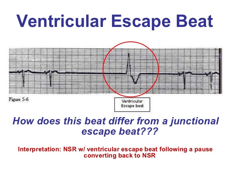
Junctional rhythm is an abnormal cardiac rhythm caused when the AV node or His bundle act as the pacemaker. Idioventricular rhythm is a cardiac rhythm caused when ventricles act as the dominant pacemaker. So, this is the key difference between junctional and idioventricular rhythm. Junctional rhythm can be without p wave or with inverted p wave, while p wave is absent in idioventricular rhythm.
Is junctional rhythm a bad thing?
famousfaqs. Is a junctional rhythm bad? Even in the setting of acute MI, junctional rhythms are usually considered benign and require no treatment. However, in certain patients the loss of AV synchrony during a junctional rhythm will result in myocardial ischemia, heart failure, or hypotension.
What are the symptoms of junctional rhythm?
Those who do have symptoms feel the following:
- Fainting spells
- Dizziness
- Heart palpitations
- Fatigue
- Feeling lightheaded
- Shortness of breath
Why are P waves inverted in junctional rhythm?
Why is the P wave inverted or not visible in junctional rhythms? The atria will be activated in the opposite direction, which is why the P-wave will be retrograde. In most cases, the P-wave is not visible because when impulses are discharged from the junctional area, atria and ventricles are depolarized simultaneously and ventricular depolarization (QRS) dominates the ECG.
Is AIVR concerning?
In conclusion, AIVR is the most frequent arrhythmia occurring during PPCI in patients with ST-elevation myocardial infarction. However, it is not a marker of successful reperfusion but is associated with extensive myocardial damage and delayed microvascular reperfusion.

What is a junctional rhythm?
A junctional rhythm is where the heartbeat originates from the AV node or His bundle, which lies within the tissue at the junction of the atria and the ventricle. Generally, in sinus rhythm, a heartbeat is originated at the SA node.
What is another name for idioventricular rhythm?
Idioventricular rhythm is very similar to ventricular tachycardia, except the rate is less than 60 bpm and is alternatively called a "slow ventricular tachycardia." When the rate is between 50 to 100 bpm, it is called accelerated idioventricular rhythm.
How do you identify idioventricular rhythm?
0:181:47Idioventricular Rhythm ECG - EMTprep.com - YouTubeYouTubeStart of suggested clipEnd of suggested clipThe rate of an idioventricular rhythm will be generally between 20 and 40 beats per minute theMoreThe rate of an idioventricular rhythm will be generally between 20 and 40 beats per minute the rhythm will be regular. There will be no P waves. And therefore no PR interval. And the QRS is are going
How would you differentiate a junctional escape rhythm at 40 bpm from and idioventricular rhythm at the same rate?
Idioventricular rhythm is a cardiac rhythm caused when ventricles act as the dominant pacemaker. So, this is the key difference between junctional and idioventricular rhythm. Junctional rhythm can be without p wave or with inverted p wave, while p wave is absent in idioventricular rhythm.
What is a junctional rhythm look like?
If you have a junctional rhythm, a small wave called a “P wave” is either inverted (upside down) or missing on your EKG. An EKG can often diagnose a junctional rhythm.
Is idioventricular rhythm shockable?
Non-shockable rhythms included asystole, pacing, slow VT, idioventricular rhythms, sinus and atrial based rhythms, some of which contained ventricular ectopic activity of differing grades.
Is junctional and idioventricular the same?
junctional. Both of these rhythms start in the wrong part of your heart, but they're in different places. Idioventricular rhythm starts in your ventricles or lower chambers. Junctional rhythm begins at the junction of your upper and lower heart chambers.
What causes an idioventricular rhythm?
Causes of Accelerated Idioventricular Rhythm (AIVR) Drug toxicity, especially digoxin, cocaine and volatile anaesthetics such as desflurane. Electrolyte abnormalities. Cardiomyopathy, congenital heart disease, myocarditis.
Which characteristics describe junctional rhythms?
Junctional rhythm can be diagnosed by looking at an ECG: it usually presents without a P wave or with an inverted P wave. Retrograde P waves refers to the depolarization from the AV node back towards the SA node.
Will atropine work on a junctional rhythm?
If the junctional rhythm is due to digitalis toxicity, then atropine, digoxin immune Fab (Digibind), or both may be necessary. In refractory cases of symptomatic digitalis toxicity that results in junctional tachycardia and causes severe symptoms, then intravenous phenytoin can be used.
What is the distinguishing factor between junctional tachycardia and junctional escape rhythms?
What is the distinguishing factor between junctional tachycardia and junctional escape rhythms. The heart rate is faster in junctional tachycardia. In accelerated junctional rhythms, why is it unlikely that patients will show signs and symptoms of low cardiac output? The rate is the same as normal sinus rhythm.
Can a junctional rhythm have a wide QRS?
If the QRS complex is wide, an accelerated junctional rhythm resembles an accelerated ventricular rhythm. The rate of the ectopic ventricular rhythm is usually 70 to 110 beats/min.
What does V paced mean?
Ventricular pacingVentricular pacing refers to the electrical stimulation provided to the ventricles of the heart by a pacemaker. It's intended to regulate the heart rate in individuals with abnormally slow heart rhythm.
What is accelerated junctional rhythm?
Accelerated junctional rhythm is a result of enhanced automaticity of the AVN that supersedes the sinus node rate. During this rhythm, the AVN is firing faster than the sinus node, resulting in a regular narrow complex rhythm.
What is a ventricular bigeminy?
The term “ventricular bigeminy” refers to alternating normal sinus and premature ventricular complexes. Three or more successive premature ventricular complexes are arbitrarily defined as ventricular tachycardia.
Is junctional tachycardia a ventricular rhythm?
Is junctional tachycardia an SVT? Yes, junctional tachycardia is a type of SVT, or supraventricular tachycardia. An SVT is a fast heart rhythm (tachycardia) that starts in the upper chambers of your heart, above your ventricles (supraventricular).
What is the rate of idioventricular rhythm?
Idioventricular rhythm is similar to ventricular tachycardia, except the rate is less than 60 bpm and is alternatively called a 'slow ventricular tachycardia.' When the rate is between 50 to 110 bpm, it is referred to as accelerated idioventricular rhythm. [1]
What is idioventricular rhythm management?
Management principles of idioventricular rhythm involve treating underlying causative etiology such as digoxin toxicity reversal if present, management of myocardial ischemia, or other cardiac structural/functional problems. [4][5]
What is the rate of an ectopic pacemaker?
Accelerated idioventricular rhythm (AIVR) results when the rate of an ectopic ventricular pacemaker exceeds that of the sinus node with a rate of around 50 to 110 bpm and often associated with increased vagal tone and decreased sympathetic tone. It is a hemodynamically stable rhythm and can occur after a myocardial infarction during the reperfusion phase. [2]
What is the BPM rate for accelerated idioventricular rhythm?
Rate 50 to 110 bpm for accelerated idioventricular rhythm
Does beta blocker decrease idioventricular rhythm?
However, in reperfusion post-myocardial ischemia and cardiomyopathy, the use of beta-blockers has not shown to decrease the risk of occurrence of idioventricular rhythm. [12]
Is idioventricular rhythm asymptomatic?
The signs and symptoms for the idioventricular or accelerated idioventricular rhythm are variable and are dependent on the underlying etiology or causative mechanism leading to the rhythm. In most cases, the patient remains completely asymptomatic and are diagnosed during cardiac monitoring. Infrequently, patients can have palpitations, lightheadedness, fatigue, and even syncope.
Can accelerated idioventricular rhythm be seen in athletes?
Accelerated Idioventricular rhythm is also be rarely seen in patients without any evidence of cardiac disease. The mechanism involves a decrease in the sympathetic but an increase in vagal tone. It can also present in athletes. [7]
Why do all beats and rhythms arising in the ventricles display discordant ST-T segments?
Because the ventricular depolarization is abnormal, the repolarization will also be abnormal (read about secondary ST-T changes on ECG ). Hence, all beats and rhythms arising in the ventricles will display discordant ST-T segments, meaning that the QRS complex and the ST-T segment will have opposite directions. Figure 1 provides an example.
What are the mechanisms that cause ventricular arrhythmias?
The usual mechanisms are responsible for all ventricular rhythms. Increased automaticity (in His-Purkinje fibers), abnormal automaticity (in contractile myocardium), re-entry (anywhere) or triggered activity (anywhere) may all cause ventricular arrhythmias. Indeed, any cell type in the ventricles may cause ventricular arrhythmias.
What is the ventricular rate?
Ventricular rhythm exists if 3 or more consecutive beats have a ventricular origin. The ventricular rate is between 20 to 40 beats per minute and the rhythm is regular. There is always secondary ST-T changes, meaning that the ST-T segment is discordant ( Figure 1 ).
Is ventricular rhythm reliable?
Importantly, ventricular rhythm is not a reliable rhythm as it may cease working. Figure 1 exemplifies a ventricular rhythm. Accelerated ventricular rhythm (idioventricular rhythm) is a rhythm with rate at 60–100 beats per minute. As in ventricular rhythm the QRS complex is wide with discordant ST-T segment and the rhythm is regular (in most cases).
Is idioventricular rhythm a prognostic marker?
As mentioned in the previous paragraph, idioventricular rhythm is very typical during reperfusion and in that scenario it is a good prognostic marker because it signals that coronary blood flow has been restored. Note that in that setting the idioventricular rhythm may appear with varying QRS morphologies (i.e multifocal ventricular complexes). In virtually all cases (particularly in myocardial ischemia) idioventricular rhythm is benign and does not demand treatment. It does not progress to ventricular tachycardia or ventricular fibrillation and it does not affect cardiac output to the point of hemodynamic compromise.
What is junctional rhythm?
Definition. Junctional rhythm describes a heart -pacing fault where the electrical activity that initiates heart muscle contraction starts in the wrong region. Heart rhythm is the result of electrical impulses sent from the pacemaker cells of the sinoatrial node (SAN) at the top of the right atrium. If the SAN fails to fire, an area located ...
What are the three types of junctional rhythm?
In order of ascending beats per minute (bpm), these are junctional rhythm (or junctional escape rhythm), accelerated junctional rhythm, and junctional tachycardia.
Why does the heart not contract with the AVN?
You might wonder why the heart doesn’t receive two orders to contract by both the SAN and the AVN. Well, the atria are extremely well insulated from the ventricles; this means that a signal from the sinoatrial node can’t make the ventricles contract without the assistance of the AVN. The AVN continues the chain of depolarization from the atria, through the bundle of His and into the ventricles. This is a lot more work than the sinoatrial node has to do (creating a larger wave on the ECG) and takes a little more time. The pause between SAN and AVN firing is therefore extremely important, as this allows the atria to empty via gravity and contraction, but also makes sure the ventricles have enough time to fill.
What is the term for a tachycardia that is accelerated?
Junctional tachycardia is also known as automatic or paroxysmal junctional tachycardia. We can describe it simply by saying it is a form of SVT where the over-rapid pacing of the AV junction overrides a slower rate of firing in the SAN. Accelerated junctional rhythm is usually seen in adults with heart disease or who are or have recently experienced acute myocardial infarction.
What is the normal sinus rhythm?
Normal sinus rhythm (NSR) originates at the sinoatrial node at an average rate of 60 to 100 beats per minute (bpm). If you feel your pulse, chances are you will feel a sinus rhythm.
Which type of rhythm is the same as sinus rhythm?
Junctional Rhythm vs Sinus Rhythm. Junctional rhythm and sinus rhythm have almost the same result – both types send electrical impulses through specialized heart muscle ( cardiac muscle) to force certain areas of the heart to contract at certain times. In which order these muscles contract is extremely important – from the top to the bottom ...
How to measure cardiac rhythm?
Cardiac rhythm can be observed by way of an electrocardiogram (ECG). You will have seen the ECG symbol on many medical business logos. Both heart rate and heart activity are measured on an electrocardiogram that maps most of the electrical activity of the heart but not all of it.
How many beats per minute is a junctional rhythm?
Junctional escape rhythm is a regular rhythm with a frequency of around 40–60 beats per minute.
What is the treatment for junctional rhythm?
Symptomatic junctional rhythm is treated with atropine. Doses and alternatives are similar to management of bradycardia in general.
What is the most common rhythm in the AV node?
The most common rhythm arising in the AV node is junctional rhythm , which may also be referred to as junctional escape rhythm. Junctional tachycardia is less common. Basic knowledge of arrhythmias and cardiac automaticity will facilitate understanding of this article.
What is the vagal tone of a well trained athlete?
Well-trained athletes may have very high Vagal tone which lowers the automaticity in the sinoatrial node to the point where cells in the AV-junction establishes an escape rhythm. This is asymptomatic and benign.
What is the primary objective of junctional tachycardia?
Treatment of junctional tachycardia. The primary objective is to treat the underlying cause and/or eliminate provocative medications. Electrical cardioversion is ineffective and should be avoided (electrical cardioversion may be pro-arrhythmogenic in patients on digoxin).
What happens when cells in bundle of His are not reached by the atrial impulse?
In such scenarios, cells in the bundle of His (which possess automaticity) will not be reached by the atrial impulse and hence start discharging action potentials and an escape rhythm. This will also manifest as a junctional escape rhythm on the ECG.
Where is the block located in a heart block?
During complete heart block (third-degree AV-block) the block may be located anywhere between the atrioventricular node and the bifurcation of the bundle of His. If there are cells (with automaticity) distal to the block, an escape rhythm may arise in those cells. For example, consider a complete block located in the atrioventricular node.
What is a slow ventricular rhythm?
An idioventricular rhythm is frequently referred to as a “slow ventricular tachycardia” for this reason. When the ventricular rate is between 60 and 100 bpm, it is referred to as an accelerated idioventricular rhythm. This is a hemodynamically stable rhythm that occurs commonly after myocardial infarctionand no treatment is needed.
What is a ventricular rate of 60 BPM?
When the ventricular rate is between 60 and 100 bpm, it is referred to as an accelerated idioventricular rhythm. This is a hemodynamically stable rhythm that occurs commonly after myocardial infarction and no treatment is needed.
