What is the inferior end of the heart called?
The base of the heart is located at the level of the third costal cartilage, as seen in Figure 1. The inferior tip of the heart, the apex, lies just to the left of the sternum between the junction of the fourth and fifth ribs near their articulation with the costal cartilages.
What is the tip of the heart called?
The apexThe apex is the pointed tip of the heart. It is located on the lower portion of the heart (left ventricle).
Which end of the heart is?
Video Solution: Which end of the heart is broader and which end is narrow? UPLOAD PHOTO AND GET THE ANSWER NOW! Solution : Heart is wider at the anterior end and narrow at the posterior end....QuestionChapter NameTransportation - The Circulatory SystemSubjectBiology (more Questions)Class10thType of AnswerVideo4 more rows•Jun 27, 2022
What are the 4 surfaces of the heart?
The heart has five surfaces: base (posterior), diaphragmatic (inferior), sternocostal (anterior), and left and right pulmonary surfaces.
What are the two sides of the heart called?
Muscular walls, called septa or septum, divide the heart into two sides. On the right side of the heart, the right atrium and ventricle work to pump oxygen-poor blood to the lungs. On the left side, the left atrium and ventricle combine to pump oxygenated blood to the body.
What are the parts of the heart?
The heart is made up of four chambers: two upper chambers known as the left atrium and right atrium and two lower chambers called the left and right ventricles. It is also made up of four valves: the tricuspid, pulmonary, mitral and aortic valves.
Where is apex of the heart?
fifth intercostal spaceThe apex (the most inferior, anterior, and lateral part as the heart lies in situ) is located on the midclavicular line, in the fifth intercostal space. It is formed by the left ventricle. The base of the heart, the posterior part, is formed by both atria, but mainly the left.
What are the 13 parts of the heart?
Anatomy of the heartLeft atrium and auricle. Left atrium. Left auricle.Right atrium and auricle. Right atrium. Right auricle.Interventricular septum and septal papillary muscles. Interventricular septum. ... Right ventricle and papillary muscles. Right ventricle. ... Left ventricle and papillary muscles. Left ventricle.
Where is the base of the heart located?
third costal cartilageThe base of the heart is located at the level of the third costal cartilage, as seen in Figure 16.1. 1. The inferior tip of the heart, the apex, lies just to the left of the sternum between the junction of the fourth and fifth ribs near their articulation with the costal cartilages.
What are the 5 structures of the heart?
The two atria are thin-walled chambers that receive blood from the veins. The two ventricles are thick-walled chambers that forcefully pump blood out of the heart....Chambers of the HeartRight atrium.Right ventricle.Left atrium.Left ventricle.
How do you remember the anatomy of the heart?
1:094:35[IGCSE/GCSE] Heart Structure - Memorize In 5 Minutes Or Less!YouTubeStart of suggested clipEnd of suggested clipAnd that's what you should memorize first ignore everything else memorize the four main chambers.MoreAnd that's what you should memorize first ignore everything else memorize the four main chambers. The right and left atrium. And the right and left ventricle.
What is the inferior side of the heart?
The inferior or diaphragmatic surface of the heart forms a roughly straight plane or slight concavity that projects to the left and slightly inferiorly to the apex of the heart. It lies superior to the central tendon of the diaphragm and at its lateral projection, the muscular part of the left hemidiaphragm.
Is the apex the top of the heart?
Because of rotation during fetal development, the apex of the heart (tip of the cone) is at its bottom and lies left of the midline. The base is at the top, where the great vessels enter the heart and lies posterior to the sternum (Fig. 7-2).
What is a pericardium?
Summary. The pericardium is a membrane, or sac, that surrounds your heart. It holds the heart in place and helps it work properly. Problems with the pericardium include: Pericarditis - an inflammation of the sac.
What is Situs Solitus?
Situs solitus describes viscera that are in the normal position, with the stomach on the left side; in situs inversus, the positions of the abdominal organs and viscera are reversed. Dextrocardia with situs solitus occurs in an estimated 7500–29 000 living people worldwide.
Is epicardium and pericardium the same?
What is the difference between the epicardium and the pericardium? The epicardium is part of the pericardium. It is the innermost layer and is referred to as the visceral layer of the serous pericardium. The main difference is the nomenclature and the specific layers of the heart that they are describing.
What is the valve between the left atrium and ventricle?
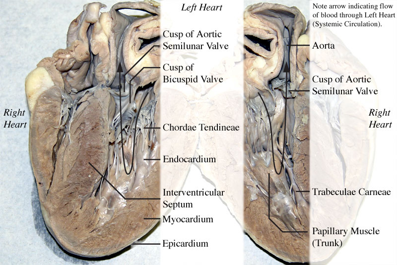
also called the bicuspid valve or mitral valve. Between left atrium and ventricle
Where is the mediastinum located?
Located in the mediastinum (space between lungs, backbone, sternum,) between the 2nd rib and the 5th intercostal space.
What is the treatment for plaque in the arteries?
Deposits of fatty materials such as cholesterol form a "plaque" in the arteries which reduces blood flow. Advanced forms are called arteriosclerosis. Treatment: angioplasty, where a catheter is inserted into the artery and a balloon is used to stretch the walls open. A bypass can also treat clogged arteries, a vein is used to replace a clogged artery, coronary bypass refers to a procedure to supply blood to the heart (the phrase "quadruple bypass" means that 4 arteries were bypassed)
What is the best place to take a pulse?
Radial artery is often used to take a person's pulse. If you lift your thumb slightly the "pocket" created is good spot to feel for pulse
Which muscle attaches to the wall of the ventricular atria?
chordae tendineae, which attach to the wall by papillary muscles. This prevents the valves from being pushed up into the atria during ventricular systole.
Which part of the brain controls the heart rate?
Controlled by the cardiac center within the medulla oblongata. The cardiac center signals heart to increase or decrease its rate according to many factors that the brain constantly monitors
How does removing a gat build up in the neck help prevent stroke?
By removing the gat and cholesterol build up inside the artery, adequate blood flow is restored, which can help prevent stroke. Blockages of carotid arteries in the neck are responsible for more than half of all strokes
What happens when you have diastolic heart failure?
Rehabilitation by Stage . If you have diastolic heart failure, your left ventricle has become stiffer than normal. Because of that, your heartcan't relax the way it should. When it pumps, it can't fill up with bloodas it's supposed to. Because there's less blood in the ventricle, less blood is pumped out to your body.
What is the term for the amount of blood flowing to your heart muscle that is blocked or less than normal?
Coronary artery disease: The amount of blood flowing to your heart muscle is blocked or less than normal. Find out more about the different types of cardiovascular diseases.
Why is diastolic heart failure more common as you get older?
So diastolic heart failure is more common as people get older. Other than normal aging, the most common causes are: High blood pressure : If you have it, your heart has to work harder to pump more blood through your body.
Why is my left ventricle stiff?
Because of that, your heart can't relax the way it should. When it pumps, it can't fill up with blood as it's supposed to. Because there's less blood in the ventricle, less blood is pumped out to your body.
What causes the wall of the heart to thicken?
Diabetes: The disease can cause the wall of your heart to thicken. That makes it stiffen. Read more on how diabetes affects your heart. Coronary artery disease: The amount of blood flowing to your heart muscle is blocked or less than normal.
Can diastolic heart failure be cured?
Although diastolic heart failure can't be cured, treatment can help ease symptoms and improve the way your heart pumps.
What is the Clavicle Bone
Clavicle, commonly known as collarbone, is a slender, S-shaped, modified long bone located at the base of the neck. It is the only long bone of the body that lies horizontally.
Where is the Clavicle Located
As stated, clavicle is located at the base of the neck and across the upper part of the ribcage. It sits between the shoulder blade ( scapula) and breastbone ( sternum ), connecting the pectoral girdle or shoulder girdle to the axial skeleton. Clavicle is the only bone that connects the axial skeleton to the appendicular skeleton.
Anatomy – Parts of the Clavicle Along With its Bony Landmarks
Being a long bone, clavicle has two ends, sternal and acromial end. The region in between the two ends is known as shaft.
Articulations
Sternoclavicular Joint: It is a synovial joint that is formed between the sternal end of the clavicle and the manubrium of the sternum.
Muscle Attachments
There is a total of six muscles that are attached to the clavicle. Out of these six, four muscles are attached to the sternal end or medial two-third of the clavicle, whereas two muscles are attached to the acromial end or lateral third of the clavicle.
Left and Right Clavicle – How to Identify
Here’s a quick way to distinguish between the left and right clavicle.
What is the condition where the heart points to the right side of the chest?
Dextrocardia is a rare heart condition in which your heart points toward the right side of your chest instead of the left side. Dextrocardia is congenital, which means people are born with this abnormality. Less than 1 percent. of the general population is born with dextrocardia.
Where is the heart located in the body?
If you have isolated dextrocardia, your heart is located on the right side of your chest, but it has no other defects. Dextrocardia can also occur in a condition called situs inversus. With it, many or all of your visceral organs are on the mirror-image side of your body. For example, in addition to your heart, your liver, spleen, ...
Why does dextrocardia cause intestinal malrotation?
This is because dextrocardia can sometimes result in a condition called intestinal malrotation, in which your gut doesn’t develop correctly. For that reason, your doctor will watch out for an abdominal obstruction, also called bowel or intestinal obstruction. An obstruction prevents waste from leaving your body.
Why do we need to treat dextrocardia?
Dextrocardia must be treated if it prevents vital organs from functioning properly. Pacemakers and surgery to repair septal defects can help the heart work normally.
What happens if you have dextrocardia?
If you have dextrocardia, you may have other heart, organ, or digestive defects related to your anatomy. Surgery can sometimes correct these problems.
Why does my heart point the wrong way?
Sometimes, your heart develops pointing the wrong way because other anatomical problems exist. Defects in your lungs, abdomen, or chest can cause your heart to develop so that it’s shifted towards the right side of your body.
Can dextrocardia be isolated?
Isolated dextrocardia usually causes no symptoms. The condition is usually found when an X-ray or an MRI of your chest shows the location of your heart on the right side of your chest.
What is the distal end of the clavicle?
The distal end, which is furthest from the middle of your body, is the end that connects to your shoulder. The proximal end, which is closest to the middle of your body, connects to your breastbone, or sternum.
What is the difference between proximal and distal?
In anatomy in general, distal means farther away from the attachment or origin of an organ, and proximal means closer to the attachment or origin —both of them usually implying relative to something else. The stomach is distal to the esophagus and the shoulder is proximal to the elbow, for example.
Where is the scapula located?
Located above the first rib it acts as a strut to keep the scapula in place so that harm can hang freely.
Which bone is most often fractured?
The clavicle is said to be the most often fractured bone of the body, so radiologists and orthopedic surgeons have frequent occasions to refer to a fracture site as being at the proximal or distal end. Most fractures are mid-clavicular, though, since that’s the most fragile part of the clavicle.
What is the term for the death of a heart muscle?
Heart Attack. Also called myocardial infarction, this happens due to reduced blood flow through heart blood vessels. This leads to the death of heart muscle cells and cause permanent damage. The pain you experience in a heart attack is quite similar to angina, but it will be more severe in case of myocardial infarction.
What side of the chest is the heart located?
Do you think your heart is on the left side of your chest? Most people think the same, but the truth is that your heart is located between your right and left lung. It means your heart is in middle of your chest, with a slight tilt towards the left. Located in the front and middle of your chest, your heart is no bigger than the size of your clenched fist. A wall separates the left and right of your heart, and each of these sides has a small chamber called atrium. The chamber then leads into a large pumping chamber known as ventricle. Your heart has four chambers, called right ventricle and right atrium, and then left atrium and left ventricle.
What About Pain on the Right Side of Chest?
However, if you experience pain on the right side of your chest, this is usually not associated with heart pain. It is not heart pain especially when it gets worse with taking a deep breath. The only way to associate your pain on the right side with your heart is to link the pain with the outer sac surrounding the heart. This happens only in rare cases, but your pain could be due to the inflammation of the outer lining of your lungs. This may happen due to pneumonia if you're also experiencing fever and shortness of breath. A trauma to your chest may also lead to severe chest pain.
What is CAD in medical terms?
Coronary Artery Disease (CAD) It refers to a condition caused by a blockage in the heart blood vessels. This blockage reduces oxygen and blood flow to the heart muscle and causes pain known as angina. Though it's a heart disease, it doesn't cause any permanent damage to your heart.
What is the term for a heart attack that causes fever, heartbeat, and fatigue?
Myocarditis. This refers to heart muscle inflammation that may also cause fatigue, fever, trouble breathing, and fast heartbeat. The pain in myocarditis may feel like a heart attack, there is usually no blockage involved in arteries or vessels.
Why is it important to know which side of your heart is on?
If it's on the side of your heart, it could be because you're having a heart attack. Knowledge about the location of your heart will prove beneficial in many cases. Keep reading to find out more about why it matters to learn which side is your heart on.
How do you know if you have a heart attack?
Unlike what you see on TV, not all heart attacks will begin with crushing chest pain that hits you out of nowhere. About one-third of the patients who had heart attacks never experienced any chest pain. It means the symptoms of heart attack will vary from person to person. Some people may experience a few symptoms, while others may feel excruciating pain when they have a heart attack. Research shows heart attacks can start slowly with mild, bearable pain, whereas people with high blood sugar may not experience any symptoms at all. Women may experience symptoms like nausea, shortness of breath, unusual tiredness, vomiting, and pain in the back, jaw, and shoulder. The most common symptoms include the following:
