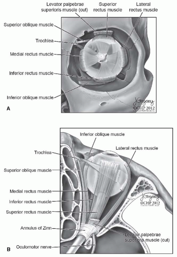
What is the annulus of Zinn used for?
The annulus of Zinn, also known as the annular tendon or common tendinous ring, is a ring of fibrous tissue surrounding the optic nerve at its entrance at the apex of the orbit. It is the common origin of the four rectus muscles (extraocular muscles). It can be used to divide the regions of the superior orbital fissure.
What muscles arise from the annulus of Zinn?
The annulus of Zinn serves as the origin of six of the seven extraocular muscles (Fig. 50-2 ). Superiorly, the superior rectus arises from the annulus, which at this point is fused with the dura of the optic nerve. The levator palpebrae arises medial and superior to the superior rectus muscle but remains intimately associated with it.
Why is the extraocular muscle innervation so complex?
One explanation for the extremely complex innervation of the extraocular muscles lies in the high functional importance of the eyes. The levator palpebrae superioris takes its origin from the lesser wing of the sphenoid just superior to the annulus of Zinn.
What is the thinnest extraocular muscle in the eye?
The lateral rectus muscle is the thinnest extraocular muscle (9.2 mm wide). The superior rectus muscle is loosely attached to the levator palpebrae superioris muscle. When the eye is hypotropic, a pseudoptosis may be present because the upper eyelid will follow the superior rectus muscle.

Which extraocular eye muscle does not originate at the common tendinous ring?
The superior obliqueThe superior oblique is one of the two noteworthy oblique extraocular muscles. These muscles are unique in that they do not originate from the common tendinous ring, have an angular attachment to the eyeball, and they attach to the posterior aspect of the eyeball.
What goes through the annulus of Zinn?
The common tendinous ring, also known as the annulus of Zinn, or annular tendon, is a ring of fibrous tissue surrounding the optic nerve at its entrance at the apex of the orbit. It is the common origin of the four recti muscles of the group of extraocular muscles.
Which extrinsic eye muscle does not originate from the tendinous ring around the opening of the optic canal?
The superior oblique muscle originates from the body of sphenoid bone, medial to the origin of the levator palpebrae superioris muscle and superomedial to the optic canal. In contrast to the other extraocular muscles, superior oblique and inferior oblique do not originate from the common tendinous ring.
Where do the extraocular muscles originate?
The origin is the sphenoid bone. Other notes: The superior, inferior, medial and lateral recti muscles originate from a shared tendinous ring on the posterior wall of the eye and insert on the anterior region of the eyeball, which is just beyond the visible sclera (“white of the eye”).
Which of the following structures pass through the superior orbital fissure outside of the annulus of Zinn?
Correct Answer: Ophthalmic artery The lacrimal, frontal and trochlear nerves, as well as the ophthalmic vein, pass through the superior orbital fissure outside of the annulus of Zinn.
What is the function of the superior rectus muscle?
The superior rectus has a primary action of elevating the eye, causing the cornea to move superiorly. The superior rectus originates from the annulus of Zinn and courses anteriorly and superiorly over the globe, making an angle of 23 degrees with the visual axis.
Which one of the following is not an extrinsic eye muscle?
Answer and Explanation: The c) ciliary is not an extrinsic muscle of the eye. The six muscles that control the movement of the eye include the lateral rectus, medial rectus, superior rectus, inferior rectus, superior oblique and inferior oblique.
What is the origin of the rectus muscles of the eye?
The four recti muscles all arise from a connective tissue ring called the common tendinous ring (annulus of Zin). This is located at the apex of orbit, surrounding the optic canal. Respectively, the recti muscles insert onto the superior, inferior, medial and lateral sides of the eyeball.
What are the 4 extrinsic muscles of the eye?
The extraocular muscles are the six muscles that control movement of the eye (Superior rectus, Inferior rectus, Lateral rectus, Medial rectus, Superior oblique and Inferior oblique) and one muscle that controls eyelid elevation (levator palpebrae).
Which extraocular muscle takes origin from the floor of orbit?
Extraocular Muscle Origins The inferior oblique muscle arises from the medial orbital floor adjacent to the lacrimal fossa. The levator palpebrae superioris originates from the lesser wing of the sphenoid bone.
Where does the levator palpebrae superioris originate?
the sphenoid boneThe levator palpebrae superioris muscle origin is the periosteum of the lesser wing of the sphenoid bone, superior to the optic foramen. The muscle travels anteriorly along the superior aspect of the orbit superior to the superior rectus muscle.
What is the origin of the superior oblique muscle of the eye?
The superior oblique muscle has its origin on the lesser wing of the sphenoid bone, medial to the optic canal near the frontoethmoid suture. The muscle courses forward and passes through the trochlea, a U-shaped piece of cartilage attached to the orbital plate of the frontal bone (see Figure 10-7).
What muscle originates from annulus Zinn?
Extraocular Muscles The four rectus muscles (each about 3–4 cm long) originate from the annulus of Zinn at the orbital apex, which is contiguous with the dura surrounding the optic nerve and the periorbita.
What passes through the tendinous ring of the orbit?
The tendinous ring straddles the lower, medial part of the superior orbital fissure. It attaches to a tubercle on the greater wing of the sphenoid bone (at the margin of the superior orbital fissure). Through it (from superior to inferior) pass: superior division of the oculomotor nerve (CN III)
What hole does the optic nerve pass through?
The optic foramen, the opening through which the optic nerve runs back into the brain and the large ophthalmic artery enters the orbit, is at the nasal side of the apex; the superior orbital fissure is a larger hole through which pass large veins and nerves.…
Where is the upper orbital fissure and what passes through it?
It lies between the lesser and greater wings of the sphenoid bone. It allows for many structures to pass, including the oculomotor nerve, the trochlear nerve, the ophthalmic nerve, the abducens nerve, the ophthalmic vein, and sympathetic fibres from the cavernous plexus.
How many extraocular muscles are there in a cow?
There are 6 of these extraocular muscles that control eye movement (cows only have 4 of these), and one muscle that controls eyelid elevation. The position of the eye at the time of muscle contraction is what determines how the 6 muscles of the orbit are engaged. Four of the 6 extraocular muscles controls movement in the cardinal directions: north, ...
What muscle inserts on the anterior superior surface of the eye?
This muscle inserts on the anterior, superior surface of the eye. The origin is the Annulus of Zinn. Rectus muscles are “straight” muscles.
What muscles control eye movement?
Extraocular muscles. Extraocular muscles are also referred to as the extrinsic (arising externally) or muscles of the orbit. There are 6 of these extraocular muscles that control eye movement (cows only have 4 of these), and one muscle that controls eyelid elevation. The position of the eye at the time of muscle contraction is what determines how ...
What is the difference between adduction and abduction?
This means that abduction refers to looking away from the midline (nose) of the face, whereas adduction is looking toward the midline.
Which muscle is responsible for elevation?
In the neutral position, this muscle is responsible for elevation, incyclotorsion and adduction (inward, rotational movement). During adduction, the superior rectus is responsible for intorsion, adduction and elevation. During abduction, this muscle is responsible for elevation. This muscle inserts on the anterior, superior surface of the eye. The origin is the Annulus of Zinn. Rectus muscles are “straight” muscles.
Which muscles control the eye movement in the cardinal directions?
The 4 extraocular muscle s that control eye movement in the cardinal directions (along with their functions) are the superior rectus, inferior rectus, lateral rectus and medial rectus muscles. Extraocular muscles and orbit in a cadaver.
What is the purpose of a clinical eye exam?
The purpose of the clinical eye exam is to investigate whether or not the extraocular eye muscles are working and moving properly. All 6 of the muscles described above can be tested by drawing a large letter “H” in the air with a finger or pen in front of a patient’s face and having them follow the tip of the finger or pen with their eyes while keeping their head stationary .
What is the superior rectus?
The superior rectus has fascial attachments to the superior oblique tendon and the levator palpebrae muscle. If the attachments to the levator palpebrae are not severed during recession or resection of the superior rectus, eyelid fissure changes may occur. Connective tissue attachments between the inferior rectus and the inferior oblique may assist the surgeon in locating a “lost” inferior rectus muscle.
How many rectus muscles are there?
Rectus muscles. There are four striated rectus muscles arising from the annulus of Zinn in the apex of the orbit. Each is 40 mm in length and inserts on the sclera anterior to the equator of the globe.
What is the annulus of Zinn?
Muscle Cone and Annulus of Zinn. The annulus of Zinn serves as the origin of six of the seven extraocular muscles (Fig. 50-2 ). Superiorly, the superior rectus arises from the annulus, which at this point is fused with the dura of the optic nerve. The levator palpebrae arises medial and superior to the superior rectus muscle ...
How many extraocular muscles are there?
Extraocular muscles. The six extraocular muscles are striated with unique properties. 16 The four rectus muscles (each about 3–4 cm long) originate from the annulus of Zinn at the orbital apex, which is contiguous with the dura surrounding the optic nerve and the periorbita.
How to correct mild ptosis?
Cases of mild ptosis can be corrected with a external levator resection and advancement, or Müllerectomy. For patients with severe ptosis with maintained levator excursion, levator advancement is the procedure of choice. Severe ptosis with absent levator function will usually require a frontalis sling procedure. Secondary correction of severe injuries should be delayed until the scar has remodeled 6–12 months post injury. 55,56
What muscle is anteriorly and laterally attached to the sclera?
The inferior rectus muscle courses anteriorly, laterally, and inferiorly to insert on the sclera. The inferior rectus forms a 23° angle with the visual axis when the globe in the primary gaze position. It has fascial attachments to the inferior oblique muscle and the lower eyelid retractors ( Fig. 85.5 ).
Where are the rectus muscles located?
This oval band of connective tissue is continuous with the periorbita and is located at the apex of the orbit anterior to the optic foramen and the medial part of the superior orbital fissure.
What is the definition of rapid eye movements?
Quick, voluntary, simultaneous movements of both eyes in the same direction; fixation, refixation, and rapid eye movements.
Which CN innervates the SO4?
LR6 means the LR is innervated by the 6th CN, the abducens nerve; SO4 means the SO is innervated by the 3rd CN, the oculomotor nerve.
Where does the IR wrap go?
Wraps under the eye and travels rearward and temporally, passing over the IR muscle, and up under the LR muscle, and attaches posterior to the equator on the temporal side of the eye.
Where is the sclera located?
To the sclera on top of the eye, anterior to the equator, approximately 7.7 mm from the limbus.
What is the parent structure of the rectus inferior?
Some sources distinguish between these terms more precisely, with the anulus tendineus communis being the parent structure, divided into two parts: a lower, the ligament or tendon of Zinn, which gives origin to the rectus inferior, part of the rectus internus, and the lower head of origin of the rectus lateralis.
What is the upper band of the rectus superior?
This upper band is sometimes termed the superior tendon of Lockwood.
What is the annulus of Zinn?
The annulus of Zinn, also known as the annular tendon or common tendinous ring, is a ring of fibrous tissue surrounding the optic nerve at its entrance at the apex of the orbit. It is the common origin of the four rectus muscles of the group of extraocular muscles .
What is the vascular structure of the optic nerve called?
The arteries surrounding the optic nerve are sometimes called the "circle of Zinn-Haller" ("CZH"). This vascular structure is also sometimes called "circle of Zinn". The following structures pass through the tendinous ring (superior to inferior): Superior division of the oculomotor nerve (CNIII) Nasociliary nerve (branch of ophthalmic nerve)
Who is the Zinn zonule named after?
It is named for Johann Gottfried Zinn. It should not be confused with the zonule of Zinn, though it is named after the same person.
