Proteoglycans
- Astrocyte–neuron interactions in synaptic development. Another class of proteoglycans, the chondroitin sulfate proteoglycans (CSPGs), has roles in regulating the stability of AMPA glutamate receptors at synapses.
- Cellular Components of Nervous Tissue. Patrick R. Hof, ... ...
- Meninges and vasculature
What is the structure and function of proteoglycans?
Proteoglycans are glycosylated proteins which have covalently attached highly anionic glycosaminoglycans. Many forms of proteoglycans are present in virtually all extracellular matrices of connective tissues. The major biological function of proteoglycans derives from the physicochemical characteris …
What are proteoglycans in the extracellular matrix?
Proteoglycans are a major component of the animal extracellular matrix, the "filler" substance existing between cells in an organism. Here they form large complexes, both to other proteoglycans, to hyaluronan, and to fibrous matrix proteins, such as collagen.
Why are proteoglycans used as lubricants in cells?
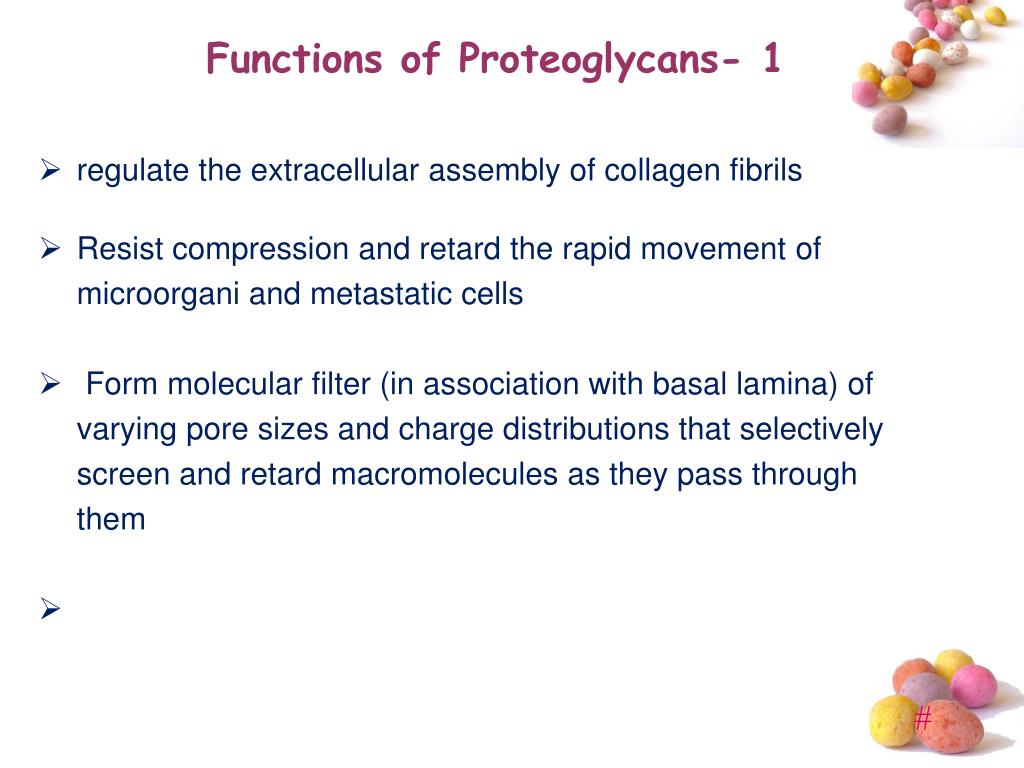
◆ Some proteoglycans either due to the core protein or the attached glycosaminoglycan chains can serve as lubricants. ◆ Proteoglycans are required for the organization of the basement membrane and play an important role in cell proliferation and differentiation.
What are the different types of proteoglycans?
These glycosaminoglycans give rise to a number of proteoglycans like decorin, biglycan, aggrecan, neurocan, testican, fibromodulin, lumican, etc. Proteoglycans form large complexes with other proteoglycans, fibrous proteins (like collagen), and other components (hyaluronan) of the extracellular matrix.

What is the structure and function of proteoglycans?
D. Proteoglycans are ubiquitous molecules that function as critical components of the extracellular matrix. These proteins are composed of glycosaminoglycan chains that are covalently attached to a protein core.
What is the function of proteoglycans quizlet?
Proteoglycans are part of the extracellular matrix; they provide structure, viscosity and lubrication, and adhesiveness.
What is the function of proteoglycan in ground substance?
The proteoglycans perform many other functions, to do with regulation of enzyme and growth factor activity, too numerous to mention here (see Molecular Biology of the Cell if you want to know more). One important proteoglycan is 'aggrecan' (shown in the diagram above) which is a major consituent of cartilage.
What is the role of proteoglycans in the stroma?
Dermatan Sulfate Proteoglycans (DSPGs) DSPGs present in the corneal stroma are involved in controlling the interfibrillar spacing and lamellar adhesion properties of corneal collagens (Maccarana et al., 2009).
What are proteoglycans quizlet?
a class of glycoproteins composed of many long, linear chains of glycosaminoglycans (GAGs) covalently linked to a core protein.
What is the difference between proteoglycans and glycosaminoglycans?
The key difference between proteoglycans and glycosaminoglycans is that proteoglycans are organic compounds containing a protein bound to a mucopolysaccharide whereas glycosaminoglycans are mucopolysaccharides containing a number of disaccharide repeating units.
What does proteoglycan mean?
Listen to pronunciation. (PROH-tee-oh-GLY-kan) A molecule that contains both protein and glycosaminoglycans, which are a type of polysaccharide. Proteoglycans are found in cartilage and other connective tissues.
Where are proteoglycans?
Proteoglycans are found in the extracellular matrix, plasma membrane of cells, and intracellular structures. Matrix proteoglycans such as perlecan, collagen XVIII, and agrin are found in the basal laminal of cells, and decorin, biglycan, and versican are found in the interstitial spaces of the lungs.
What are proteoglycans in biochemistry?
Proteoglycans (mucoproteins) are formed of glycosaminoglycans (GAGs) covalently attached to the core proteins. They are found in all connective tissues, extracellular matrix (ECM) and on the surfaces of many cell types.
What are the functions of glycosaminoglycans?
In conclusion, glycosaminoglycans (GAGs), have widespread functions within the body. They play a crucial role in the cell signaling process, including regulation of cell growth, proliferation, promotion of cell adhesion, anticoagulation, and wound repair.
What is collagen function?
Collagen is protein molecules made up of amino acids. It provides structural support to the extracellular space of connective tissues. Due to its rigidity and resistance to stretching, it is the perfect matrix for skin, tendons, bones, and ligaments.
What are the functions of fibroblasts?
A fibroblast is a type of cell that contributes to the formation of connective tissue, a fibrous cellular material that supports and connects other tissues or organs in the body. Fibroblasts secrete collagen proteins that help maintain the structural framework of tissues.
What is the difference between glycoprotein and proteoglycan?
Glycoproteins are proteins to which carbohydrates are covalently linked through glycosidic bonds. Proteoglycans are a subclass of glycoproteins with distinctive features of carbohydrate structure.
What are proteoglycans examples?
Proteoglycans are a type of glycoproteins present in the body, especially in connective tissues, bone and cartilage, and cell surfaces. Examples of proteoglycans are versican (a large chondroitin sulfate proteoglycan), perlecan, neurocan, aggrecan, brevican, fibromodulin, and lumican.
Why can proteoglycans hold so much water?
Proteoglycans (PGs) are one type of non-collagenous proteins in the extracellular matrix of bone, which primarily contain a core protein and glycosaminoglycans (GAGs). GAGs are highly polar and negatively charged, thus having a strong tendency in attracting water molecules into the matrix.
What does ground substance typically include?
Ground substance is primarily composed of water and large organic molecules, such as glycosaminoglycans (GAGs), proteoglycans, and glycoproteins. GAGs are polysaccharides that trap water, giving the ground substance a gel-like texture.
What is the function of proteoglycans?
Function of proteoglycans in the extracellular matrix. Proteoglycans are glycosylated proteins which have covalently attached highly anionic glycosaminoglycans. Many forms of proteoglycans are present in virtually all extracellular matrices of connective tissues.
Where are proteoglycans found?
Many forms of proteoglycans are present in virtually all extracellular matrices of connective tissues. The major biological function of proteoglycans derives from the physicochemical characteristics of the glycosaminoglycan component of the molecule, which provides hydration and swelling pressure to the tissue enabling it to withstand compressional ...
What are the two major families of cell surface heparan sulfate proteoglycans?
The two major families of cell surface heparan sulfate proteoglycans are syndecans and glypicans. These two families bind to several growth factors and matrix proteins and are involved in various signal transduction pathways implicated in the proliferation of cells and cell shape changes.
What are the proteins in the proteoglycan family?
These small core proteins include decorin, biglycan, fibromodulin, etc. Heparan sulfate proteoglycans are present in the extracellular or basement membrane and are related to the heparan sulfate proteoglycan secreted by ...
What are proteoglycans made of?
These proteins are composed of glycosaminoglycan chains that are covalently attached to a protein core.
What are the roles of heparan sulfate?
In one study, the role of heparan sulfate proteoglycans was described during the critical stages of development, including generation and differentiation of neurons, axonal guidance, synapse development, etc.
What is the elongation of glycosaminoglycan chains?
The elongation of the chain is initiated by xylosylation of specific serine residues. The serine units that are susceptible to xylosylation occur in the specific tetrapeptide sequence that is preceded by a few acidic residues. Synthetic peptides that contain this sequence have been shown to be adequate substrates for xylosylation in vitro.
What are syndecans?
Syndecans are transmembrane proteins that are linked to the cell membrane using glycosylphosphatidylinositol (GPI) lipid anchors. There are four known mammalian syndecan proteins. While the structure of these proteins is quite similar to several shared cytoplasmic, juxtamembrane and transmembrane domains, they also have distinct regions and distributions inside cells. Both conserved and divergent protein partners have roles in the cellular and developmental functions of proteoglycans.
What is the function of proteoglycans?
Proteoglycans act as polysaccharides rather than proteins as 95% of their weight is composed of glycosaminoglycan. The glycosaminoglycan chains consist of alternating hexosamine and hexuronic acid or galactose units. There are also glycopeptide linkage regions that connect the polysaccharide chains to the core proteins that contain N- and/or O-linked oligosaccharides.
What are the two proteins that are found in bones?
Decorin and biglycan are predominantly present in bones and contain either chondroitin sufate or dermatan sulfate. These proteoglycans are required in all phases of bone development such as proliferation of cells, deposition of matrix, and mineral deposition of cells. Biglycan is required for proper formation of collagen fibers and bone production ...
What are the four types of glycosaminoglycans?
There are four basic types of glycosaminoglycans: chondroitin sulfate (CS), heparan sulfate, dermatan sulfate (DS), and keratan sulfate (KS). These glycosaminoglycans give rise to a number of proteoglycans like decorin, biglycan, aggrecan, neurocan, testican, fibromodulin, lumican, etc. Proteoglycans form large complexes with other proteoglycans, ...
How many proteoglycans are in cartilage?
The extracellular matrix of cartilage contains five well-characterized proteoglycans. Out of all the proteoglycans present, aggrecan is the most abundant. It has more than hundred chondroitin sulfates and keratan sufaltes. They interact with molecules of hyaluronan to form large aggregates.
Why do different proteoglycans arise?
These glycosaminoglycans impart a negative charge on proteoglycans. Different proteoglycans arise due to the different glycosaminoglycans that are attached to it. Proteoglycans can be classified on the basis of glycosaminoglycans they possess.
What percentage of proteoglycans are glycosaminoglycans?
Glycosaminoglycans make about 95% of the weight of proteoglycans; due to this, proteoglycans resemble more of polysaccharides rather than proteins.
What are glycosaminoglycans?
Glycosaminoglycans are long, unbranched molecules that contain repeating disaccharide units of a uronic acid such as glucuronic acid (GlcA) or iduronic acid. and an amino sugar (either N-acetylglucosamine, or N-acetylgalactosamine). These glycosaminoglycans impart a negative charge on proteoglycans. Different proteoglycans arise due ...
What are the two basic molecules of proteoglycans?
Proteoglycans are composed of two basic molecules, core protein and glycosaminoglycans. The core protein may contain serine residues; these residues act as a point of attachment to which different glycosaminoglycans attach. The glycosaminoglycans attach to the core proteins perpendicularly and give rise to a brush-like structure. Their attachment in most but not all proteoglycans is through a tetrasaccharide linker that consist of glucuronic acid (GlcA), two galactose (Gal) and a xylose (Xyl) residue via glycosidic bonds. And some glycosaminoglycans are linked to the protein core of proteoglycans through a trisaccharide linkage that lacks the GlcA residue.
What is the inability to break down proteoglycans?
An inability to break down the proteoglycans is characteristic of a group of genetic disorders , called mucopolysaccharidoses. The inactivity of specific lysosomal enzymes that normally degrade glycosaminoglycans leads to the accumulation of proteoglycans within cells. This leads to a variety of disease symptoms, depending upon the type of proteoglycan that is not degraded. Mutations in the gene encoding the galactosyltransferase B4GALT7 result in a reduced substitution of the proteoglycans decorin and biglycan with glycosaminoglycan chains, and cause a spondylodysplastic form of Ehlers-Danlos syndrome.
What are the different types of proteoglycans?
Types include: Certain members are considered members of the "small leucine-rich proteoglycan family" (SLRP). These include decorin, biglycan, fibromodulin and lumican .
What is a proteoglycan?
Proteoglycan. Not to be confused with peptidoglycan, glycoprotein, or glycopeptide. Proteoglycans are proteins that are heavily glycosylated. The basic proteoglycan unit consists of a "core protein " with one or more covalently attached glycosaminoglycan (GAG) chain (s).
What is the combination of collagen and proteoglycans?
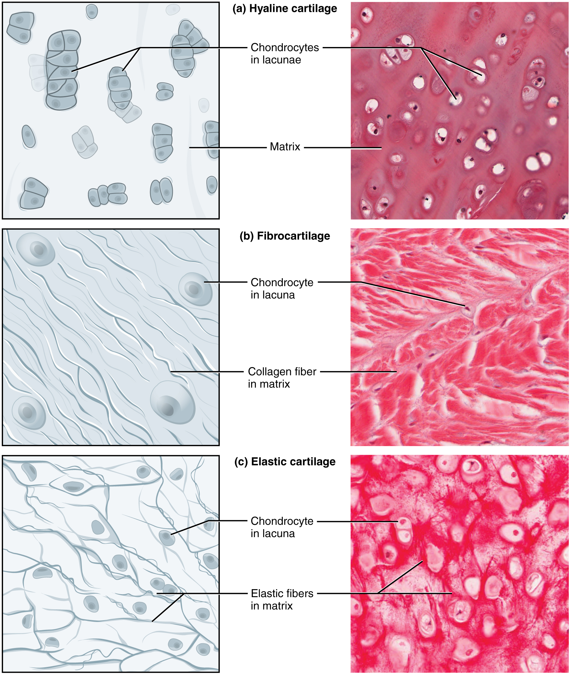
The combination of proteoglycans and collagen form cartilage, a sturdy tissue that is usually heavily hydrated (mostly due to the negatively charged sulfates in the glycosaminoglycan chains of the proteoglycans).
What is the difference between glycoproteins and proteoglycans?
Distinction between proteoglycans and glycoproteins. A glycoprotein is a compound containing carbohydrate (or glycan) covalently linked to protein. The carbohydrate may be in the form of a monosaccharide, disaccharide (s). oligosaccharide (s), polysaccharide (s), or their derivatives (e.g. sulfo- or phospho-substituted).
What is the function of proteoglycans?
Function. Proteoglycans are a major component of the animal extracellular matrix, the "filler" substance existing between cells in an organism. Here they form large complexes, both to other proteoglycans, to hyaluronan, and to fibrous matrix proteins, such as collagen.
What are the chains of carbohydrate?
The chains are long, linear carbohydrate polymers that are negatively charged under physiological conditions due to the occurrence of sulfate and uronic acid groups. Proteoglycans occur in connective tissue .
What is the GPC3 gene?
One exception is glypican ( Gpc) 3, a gene encoding a heparan sulfate proteoglycan linked to the cell surface via a glycosyl-phosphatidylinositol anchor. Gpc3 knockout mice display enhanced ureteric branching and dysplastic kidneys. 220Gpc3 is expressed in the ureteric bud cells and modulates BMP and FGF action on the ureteric bud. 220 A potent angiogenesis inhibitor, endostatin, a breakdown product of extracellular proteoglycan, collagen XVIII, inhibits ureteric bud branching morphogenesis. 35 Interestingly, this action is likely to be mediated through its binding to glypicans. 35
What are the components of extracellular matrix?
Proteoglycans are important components of extracellular matrices (ECM) and have multiple functions that depend on both their protein and carbohydrate constituents. They form a special class of glycoproteins with attached long unbranched and highly charged glycosaminoglycan chains [1, 2 ]. These chains are strongly hydrophilic and dominate the physical properties of proteoglycans, but the proteins to which they are attached are quite diverse in structure and form several distinct protein families. Proteoglycans are most abundant in those tissues where the ECM is highly hydrated. Cartilage and bone are tissues that both contain large expanded ECM, although the composition of the two tissues are strikingly different. Cartilage contains 70–75% (w/w) water and it has a high fibrillar collagen content (∼20% w/w) and also a high proteoglycan content (5–7% w/w). In contrast, bone matrix is mainly mineral, reinforced with fibrillar collagens (∼5% w/w), it contains only 10% (w/w) water and it has a correspondingly low proteoglycan content (∼0.1% w/w). This contrast in composition is also reflected in the proteoglycans present in the tissue. In cartilage the major proteoglycan is aggrecan, which forms supramolecular aggregates and is important in expanding and hydrating the matrix. It contains mainly chondroitin sulfate and increasing amounts of keratan sulfate with age. There also lesser amounts of the lower-molecular-weight leucine-rich proteoglycans, including decorin, fibromodulin, and lumican, which are collagen fibril-associated, and also biglycan. These also contain chondroitin sulfate, but with varying degrees of epimerization to dermatan sulfate. In bone there is no aggrecan and the collagen fibril associated proteoglycans and biglycan are the predominant forms. The cells of bone and cartilage also contain some cell-surface integral membrane proteoglycans, which predominantly contain heparan sulfate. These are very important in cell–matrix interactions and cell signaling, and control many aspects of chondrocyte and osteoblast function, but as the cell density is low they do not contribute to a large fraction of the tissue content of proteoglycans.
What are the proteins in the ECM?
A proteoglycan consists of a core protein having a variable number of GAG side chains [21] ( Table 14.1). The ECM proteoglycan superfamily includes the small leucine-rich repeat (LRR) family, the lectican family, the collagen family, and others. Members of these families present in the brain ECM include phosphacan, agrin, versican (also known as PG-M), neurocan, and brain-enriched hyaluronan-binding (BEHAB [22]; also known as brevican), and the vascular basement membrane proteoglycan perlecan. In brain tumors, changes in the expression of several ECM proteoglycans are observed within the tumor-brain adjacent ECM and the tumor vasculature.
What are proteoglycans?
Proteoglycans also are commonly referred to in the older literature as connective tissue mucins or mucopolysaccharides. These molecules are large glycoconjugate complexes that are found in high concentrations within the extracellular matrix of connective tissues. Proteoglycans are highly glycosylated and, in many cases, 90–95% of the molecular weight of the typical proteoglycan is due to the carbohydrate components.
Which sarcomas express glycosaminoglycans?
Glycosaminoglycans and proteoglycans are expressed by a number of different sarcomas. Hyaluronic acid and chondroitin sulfates in particular may be found in high concentrations in myxoid chondrosarcomas as well as the myxoid variants of liposarcoma and malignant fibrous histiocytoma ( Tighe 1963; Kindblom & Angervall 1975; Weiss & Goldblum 2001 ). In addition, proteoglycans may be observed in the stromal components of sarcomas as well as some carcinomas.
Where are proteoglycans found?
Proteoglycans are mostly found at the cell surface or in the extracellular matrix. In embryonic kidneys, sulfated proteoglycans are concentrated around the tip of the ureteric bud, and perturbation of their synthesis by β-D-xyloside results in the inhibition of ureteric bud branching morphogenesis. 217,218 This perturbation also abolishes the expression of Wnt11 at the tip of the ureteric bud. 219 As described previously, loss of Wnt11 expression at the tip of the ureteric bud can lead to loss of Gdnf expression in the metanephric mesenchyme. However, the linking mechanism between inhibition of sulfated proteoglycan and the loss of Wnt11 expression is unclear. Along the same lines, genetic inactivation of an enzyme, heparan sulfate 2-O-sulfotransferase, involved in heparan sulfate proteoglycan synthesis results in kidney agenesis. 53 In this case, the ureteric bud forms from the Wolffian duct, but subsequent invasion of the metanephric mesenchyme is perturbed. As for the mesenchyme, initial specification of the metanephric mesenchyme appears intact, but subsequent mesenchymal condensation is affected. Consistent with the in vitro result described above, heparan sulfate biosynthesis perturbation results in loss of Wnt11 expression and reduced Gdnf expression. Given the fact that many growth factors that regulate branching morphogenesis of the ureteric bud are heparin (a heavily sulfated form of GAG chain)-binding, it is likely that global inhibition of GAG chain synthesis or its sulfation could compromise actions of these heparin-binding growth factors.
Which amino acids are covalently bound to the protein core of the proteoglycan?
The glycosaminoglycan chains are covalently bound to a protein core of the proteoglycan via the side chain of the amino acids serine or threonine ( O -glycosidic linkage) and to a lesser extent to asparagine ( N -glycosidic linkage). The number of glycosaminoglycan chains varies greatly among different proteoglycans. While some proteoglycans may contain only one or two glycosaminoglycans per protein core, other proteoglycans may contain as many as 100 glycosaminoglycans ( Varki et al 1999 ). An exception to this structural motif is hyaluronic acid which does not contain a covalently bound protein core ( Mason et al 1982 ).
What is the role of polyanionic sGAG in cartilage?
The polyanionic sGAG moieties of aggrecan molecules, when restrained by the COL network, provide cartilage with a high osmotic pressure, a high tendency to retain water, and a high resistance to fluid flow.
What is the nidogen in the basement membrane?
The proteoglycan nidogen (also known as entactin) is also a ubiquitous component of basement membranes with a molecular weight of ~150 kD ( Yurchenco, 2011 ). There are two nidogen family members, Nidogen 1 and Nidogen 2. Expression of mRNA encoding nidogens is predominantly associated with cells of mesenchymal origin whereas nidogen proteins are found within basement membranes ( Breitkreutz et al., 2013 ). Therefore nidogens provide another example of the mesenchymal contribution to basement membranes that form adjacent to epithelial cells. Expression patterns and protein localization patterns of nidogen 1 and 2 are overlapping ( Breitkreutz et al., 2013 ). Transgenic mice that lack either nidogen 1 or lack nidogen 2 expression displayed mild phenotypes with no apparent alterations in basement membranes, presumably due to compensation from continued expression of the alternate nidogen. However, mice with abrogated expression of both nidogen 1 and 2 display perinatal lethality with defects in lung and heart basement membranes ( Bose et al., 2006 ). Interestingly, other basement membranes in the double nidogen null mice exhibited apparently normal morphology including the dermal–epidermal junction of the skin. Thus, different basement membranes in the body appear to have different dependencies upon the presence of nidogen for their integrity.
What is the PG content of cartilage?
The PG content in cartilage, together with the COL network, is the major determinant of the tissue's compressive properties. 11,12 PG aggregates are large molecules formed as aggrecan monomers noncovalently associated with hyaluronan and stabilized by link protein. Each aggrecan monomer has a large number of sulfated glycosaminoglycan (sGAG) chains of chondroitin sulfate and keratan sulfate. The polyanionic sGAG moieties of aggrecan molecules, when restrained by the COL network, provide cartilage with a high osmotic pressure, a high tendency to retain water, and a high resistance to fluid flow. The swelling pressure exerted by the PG contributes to a high resistance to compression and the tensile stress of COL network. 13
What is phosphacan KS?
Phosphacan is a chrondroitin sulfate or keratin sulfate (phosphacan-KS) proteoglycan of nervous tissue, grouped in the “Other” category ( Table 14.1 ). It is the extracellular domain of the receptor-type tyrosine phosphatase adhesion molecule ( Table 14.2 ). Like neurocan, it is present in normal brain and oligodendrogliomas; but expression is lost in astrocytic tumors [ 28 ]. It has a high affinity for fibroblast growth factor-2 (FGF2) [ 21 ], and like phosphacan, can bind NCAM and NgCAM, suggesting influences on growth and adhesion, respectively. In addition, it can bind to TN-C, and inhibits adhesion of C6 glioma cells to this matricellular protein [ 29 ].
What is the function of nidogens?
In studies by Behrens et al. (2012) linking of the laminin and collagen IV networks was dependent upon the heparan sulfate proteoglycan perlecan whereas nidogens were found to associate within both laminin and collagen IV networks but not as a cross-bridge between the two. Nidogens also function to sequester and distribute growth factors in the basement membrane. For example, the growth factor FGF-8 demonstrated altered localization in nidogen double null animals and this was associated with syndactyly and aberrant limb development in these mice ( Bose et al., 2006 ).
What are the members of the ECM family?
Members of these families present in the brain ECM include phosphacan, agrin, versican (also known as PG-M), neurocan, and brain-enriched hyaluronan-binding (BEHAB [ 22 ]; also known as brevican), and the vascular basement membrane proteoglycan perlecan.
What is the proteoglycan superfamily?
The ECM proteoglycan superfamily includes the small leucine-rich repeat (LRR) family, the lectican family, the collagen family, and others. Members of these families present in the brain ECM include phosphacan, agrin, versican (also known as PG-M), neurocan, and brain-enriched hyaluronan-binding (BEHAB [ 22 ]; also known as brevican), and the vascular basement membrane proteoglycan perlecan. In brain tumors, changes in the expression of several ECM proteoglycans are observed within the tumor-brain adjacent ECM and the tumor vasculature.
