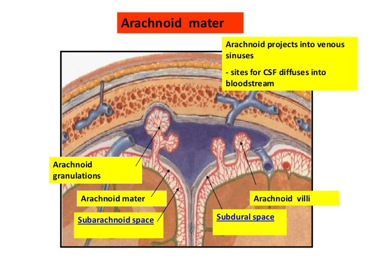
What is the function of the arachnoid?
Recommended article: "Parts of the human brain (and their functions)" The arachnoid, along with the dura and pia mater, is one of the three meninges. These are membranes that protect our brain and spinal cord from injuries from outside and that play an important role in our survival.
What is the arachnoid barrier layer?
Arachnoid or arachnoid barrier layer It corresponds to the part of the arachnoid that is in contact with the dura mater. Its cells are closely knit and barely allow the passage of interstitial fluid, being the most resistant part of the arachnoid.
Where is the arachnoid layer attached to the meninges?
It is interposed between the two other meninges, the more superficial and much thicker dura mater and the deeper pia mater, from which it is separated by the subarachnoid space. The delicate arachnoid layer is attached to the inside of the dura and surrounds the brain and spinal cord.
What is the ventral layer of the arachnoid membrane?
The ventral layer of arachnoid membrane, on the other hand, is a direct anterior extension of this arachnoid envelope that the dorsal layer forms over the pineal region. CSF circulates in the subarachnoid space (between arachnoid and pia mater).

What does the arachnoid layer do?
Three layers of membranes known as meninges protect the brain and spinal cord. The delicate inner layer is the pia mater. The middle layer is the arachnoid, a web-like structure filled with fluid that cushions the brain.
What is the function of the arachnoid mater quizlet?
*Acts as a barrier and aids in production of CSF. *The term arachnoid refers to blood vessels spider web-like appearance.
Where is the arachnoid layer?
Your arachnoid mater, the middle layer of your meninges, lies directly below your dura mater. It's a thin layer that lays between your dura mater and pia mater. It doesn't contain blood vessels or nerves.
How does the arachnoid protect the brain?
Additionally, all cerebral arteries and veins are located in this space. The outer surface of the arachnoid attaches to the dura mater forming a barrier that prevents the leakage of CSF into the subdural space.
What is arachnoid mater quizlet?
arachnoid mater. a thin layer of loose connective tissue attached to the inner surface of the dura mater. Main function of the arachnoid mater. - to circlate CSF. Subarachnoid space.
What are the characteristics of arachnoid mater?
The arachnoid mater, named for its spiderweb-like appearance, is a thin, transparent membrane surrounding the spinal cord like a loosely fitting sac. Continuous with the cerebral arachnoid above, it passes through the foramen magnum and descends caudally to the S2 vertebral level.
What is the arachnoid space?
Anatomically, the subarachnoid space exists between the arachnoid mater externally and pia mater internally. A network of fine delicate connective tissue called trabeculae connects these two layers and gives this space its characteristic spider web appearance.
What keeps the brain in place?
The meninges help to anchor the CNS in place to keep, for example, the brain from moving around within the skull. They also contain cerebrospinal fluid (CSF), which acts as a cushion for the brain and provides a solution in which the brain is suspended, allowing it to preserve its shape.
What are the layers that make up the meninges protective layer of the brain list down what can be seen in the in those parts?
Meninges LayersDura Mater. This outer layer connects the meninges to the skull and vertebral column. ... Arachnoid Mater. This middle layer of the meninges connects the dura mater and pia mater. ... Pia Mater. This thin inner layer of the meninges is in direct contact with and closely covers the cerebral cortex and spinal cord.
What is the arachnoid mater?
a·rach·noid mat·er. [TA] a delicate fibrous membrane forming the middle of the three coverings of the central nervous system. In life, the arachnoid (specifically the arachnoid barrier cell layer) is tenuously attached to the externally adjacent dura mater (specifically the dural border cell layer), and no natural space occurs at ...
What is the name of the filaments that extend from the deep surface of the arachnoid mater?
The arachnoid mater is named for the delicate, spiderweblike filaments that extend from its deep surface, through the cerebrospinal fluid of the subarachnoid space, to the pia mater. See: cranial arachnoid mater, spinal arachnoid mater. See also: leptomeninx.
Where is the weblike membrane of the brain located?
The increasingly preferred term for the weblike membrane covering the brain that lies between the outer (and much thicker) dura mater and the deeper pia mater, from which it is separated by the subarachnoid space through which CSF flows and is absorbed by the arachnoid granulations.
Which membrane encases the brain and spinal cord?
One of three membranes that encase the brain and spinal cord. The arachnoid mater is the middle membrane.
Is the dura mater attached to the arachnoid?
In life the arachnoid (specifically the arachnoid barrier cell layer) is tenuously attached to the externally adjacent dura mater (specifically the dural border cells) and there is no naturally occurring space at the dura-arachnoid interface. Thus, in a spinal puncture, dura mater and arachnoid are penetrated simultaneously as if a single layer.
Can arachnoid cysts be left untreated?
A. An arachnoid cyst that leads to symptoms usually needs treatment. Mild symptoms as you suggested are ok to left untreated however gradual onset of new symptoms may arise such as seizures, paralysis and other complications, therefore once symptoms occur one should consider treatment.
Can a spinal puncture cause arachnoid and dura mater to separate?
Thus, in a spinal puncture, dura mater and arachnoid are penetrated simultaneously as if a single layer. Separation of the arachnoid mater from the dura mater (usually through the dural border cell layer) may result from traumatic or pathologic processes creating what is commonly, but quite incorrectly, called a subdural hematoma.
What is the function of the arachnoid?
Despite being relatively fragile, the arachnoid together with the rest of the meninges allow the brain and spinal cord to be protected against blows and injuries, as well as contamination and infection by harmful agents.
What is the function of the arachnoid barrier layer?
The cells of the arachnoid barrier layer project towards the pia mater, forming a network that crosses the subarachnoid space which in turn forms a network or mesh that actually gives the meninge its name (due to its resemblance to a spider's web). Within these projections we find net fibers, anchor fibers and microfibers. The exact function of the trabeculae is not yet fully known, although it is speculated that they are capable of perceiving the pressure caused by cerebrospinal fluid.
How is the arachnoid separated from the dura?
Although they are in close contact, the arachnoid is separated from the dura by means of the subdural space, which is more than a space, a thin layer of cells between which is interstitial fluid. With respect to the pia mater, it is separated from it by the subarachnoid space, and in turn connects with it by means of the arachnoid trabeculae.
What is the subarachnoid space?
Although more than part of the arachnoid is a space located between its laminae, the subarachnoid space is one of the most important parts of the arachnoid. This is so because it is through it that the cerebrospinal fluid passes. In this space we can also find a series of important cerebral pits and cisterns in which cerebrospinal fluid accumulates and which allow its distribution.
What causes a swollen subarachnoid space?
Occurs when due to illness or injury (such as a head injury), blood enters and floods the subarachnoid space. It can be deadly. Headache, altered consciousness, and gastrointestinal problems such as nausea and vomiting are common.
What protects the central nervous system?
The meninges are a series of membranes that together with the skull and spinal column protect the central nervous system, so that minor blows or injuries can alter its operation or destroy it completely.
Where is the orbital subarachnoid space?
In addition to the brain itself, an orbital subarachnoid space can be found that surrounds the optic nerve.
What is the specialized function of arachnoid projections?
Specialized projections of arachnoid, termed pacchionian granulations, are projections of the arachnoid into the dura with the specialized function of transmitting CSF. The granulations are composed of clusters of arachnoid villi.
What is the arachnoid mater?
The arachnoid (Gk. spider) is a delicate fibrocellular layer beneath the dura (separated by potential subdural space) that is connected to the pia mater covering the brain by numerous fibrocellular bands that cross the cerebrospinal fluid-filled subarachnoid space.
How do arachnoid villi work?
One theory suggests the arachnoid villi work with their relatively freely permeable membranes as a one-way valve system utilizing hydrostatic pressure differences between the CSF and venous system as the operating principle. Alternatively, electron microscopy studies in animals suggest free access between the subarachnoid space and dural sinuses on the basis of endothelial tubules within the arachnoid granulations. It is clear that fluid injected into the subarachnoid space passes into the granulations and villi and then into the dural venous sinuses.
What are the layers of the arachnoid villi?
Human arachnoid villi are composed of five layers: endothelial layer, fibrous capsule, arachnoid cell layer, cap cells, and central core. The outermost layer, an endothelial lining has a pivotal role in the absorption process of CSF, and displays a number of micropinocytotic vesicles, intracytoplasmic vacuoles, and villous projections.
What is the relationship between the arachnoid and the pons?
At the level of the pons, it is thicker, more opaque, and widely separated from the pia. The arachnoid unites with the pia at the level of the pituitary fossa. The pia and the arachnoid communicate with each other via fine connective tissue cellular septa that cross the subarachnoid space.
What is the arachnoid that protrudes and bulges into the resection cavity?
The arachnoid that protrudes and bulges into the resection cavity after adenoma removal is often covered by adenoma or compressed gland tissue and should be distinguished from septations within the adenoma. Some pituitary surgeons recommend perforating this layer for differentiation; this procedure is not necessary as iMRI will identify and differentiate the two structures reliably. The arachnoid bulging into the resections cavity usually covers more lateral or posterior portions of residual adenoma that may be difficult to detect either by microscope or endoscope. The iMRI visualizes reliably these hidden adenoma residuals, which then can be removed in a targeted manner (Figures 8-11 and 8-12 ).
How to remove a tumor from the arachnoid?
The meatal part of the tumor is gently retracted backward. Small hooks or fine microscissors are used to free the facial nerve from the arachnoid fibers that bind the nerve to the tumor. Because cutting of the arachnoid occurs along the facial nerve, the surgeon must have identified the inferior and the superior edges of the nerve accurately. Usually, it is relatively easy to develop the dissection plane between the facial nerve and the tumor in the internal auditory canal, but difficulties often arise at the porus. Around the entire circumference of the porus, dural adhesions to the tumor make dissection of the facial nerve from the tumor difficult. The exact position of the facial nerve in the porus must be established before the adhesions between the dura and the tumor are removed. Inferiorly, freeing of the tumor from the porus is simpler because damage to the cochlear nerve is insignificant. Superiorly, the facial nerve may be at risk. At times, it is difficult to isolate the facial nerve at the porus. In these cases, it is wise to carry out a partial tumor removal, identify the facial nerve medially, and follow the nerve laterally until the porus is reached. During this work, the surgeon must be careful not to push the tumor forward or medially, because stretching the facial nerve, especially at the porus level, can damage the nerve. Early mobilization of the tumor from the internal auditory canal has the advantage that the landmarks are well defined and are not obscured with blood, as tends to happen later in the surgical dissection.
